Introduction
External auditory canal cholesteatoma is a rare disease entity with an uncertain aetiology. Few case series have been reported in the literature, and current management is highly variable between centres. The locally erosive and indolent nature of cholesteatoma in the bony external ear canal gives rise to complications resulting from erosion into adjacent structures, such as the mastoid and the temporomandibular joint.
This study aimed to describe the clinical features of a series of patients treated for cholesteatoma of the external ear canal. We report our experience of operative management in these patients, who underwent bony meatoplasty.
Materials and methods
The majority of patients in the series reviewed were referred to otolaryngology by their general practitioner with a history of unilateral otalgia and otorrhoea, for which they had received an (often unsuccessful) trial of conservative antimicrobial medical therapy prior to referral. Otoscopy most frequently showed erosion of the inferior aspect of the bony canal wall, with accumulated keratin and bony sequestrum. A clinical diagnosis of external auditory canal cholesteatoma was made on the basis of these findings.
Each patient underwent pre-operative investigation with high-resolution computed tomography (CT) of the temporal bones, audiometric assessment, and examination under anaesthesia with biopsy of the ear canal lesion where necessary. Axial CT with coronal reconstruction was used to assess the extent of local involvement. These imaging studies confirmed the presence of bony canal wall erosion, with or without overlying soft-tissue attenuation material. Examination of the external auditory canal lesion under anaesthesia, and biopsy where appropriate, was used to exclude malignant (necrotising) otitis externa and squamous cell carcinoma of the external auditory canal skin. Histological analysis of biopsy specimens showed keratin, with no evidence of squamous malignancy.
In each case, the disease was at a relatively advanced stage, with erosion of the temporal bone. All cases were managed surgically in order to eradicate the disease process, preserve normal structure and function, and provide patients with a self-cleaning ear canal. Bony meatoplasty, carried out via a postauricular approach, was performed under general anaesthesia in a main operating theatre. The postauricular and ear canal skin was first infiltrated with 4 ml of 2 per cent lidocaine hydrochloride with adrenaline (1:80 000). The ear canal re-entry incision was made just lateral to the cholesteatoma sac. The cholesteatoma sac was elevated and the cholesteatoma was excised. In order to provide a smooth ear canal contour, the bony irregularities and the edge of the bony erosion were drilled out. The resulting cavity was obliterated with tragal cartilage and perichondrium grafts, with or without temporalis fascia, to achieve a smooth, self-cleaning ear canal.
The postauricular wound was closed using layers of subcutaneous vicryl with subcuticular Monocryl sutures (Ethicon, Somerville, New Jersey, USA). The ear canal was then packed with two small pieces of ribbon gauze (approximately 2 cm in length) impregnated with bismuth iodoform paraffin paste (BIPP). A simple dry gauze dressing was applied to the ear. Patients were reviewed and, if well, discharged home on day one following surgery.
Follow up in the out-patient otolaryngology clinic was arranged two weeks post-surgery for removal of the BIPP packing. At this stage, patients were given a one-week course of Sofradex ear drops (Sanofi-Aventis, Guildford, UK). Patients were reviewed again at six weeks post-surgery. Thereafter, patients were kept under regular follow up (usually with six-monthly intervals). The post-operative follow-up period for this case series ranged from 6 to 36 months (median 20.5 months).
Results
Eight cases of external auditory canal cholesteatoma were identified within the study period (Table I). The median age of patients at presentation was 46.5 years (range 14–68 years), and the male to female ratio was 1:1. The most common presenting features were non-specific unilateral otalgia and purulent otorrhoea. All patients had relatively advanced disease at presentation, with erosion of the temporal bone (Table II). The patients underwent bony meatoplasty via a postauricular approach in order to eradicate the disease process. One patient developed a second lesion post-operatively. The median follow-up period was 16 months. Three selected cases are described in detail below, with exemplar CT images and intra-operative photographs.
Summary of results

Pt = patient; y = years; M = male; F = female; GP = general practitioner
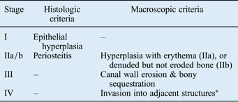
External auditory canal cholesteatoma staging criteria

*Subclasses of disease stage IV include: mastoid (M), skull base and sigmoid sinus (S), temporomandibular joint (J), and facial nerve canal (F).
Case one
A 68-year-old man was referred by his general practitioner with a two-year history of recurrent right-sided otitis externa. He was a smoker, with a background of chronic obstructive pulmonary disease, hypertension, and ischaemic heart disease with previous coronary artery bypass grafting. The otitis externa had been managed intermittently with topical antibiotics prescribed by his general practitioner. On attending the senior house officers' emergency otolaryngology clinic, the patient described right-sided otalgia and a sense of aural fullness, with associated purulent otorrhoea. Medical management with topical and systemic antibiotics, in combination with aural toilet, was unsuccessful. He was therefore referred for a senior specialist opinion.
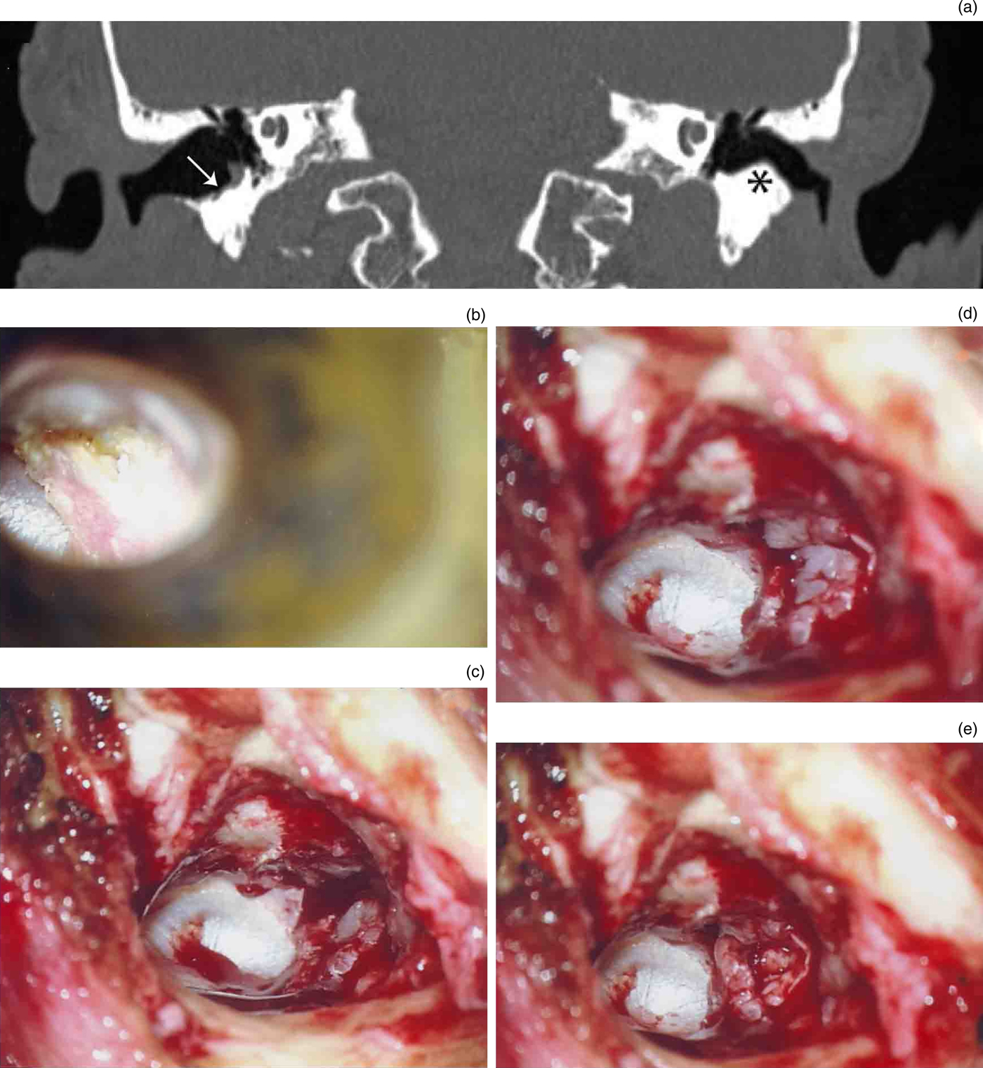
Otoscopy revealed two areas of erosion in the inferior aspect of the ear canal, which contained bony sequestra. Computed tomography confirmed the presence of abnormal soft tissue and erosion of the external auditory canal floor (Figure 1a). The appearance of the middle-ear cleft on CT scans was normal. Audiometric assessment showed moderate to severe high-frequency sensorineural hearing loss bilaterally. Examination under general anaesthesia revealed a 3 mm defect filled with cholesteatoma, with no involvement of the middle ear. There were no suspicious areas; hence biopsy was not carried out.
Case one: (a) coronal computed tomography scan, showing soft-tissue attenuation material overlying bony erosion in the right external auditory canal floor (arrow) and normal bony appearances on the left (asterisk); (b) otoscopic appearance of ear canal cholesteatoma; and intra-operative photographs, showing (c) cholesteatoma excised and bony irregularities drilled out, (d) cartilage graft in situ, and (e) perichondrium in situ.

Bony meatoplasty was performed through a postauricular approach, with an ear canal skin incision just lateral to the cholesteatoma pocket. Henle's spine was drilled out, the cholesteatoma pocket was elevated, and the bony irregularities and ridge were drilled to smooth the cavity. The cholesteatoma cavity was obliterated with a tragal cartilage and perichondrium graft (Figure 1b–e). The canal floor was well healed at the six-week follow up. He remained well six months later.
Case two
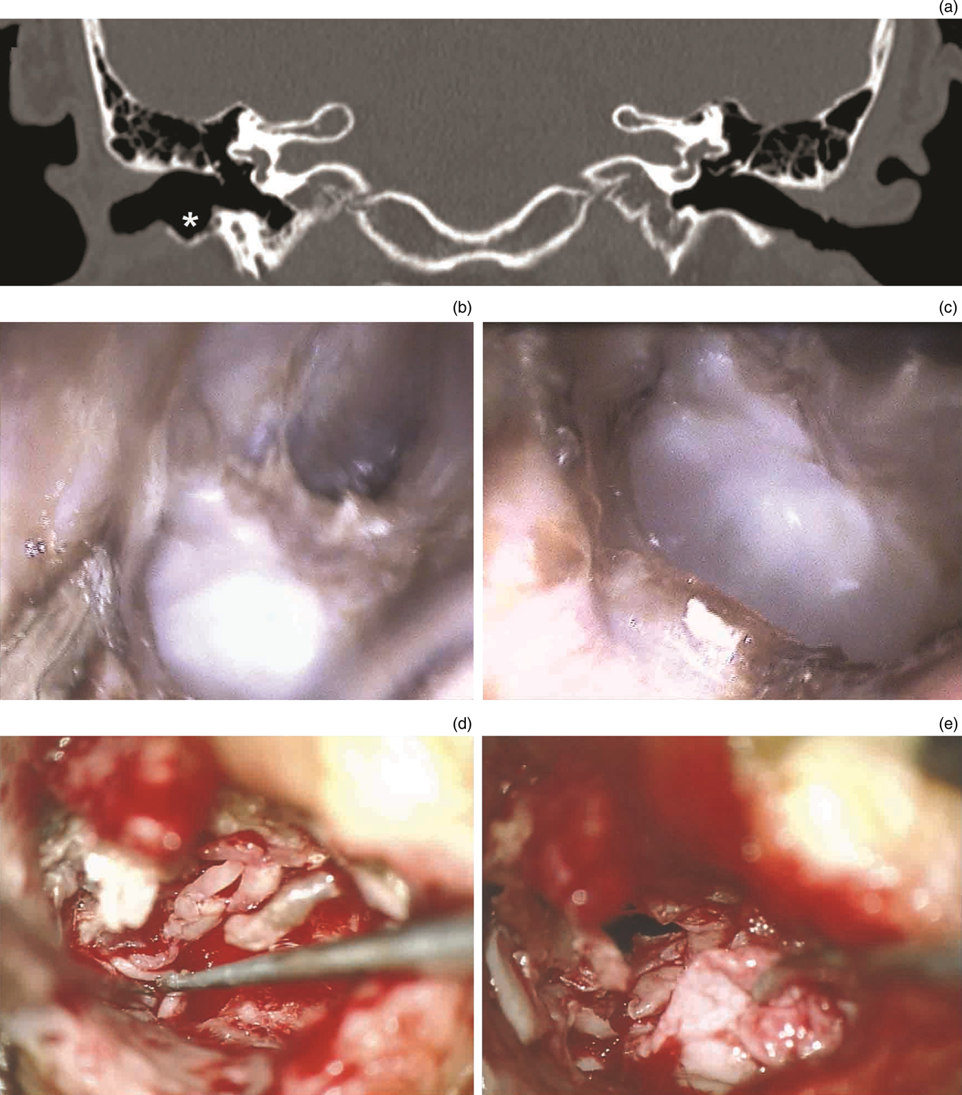
A 14-year-old boy was referred by his general practitioner with an 8-week history of right-sided otalgia and purulent otorrhoea, which was unresponsive to topical and systemic antimicrobial therapy. Initial examination in the senior house officers' emergency otolaryngology clinic showed a large aural granulation tissue polyp. After aural microsuction, the patient was given a course of Gentisone HC ear drops (Amdipharm, Basildon, UK) and followed up one week later. At his next clinic appointment, the polyp had receded and was cauterised with silver nitrate. One week later, he was found to have an underlying keratin-filled erosion of the inferior wall of the right ear canal (Figure 2b, c).
Case two: (a) coronal computed tomography scan, showing bony erosion of the right external auditory canal floor (asterisk); (b) and (c) otoscopic appearances of the cholesteatoma eroding the external auditory canal wall; and intra-operative photographs showing (d) the middle ear clear of disease, with the chorda tympani intact, and (e) cartilage palisade grafts in situ.

Computed tomography revealed bony erosion of the inferior wall of the right external ear canal, with normal appearances of the middle ear (Figure 2a). Audiometric assessment showed normal hearing bilaterally. The patient subsequently underwent bony meatoplasty. At surgery, he was found to have two cholesteatoma pockets adjacent to the tympanomastoid suture line, with no extension into the middle ear. The cholesteatoma was removed and the defects were filled with cartilage palisade grafts (Figure 2d, e).
Case three
A 46-year-old woman presented with a 6-month history of intermittent right-sided otalgia and otorrhoea. She had been treated with oral amoxicillin for otitis externa on two occasions over the preceding four months, as prescribed by her general practitioner. The patient had a background of treated hypertension. In addition, she had well controlled type II diabetes mellitus and was on oral hypoglycaemic medication; her most recent HbA1c had been reported as being 7.9 per cent. She was a non-smoker.
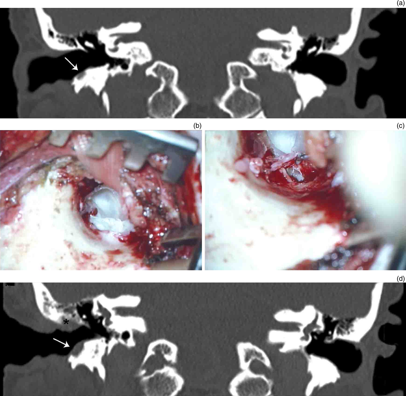
Initial assessment revealed erosion of the floor of the right external auditory canal, with an accumulation of keratin debris. Following aural toilet, tri-adcortyl ointment was applied to the erosion. Audiometric assessment showed a high-frequency sensorineural hearing loss bilaterally. Computed tomography confirmed the superficial erosion of the floor of the right ear canal, which involved the temporal bone inferiorly and posteriorly (Figure 3a).
Case three: (a) coronal computed tomography (CT) scan, showing soft-tissue attenuation mass in the right ear canal, with underlying erosion of the adjacent temporal bone (arrow); intra-operative photographs demonstrating (b) abnormal bony prominence and cholesteatoma in ear canal, and (c) a clear middle ear; and (d) coronal CT scan, showing erosion of the adjacent temporal bone anteroinferiorly (arrow) and bony erosion adjacent to the scutum superiorly (asterisk).

Biopsy of the ear canal lesion was carried out under general anaesthesia to rule out malignant (necrotising) otitis externa and squamous cell carcinoma of the external auditory canal skin. Histological analysis of the tissue specimen showed no evidence of malignancy, and was consistent with a diagnosis of external auditory canal cholesteatoma. The patient subsequently underwent right-sided bony meatoplasty and tympanotomy to exclude any middle-ear disease. At surgery, the middle ear was found to be free from cholesteatoma (Figure 3b, c). Bony meatoplasty was carried out with tragal cartilage and a perichondrium graft to the external auditory canal wall.
At follow up, the external auditory canal was initially well healed. However, one year following surgery the patient was referred back to the otolaryngology clinic by her general practitioner with increasing right-sided otalgia and otorrhoea. She was diagnosed with eczematous otitis externa. After a further six months, examination by the senior author revealed a small area of cavitation in the external auditory canal posterosuperiorly, which contained bony sequestrum. This was in a different location to her previous disease. Computed tomography was repeated. This showed thickening of the ear canal soft tissues, with bony destruction adjacent to the scutum superiorly. Anteriorly, there was involvement of temporal bone at the level of the temporomandibular joint (Figure 3d).
The patient was treated initially with oral ciprofloxacin and topical ofloxacin ear drops. Two months later, the ear canal lesion was found to be healing well. She was still under regular follow up at the time of writing.
Discussion
Aetiology
Cholesteatoma is most commonly found in the middle-ear cavity. Rarely, cholesteatoma occurs in the external auditory canal, with an estimated incidence rate of 1.2–3.7 per 1000 new otology patients.Reference Anthony and Anthony1, Reference Owen, Rosborg and Gaihede2 The annual incidence rate of external auditory canal cholesteatoma in the general population is 0.15 cases per 100 000 individuals,Reference Owen, Rosborg and Gaihede2 compared with 9.2 cases per 100 000 individuals per year for middle-ear cholesteatoma.Reference Kemppainen, Puhakka, Laippala, Sipila, Manninen and Karma3
The aetiology of external auditory canal cholesteatoma remains unclear.Reference Dubach and Hausler4, Reference Dubach, Mantokoudis and Caversaccio5 Idiopathic external auditory canal cholesteatoma is distinguished from cholesteatoma that arises secondary to a previous ear canal insult. Many authorities believe that idiopathic external auditory canal cholesteatoma results from a reduced migratory capacity of the canal epithelium, which leads to ‘keratinisation in situ’.Reference Owen, Rosborg and Gaihede2, Reference Makino and Amatsu6–Reference Naim, Linthicum, Shen, Bran and Hormann8 However, this view has recently been challenged by a study which showed no difference in the rate of epithelial migration between normal ears and those affected by external auditory canal cholesteatoma.Reference Bonding and Ravn9 In some cases, the occurrence of external auditory canal cholesteatoma has been linked to branchial arch anomalies, which result in the retention of epithelial masses and lead to cholesteatoma formation in the external auditory canal floor.Reference Dubach and Hausler4, Reference Hickey, Scott and Traub10
Epidemiologically-linked risk factors for the development of external auditory canal cholesteatoma include smoking, diabetes mellitus and repeated microtrauma (e.g. from cotton buds or hearing aids).Reference Dubach and Hausler4, Reference Dubach, Mantokoudis and Caversaccio5 Smoking and diabetes are thought to result in microangiopathic changes in the ear canal that potentially impair epithelial migration.Reference Dubach, Mantokoudis and Caversaccio5 The majority of idiopathic external auditory canal cholesteatoma occur in the inferior canal wall. This is thought to be due to the relatively poor blood supply to the skin along the inferior aspect of the canal.
None of the cases presented here had any identifiable aetiological factor; at best, they can be regarded as idiopathic external auditory canal cholesteatoma (Table I). Four of the patients were smokers, one patient was diabetic, and one wore hearing aids; the use of hearing aids may have predisposed the individual to external auditory canal cholesteatoma through repeated microtrauma to the ear canal skin.
Presenting features
A recent meta-analysis of published case series concluded that the most common presenting symptoms of idiopathic external auditory canal cholesteatoma are unilateral otorrhoea with mild to moderate otalgia.Reference Dubach, Mantokoudis and Caversaccio5 A minority of patients complained of unilateral hearing loss; or were asymptomatic, with the cholesteatoma being discovered on routine otoscopy performed for a separate indication. These findings are consistent with our experience. Six of the eight patients presented with unilateral otalgia, five of which presented with associated otorrhoea. One patient had painless otorrhoea; one patient mentioned progressive unilateral hearing loss; and one was asymptomatic, with cholesteatoma being discovered incidentally on otoscopy.
Staging and complications
The indolent and locally erosive nature of external auditory canal cholesteatoma can result in late presentation, with severe complications due to the involvement of neighbouring structures. If the cholesteatoma invades into the mastoid, it may erode the facial nerve canal, sigmoid sinus and semicircular canals.Reference Owen, Rosborg and Gaihede2, Reference Heilbrun, Salzman, Glastonbury, Harnsberger, Kennedy and Shelton7, Reference Belcadhi, Chahed, Mani and Bouzouita11–Reference Zanini, Ameno, Magaldi and Lamar17 Erosion through the anterior wall of the canal may affect the temporomandibular joint.Reference Owen, Rosborg and Gaihede2, Reference Darr and Linstrom13, Reference Garin, Degols and Delos18 Rarely, in advanced cases, extension into the posterior fossa has been reported, with resulting intracranial abscess.Reference Martin, Selesnick and Parisier15
Naim et al. Reference Naim, Linthicum, Shen, Bran and Hormann8 developed a (I–IV) staging system for external auditory canal cholesteatoma based on the extent of erosion into nearby structures (Table II). Seven of the cases reported here presented with stage III disease, in which there was erosion of the adjacent temporal bone in the inferior aspect of the external auditory canal. In one patient, the disease involved the adjacent mastoid air cells and was therefore graded as stage IV (mastoid subclass).Reference Naim, Linthicum, Shen, Bran and Hormann8 The majority of cases reported in the literature are of at least stage IIb. Reports of stage I external auditory canal cholesteatoma are very rare.Reference Dubach, Mantokoudis and Caversaccio5, Reference Naim, Linthicum, Shen, Bran and Hormann8 This is probably because the earliest stages of the disease are usually asymptomatic. As the disease progresses to involve the bony canal walls, the accumulated keratin debris and bony sequestrum become a focus for infection, which results in the typical presentation with recurrent otalgia and otorrhoea.
Computed tomography of the temporal bones has become accepted as the gold standard for staging and pre-operative planning in external auditory canal cholesteatoma.Reference Shin, Shim and Lee19 This method allows accurate evaluation of the extent of local bone erosion and examination for the involvement of adjacent structures.
Treatment
As with middle-ear cholesteatoma, the aims of treatment in external auditory canal cholesteatoma cases are the eradication of the disease with preservation of normal structure and function, and the restoration of normal epithelial migration. Conservative therapy, with frequent debridement of the keratin debris and sequestered bone, is favoured in early stage disease.Reference Dubach, Mantokoudis and Caversaccio5, Reference Belcadhi, Chahed, Mani and Bouzouita11 If these simple measures are inadequate to control otalgia and otorrhoea, or in more advanced disease (stages IIb–IV), surgical intervention is necessary. The aim of surgery is to excise the cholesteatoma and to restore a smooth, self-cleaning canal wall epithelium, usually with the aid of cartilage and fascial grafts to protect denuded structures, and fill any canal wall defect. When the mastoid air cells are invaded, a modified radical mastoidectomy may be indicated, with the tympanic membrane and ossicles left intact.Reference Piepergerdes, Kramer and Behnke20
• External auditory canal cholesteatoma is rare, with potentially serious complications
• Computed tomography is the investigation of choice for disease staging and pre-operative planning
• Most patients present with non-specific symptoms of unilateral otalgia and otorrhoea
• Bony meatoplasty with cartilage graft repair is effective in the definitive management of this disease
All our patients underwent bony meatoplasty via a postauricular approach. One patient was initially reluctant to pursue operative management as her symptoms were mild. She was therefore initially managed with topical antibiotic preparations and regular debridement in the clinic. After three years of follow up, she underwent successful curative surgery. One patient suffered further external auditory canal cholesteatoma at a separate location in the same ear canal 18 months after bony meatoplasty. This was being managed conservatively at the time of writing.
Conclusion
External auditory canal cholesteatoma is a poorly understood and rare disease entity, which presents diagnostic and therapeutic challenges. Computed tomography is the most useful investigation for disease staging and pre-operative planning. Of the eight patients reviewed, seven underwent meatoplasty for stage III external auditory canal cholesteatoma and one for stage IV (mastoid subclass) disease. In the absence of surgical intervention, close clinical monitoring with regular follow up is required to treat symptoms and monitor patients for potentially serious complications.
Acknowledgement
We would like to thank Mr Patrick Axon (consultant ENT surgeon at Addenbrooke's Hospital, Cambridge, UK) for his assistance in the management of case seven.