Introduction
The maritime nation Indonesia can be considered as one of the centres of marine biodiversity (Roberts et al., Reference Roberts, McClean, Veron, Hawkins, Allen, McAllister, Mittermeier, Schueler, Spalding, Wells, Vynne and Werner2002). With over 80,000 km, it has the second largest coastline of any country worldwide. More than 130 million people live within 50 km of the coast. Marine resources, from fisheries, aqua- and mariculture, play a major role as protein resources, and generate substantial income and commercial profit for the nation (Rückert, Reference Rückert2006; MoMAF, 2009). After the collapse of shrimp cultures (tiger prawns, Panaeus monodon) between 1992 and 1998 (Harris, Reference Harris2001) the culture of coral reef fishes of the genera Epinephelus, Lates and Cromileptes were promoted in Indonesia. As potential mariculture species, groupers (e.g. Epinephelus coioides, E. fuscoguttatus, Lates calcarifer and Cromileptes altivelis) are the main target species in Indonesia (Rimmer et al., Reference Rimmer, McBride and Williams2004).
Finfish mariculture often underlies economic losses caused by diseases and parasites (Rückert et al., Reference Rückert, Klimpel, Mehlhorn and Palm2009b). Justine et al. (Reference Justine, Beveridge, Boxhall, Bray, Moravec, Trilles and Whittington2010) recorded 146 parasite species in 28 investigated grouper species (Serranidae, Epinephelinae) from New Caledonia. For example, 23% of the then known 254 fish parasitic trypanorhynch cestodes have been recorded from Indonesian waters (Palm, Reference Palm2004). Jakob & Palm (Reference Jakob and Palm2006) recorded 38 fish parasite species from mainly deepwater fishes from Pelabuhan Ratu, southern Java coast. Fish parasitological studies in Indonesia have been intensified in recent years (e.g. Jakob & Palm, Reference Jakob and Palm2006; Rückert et al., Reference Rückert, Palm and Klimpel2008, Reference Rückert, Hagen, Yuniar and Palm2009a, Reference Rückert, Klimpel, Mehlhorn and Palmb, Reference Rückert, Klimpel and Palm2010; Palm et al., Reference Palm, Damriyasa, Linda and Oka2008, Reference Palm, Kleinertz and Rückert2011; Kleinertz, Reference Kleinertz2010), resulting in faunistical treatments, revisions and several new species descriptions (e.g. Palm, Reference Palm2000, Reference Palm2008; Rückert et al., Reference Rückert, Palm and Klimpel2008, Reference Rückert, Hagen, Yuniar and Palm2009a, Reference Rückert, Klimpel, Mehlhorn and Palmb, Reference Rückert, Klimpel and Palm2010; Bray & Palm, Reference Bray and Palm2009; Kuchta et al., Reference Kuchta, Scholz, Vlčková, Říha, Walter and Palm2009).
Palm et al. (Reference Palm, Damriyasa, Linda and Oka2008) summarized the knowledge of the zoonotic nematode genus Anisakis from Indonesia, reporting 21 different and also commercially important fish species to be infested with this potentially harmful parasite. The authors studied the scombrid Auxis rochei rochei and the carangid Decapterus russelli for larval Anisakis spp., and detected three different genotypes of Anisakis typica. Recently, this common tropical nematode species has also been identified genetically from the South-west Atlantic (Brazil; Nadler et al., Reference Nadler, D'Amelio, Dailey, Paggi, Siu and Sakanari2005), North-west Atlantic (Florida; Mattiucci et al., Reference Mattiucci, Nascetti, Dailey, Webb, Barros, Cianchi and Bullini2005) and the Mediterranean (North Africa; Farjallah et al., Reference Farjallah, Ben Slimane, Busi, Paggi, Amor, Blel, Said and D'Amelio2008). So far, all authors recorded a high number of anisakids in the studied fish and discussed their zoonotic potential. However, due to high morphological similarities in ‘sibling species’, exact species identification was not possible at that time. According to a review on anisakid parasites in fish, Klimpel & Palm (Reference Klimpel, Palm and Mehlhorn2011) recognized eight different Anisakis sibling species that could be differentiated by using molecular methods. Theisen (Reference Theisen2009) also detected Anisakis sp. from Javanese fishes (coastal zone off Segara Anakan lagoon), isolating the worms from three different commercially important fish hosts from the coastal zone off Segara Anakan lagoon.
The mangrove-fringed Segara Anakan lagoon is located in South Central Java, separated from the Indian Ocean by the rocky mountainous island of Nusakambangan (Jennerjahn et al., Reference Jennerjahn, Nasir and Pohlenga2009). For many decades the lagoon has been facing a number of environmental problems due to resource exploitation. Most important are overfishing, logging and high sediment input by the Citanduy River because of poor upland agricultural practices, agricultural runoff, potential pesticide and oil pollution (White et al., Reference White, Marosubroto and Sadorra1989; Jennerjahn et al., Reference Jennerjahn, Nasir and Pohlenga2009). Recent investigations stated a high level of water and sediment contamination in the vicinity of the oil refinery of Cilacap, and a wide spectrum of contaminants in the tissues of aquatic organisms (Dsikowitzky et al., Reference Dsikowitzky, Nordhaus, Jennerjahn, Khrycheva, Sivatharshan, Yuwono and Schwarzbauer2011). Over 50 organic contaminants were found in water, sediment and macrobenthic invertebrates from the lagoon, mostly polycyclic aromatic compounds (PACs) (Dsikowitzky et al., Reference Dsikowitzky, Nordhaus, Jennerjahn, Khrycheva, Sivatharshan, Yuwono and Schwarzbauer2011). The hydrology of the mangrove-fringed Segara Anakan lagoon has a high water mass influx. The high freshwater input, mostly from Citanduy River (Holtermann et al., Reference Holtermann, Burchard and Jennerjahn2009), is additionally governed by the tides (Jennerjahn et al., Reference Jennerjahn, Nasir and Pohlenga2009), directly linking the lagoon with the coastal ecosystem. The lagoon plays an important ecological role as nursery ground for several fish species, as it supports a large and productive mangrove ecosystem (Romimohtarto et al., Reference Romimohtarto, Hutagalung, Razak, Chou, Chua, Khoo, Lim, Paw, Silvestre, Valencia, White and Wong1991).
Palm & Rückert (Reference Palm and Rückert2009), Palm (Reference Palm and Mehlhorn2011) and Palm et al. (Reference Palm, Kleinertz and Rückert2011) developed a method to use fish parasites as biological indicators serving as biomarkers for the environmental conditions inside Segara Anakan lagoon and inside a grouper mariculture farm in the Thousand Islands, and they also discussed the potential use of grouper parasites to assess environmental change in the wild. Epinephelus areolatus is one of the most abundant grouper species in Indonesian fish markets (personal observation) and is also cultivated in commercial fish aquaculture (Heemstra & Randall, Reference Heemstra and Randall1993; Froese & Pauly, Reference Froese and Pauly2010). Data on the parasite infection of this fish species, however, are scarce. The purpose of the present study is a first detailed analysis of the metazoan fish parasite fauna of E. areolatus from Indonesia, including new host and locality records. The acquired data are used to assess the environmental status at two different sampling sites, based on the observed regional differences in the parasite fauna of this commercially important fish.
Materials and methods
Collection and examination of fish
Samples were taken within the framework of the SPICE project (Science for the Protection of Indonesian Coastal Marine Ecosystems) during the dry season of 2009. A total of 60 E. areolatus were obtained fresh from local fishermen and studied from Javanese (off Segara Anakan lagoon) (108°46′–109°03′W; 08°35′–08°48′S) and Balinese waters (114°25′53″–115°42′40″W; 08°30′40″–08°50″48″S).
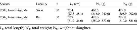
Fish were examined immediately after collection, or kept on ice and then frozen (c. − 20°C) until subsequently dissected in the laboratory. Total fish length (L T) and weight (W T), weight at slaughter (W S) and liver weight (W L,) were measured to the nearest 0.1 cm and 0.1 g (table 1) prior to the parasitological examination (see Rückert et al., Reference Rückert, Hagen, Yuniar and Palm2009a). Skin, fins, eyes, gills, mouth- and gill-cavity were studied for ectoparasites. Inner organs, such as digestive tract, liver, gall bladder, spleen, kidneys, gonads, heart and swim bladder, were separated and transferred into saline solution for microscopic examination; belly flaps and musculature were examined on a candling table. Isolated parasites were fixed in 4% borax-buffered formalin and preserved in 70% ethanol. Musculature was sliced into fillets 0.5–1 cm thick, and pressed between two Petri dishes to identify and isolate the parasites from the musculature. For identification purposes Nematoda were dehydrated in a graded ethanol series and transferred to 100% glycerine (Riemann, Reference Riemann, Higgins and Thiel1988). Anisakis spp. specimens were fixed and stored in 98% ethanol for subsequent molecular investigation. Digenea, Monogenea and Cestoda were stained with acetic carmine, dehydrated, cleared with eugenol and mounted in Canada balsam. Crustacea were dehydrated and transferred into Canada balsam. Parasite identification literature included original descriptions (for details see Palm et al., Reference Palm, Kleinertz and Rückert2011).
Sampling periods, number (n) of dissected specimens, mean length and mean weight (range in parantheses) of free-living Epinephelus areolatus, sampled in dry season (ds) 2009 from Balinese and Javanese waters.
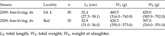
SA 4, coastline Segara Anakan lagoon.
L T, total length; W T, total weight; W S, weight at slaughter.
Parasitological parameters
Different ecological parameters were evaluated at both sampling sites, such as diversity indices like the Shannon–Wiener, Evenness and Simpson indices, fish ecological indices such as the Hepatosomatic index, and the parasitological parameters ecto-/endoparasite ratio and different prevalences of infection of metazoan parasites (see Palm & Rückert, Reference Palm and Rückert2009; Palm, Reference Palm and Mehlhorn2011; Palm et al., Reference Palm, Kleinertz and Rückert2011). Parasitological calculations were made according to Bush et al. (Reference Bush, Lafferty, Lotz and Shostak1997).
The present study applies the method of Palm & Rückert (Reference Palm and Rückert2009) and Palm et al. (Reference Palm, Kleinertz and Rückert2011) to monitor the parasite community of groupers in Indonesia. The Berger–Parker index (BP) characterizes the dominance of a respective parasite species within the sample: BP = N max/N, with N max being the number of specimens of the most dominant species in relation to the total number of collected parasites within the sample (N). The diversity of the metazoan endoparasite fauna of each fish species was determined by using the Shannon–Wiener diversity index (H′) and, in addition to Palm et al. (Reference Palm, Kleinertz and Rückert2011), the Evenness (E) of Pielou (Magurran, Reference Magurran1988). Microsporean parasites were recorded qualitatively but not considered in the ecological analysis, because it was not possible to calculate their intensity or density. Based on calculations from Palm & Rückert (Reference Palm and Rückert2009), and for comparison purposes, the ratio of ecto- to endoparasites was calculated [Ec/En ratio (R) = number of ectoparasite species/number of endoparasite species]. Species groups, which could not be further identified and might represent other recorded taxa (higher taxa such as Nematoda indet.), were omitted from the calculations (see Palm et al., Reference Palm, Kleinertz and Rückert2011). The Hepatosomatic index was calculated as a descriptor of a possible pollution impact on the fish host, which may affect increasing liver weights (W L) in relation to the total weight (W T) of the host [HIS = (W L/W T) × 100] (Munkittrik et al., Reference Munkittrik, Van der Kraak, McMaster, Portt, Van den Heuval and Servos1994). The Simpson diversity index (D) was considered to be the ‘better’ diversity index to visualize regional differences  $$\left [ D = 1/ \sum _{i = 1}^{ s }( n _{i}/ N )^{2}\right ] $$, where S= the total number of collected parasite species within the sample (ecto- and endoparasites included), N= the total number of collected parasite individuals within the sample and n i = number of specimens of a single parasite species, i.
Visual integration
The visual integration of the fish parasitological data follows Palm & Rückert (Reference Palm and Rückert2009) for the prevalence of trichodinids, ecto-/endoparasite ratio and endoparasite diversity after Shannon–Wiener, and fish parasite prevalence data according to Palm et al. (Reference Palm, Kleinertz and Rückert2011). Parameters that are herewith suggested to be useful as biological indicators (Simpson diversity index, Evenness index and Hepatosomatic index) were also applied (see Kleinertz, Reference Kleinertz2010). Values that indicate unnatural environmental conditions are orientated towards the centre of the stargraph. Values representing natural and unaffected environmental conditions are arranged towards the frame of the stargraph.
Molecular identification
Genomic DNA was isolated and purified from 20 individual Anisakis larvae using a genomic DNA extraction kit (Peqlab Biotechnology GmbH, Erlangen, Germany) according to the instructions of the manufacturer. The rDNA region comprising the internal transcribed spacer (ITS), ITS-1, 5.8S, ITS-2 and flanking sequences were amplified using the primers TK1 (5′-GGC AAA AGT CGT AAC AAG CT-3′) and NC2 (5′-TTA GTT TCT TTT CCT CCG CT-3′) (Zhu et al., Reference Zhu, Gasser, Podolska and Chilton1998, Reference Zhu, D'Amelio, Paggi and Gasser2000a, Reference Zhu, Gasser, Jacobs, Hung and Chiltonb; Shih, Reference Shih2004; Klimpel & Palm, Reference Klimpel, Palm and Mehlhorn2011) (primer by Eurofins MWG Synthesis, Ebersberg, Germany). Polymerase chain reactions (PCR) (50 μl) included 25 μl 2 × master-mix (Peqlab Biotechnology GmbH) containing deoxynucleoside triphosphates (dNTPs), MgCl2, buffer and Taq polymerase, 3 μl of each primer (5 pmol/μl), 14 μl water and 5 μl genomic DNA. Each PCR reaction was performed in a thermocycler (Biometra, Göttingen, Germany) under the following conditions: initial denaturation at 95°C for 15 min, 30 cycles of 94°C for 1 min (denaturation), 55°C for 1 min (annealing), 72°C for 1 min (extension) followed by a final extension at 72°C for 5 min. Samples without DNA were included in each PCR run. PCR products were checked on 1% agarose gels (Cambrex Bio Science, USA; www.cambrex.com/bioproducts). A 100 bp ladder marker (Peqlab Biotechnology GmbH) was used to estimate the size of the PCR products. To identify the anisakid nematodes, the PCR products were purified (EZNA Cycle-Pure Kit (Peqlab Biotechnology GmbH) and double strand sequenced (Seqlab, Göttingen GmbH, Germany). The obtained sequences were identified by BLASTN database search in GenBank and aligned with homologous sequences of Anisakis using CLUSTAL W (1.83) (Thompson et al., Reference Thompson, Higgins and Gibson1994) (see accession numbers AB432909.1 for A. typica and EU718474.1 for Anisakis sp. HC-2005 of the present study, and for the comparison data from Palm et al. (Reference Palm, Damriyasa, Linda and Oka2008) see EU346093).
Data analysis
Univariate and multivariate statistical analyses were conducted with the programs Statistica (release 6, StatSoft Inc., Tulsa, Oklahoma, USA) and Primer (release 6, Primer-E Ltd., Ivybridge, Devon, UK), respectively. Homogeneously distributed (Levene's test) and normally distributed data (Shapiro test) were tested for significant differences with the t-test or with one- or two-factorial analysis of variances (ANOVA), using Tukey's HSD test for post hoc comparisons (see also Nordhaus et al., Reference Nordhaus, Hadipudjana, Janssen and Pamungkas2009). The chi-square test was used to compare each year and site with another for all parameters showing parasite prevalence and ecto-/endoparasite ratio. All tests were considered statistically significant at P< 0.05. In order to compare the parasite community abundance, data were square-root transformed. A similarity matrix was constructed by using the Bray–Curtis similarity measure (Primer, release 6, Primer-E Ltd.). The relation between samples based on the comparison of similarity matrices was displayed by using multi-dimensional scaling (MDS). One-way analyses of similarity were applied to identify the differences in parasite species composition between the sampling sites (routine ANOSIM, values close to 1 indicate high differences and close to 0 indicate high similarity between species compositions). Routine Simper analysis was applied to test which parasite species contributed most to the shown differences between stations (Nordhaus et al., Reference Nordhaus, Hadipudjana, Janssen and Pamungkas2009).
Results
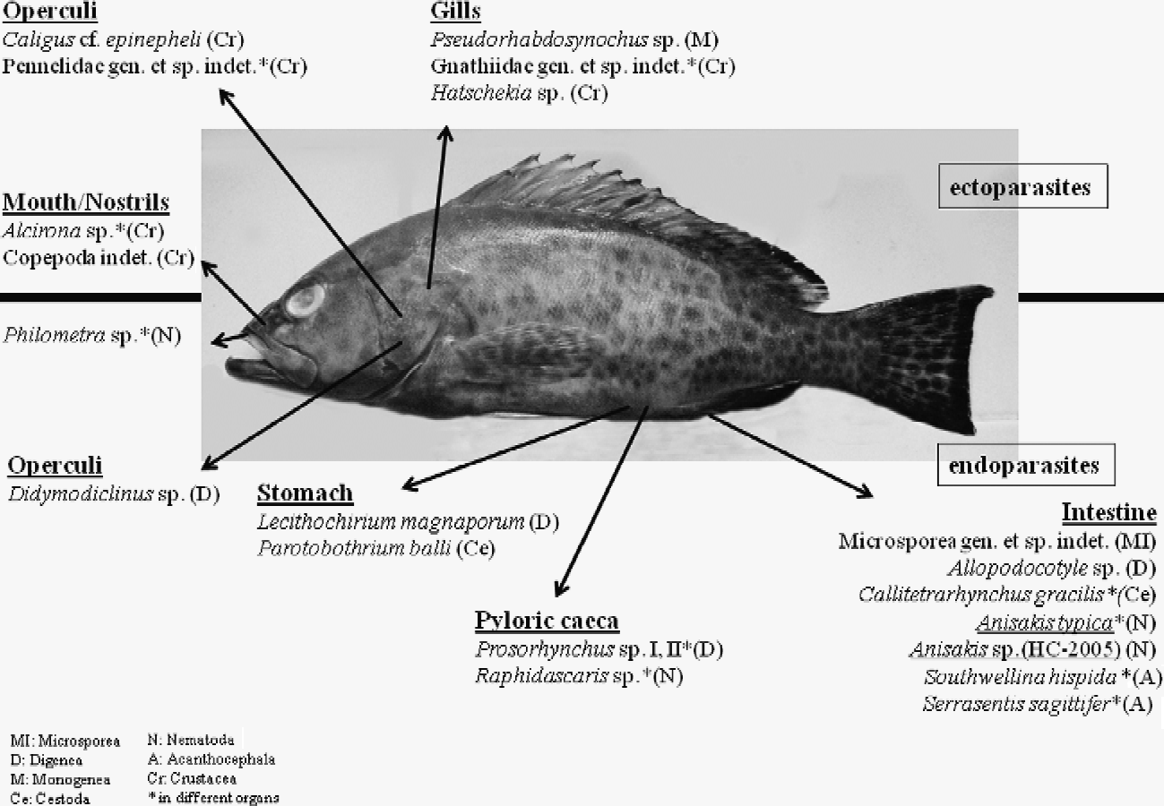
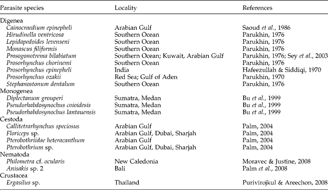
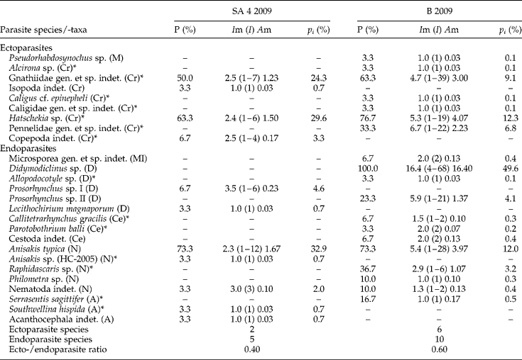
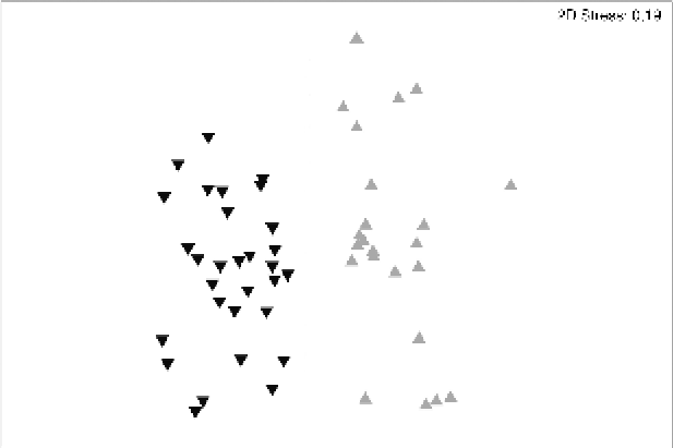
Fish parasitological studies of E. areolatus from the coastal zone off Segara Anakan lagoon and in Balinese waters revealed 21 different metazoan parasite species belonging to the taxa Microsporea (1), Digenea (5), Monogenea (1), Cestoda (2), Nematoda (4), Acanthocephala (2) and Crustacea (6) (fig. 1). Fourteen new host and locality records could be established for E. areolatus (see tables 2 and 3), mainly in Balinese waters. Furthermore, the morphologically identified Anisakis spp. specimens were confirmed as mainly A. typica and a single case of Anisakis sp. HC-2005, using molecular methods (see fig. 4). The information on prevalence, intensity, mean intensity, mean abundance and relative occurrence of the collected parasite species is summarized in table 3. To analyse the parasite composition and environmental conditions at the respective sampling sites, the ecological parameters as suggested by Palm & Rückert (Reference Palm and Rückert2009) and Palm et al. (Reference Palm, Kleinertz and Rückert2011) were considered as given in table 4. Regional differences for E. areolatus were found in terms of species composition (see fig. 2), endoparasite diversity, ecto-/endoparasite ratio, Hepatosomatic index and prevalences of infection of the metazoan parasites (see fig. 3).
‘The grouper habitat’. Given are all isolated parasite species/taxa from the investigated Epinephelus areolatus from Indonesian waters. Underlined parasites: anisakid nematodes with zoonotic potential. Above the bold, horizontal line, ectoparasites; below the line, endoparasites.

A list of parasite species previously identified from Epinephelus areolatus.
Prevalence (P), intensity (I), mean intensity (Im), mean abundance (Am) and relative occurrence (pi (%)) of the respective parasite species of Epinephelus areolatus, sampled from Javanese and Balinese waters.
SA 4, coastline Segara Anakan lagoon; B, Bali; *, new host record; A, Acanthocephala; Ce, Cestoda; Cr, Crustacea; D, Digenea; M, Monogenea; MI, Microsporea; N, Nematoda.
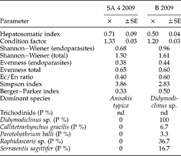
Parasitological and ecological metrics from the studied Epinephelus areolatus used as biological indicators to visualize regional environmental change.
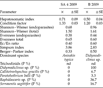
SE, standard error; SA 4, coastline Segara Anakan lagoon; B, Bali; Ec/En, ecto-/endoparasite; nd, no data.
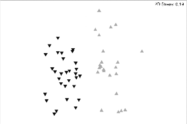
Multi-dimensional scaling (MDS) plot for the parasite community of Epinephelus areolatus from Javanese and Balinese waters during the dry season, 2009. ▾, Bali;  , coastline of Segara Anakan lagoon.
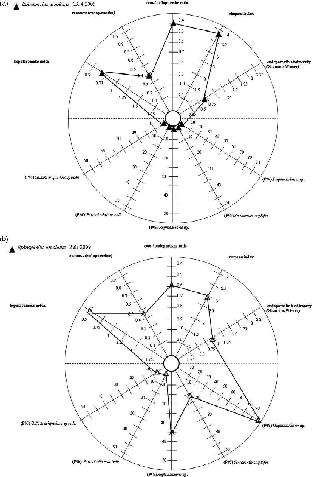
Visual indicator integration for Epinephelus areolatus from Javanese (a) and Balinese (b) waters during the dry season, 2009. SA 4, coastline Segara Anakan lagoon.
Parasite diversity and levels of infection
Epinephelus areolatus from Bali exhibited higher parasite diversity (16 taxa) compared to Segara Anakan lagoon (7 taxa) (table 3). Lower ectoparasite richness was found off Segara Anakan in contrast to Bali (2 versus 6 taxa, table 3). Endoparasite richness was twice as high in Balinese fish, with A. typica (Segara Anakan, 73% prevalence) and Didymodiclinus sp. (Bali, 100% prevalence) (Berger–Parker index of 0.33–0.50, see table 4) being the most predominant taxa. Ecto-/endoparasite ratios ranged from 0.4 to 0.6, with a lower number of ectoparasites compared to the number of endoparasites. Regional difference of the ecto-/endoparasite ratio was not significant between the two sampling sites. Endoparasite diversity of E. areolatus ranged from 0.68 (Segara Anakan) to 0.96 (Bali) for the Shannon–Wiener index. Simpson diversity index for the whole parasite community was higher for grouper parasites off Segara Anakan (3.86) compared to Bali (2.83) (table 4). Highest Evenness for the endoparasites was recorded in Bali (0.44) in contrast to Segara Anakan (0.38), whereas the values for the Hepatosomatic index were regionally contrary (highest in Segara Anakan) (0.50–0.71) (table 4), with a significant difference (ANOVA: F= 5.92, P< 0.05, post hoc test: P= 0.036).
Prevalence of infection of the digenean Didymodiclinus sp. was significantly higher from Balinese waters (100 versus 0% prevalence, table 3) (P= 0.000) compared with Javanese waters. Trypanorhynch cestodes Callitetrarhynchus gracilis and Parotobothrium balli occurred in Balinese waters (6.7 and 3.3% prevalence, table 3). Because of the low prevalence of infection, regional differences for both cestodes were not significant (P>0.05). Infection with the fish parasitic nematode Raphidascaris sp. was 36.7% in Bali (table 3). This displays a significant regional difference (P= 0.002). Infection with the acanthocephalan Serrasentis sagittifer was significantly higher in E. areolatus from Balinese waters (16.7 versus 0% prevalence, table 3) (P= 0.020).
Regional parasite composition and visual integration
Significant differences in species composition were found between the sampled E. areolatus from Balinese and Javanese coastal waters (ANOSIM: R, 0.642; P, 0.1%). Parasite species contributing most to the regional differences off Segara Anakan according to Simper analysis were A. typica (46.7%), Hatschekia sp. (34.3%) and Gnathiidae gen. et sp. indet. (18.8%), and those of Balinese waters were Didymodiclinus sp. (67.6%), Hatschekia sp. (12.5%) and A. typica (10.9%). There was a clear separation of the parasite communities at both sampling sites (fig. 2). Ten chosen bioindicators are visualized within a stargraph according to Bell & Morse (Reference Bell and Morse2003) and Palm & Rückert (Reference Palm and Rückert2009) (fig. 3).
Molecular nematode identification
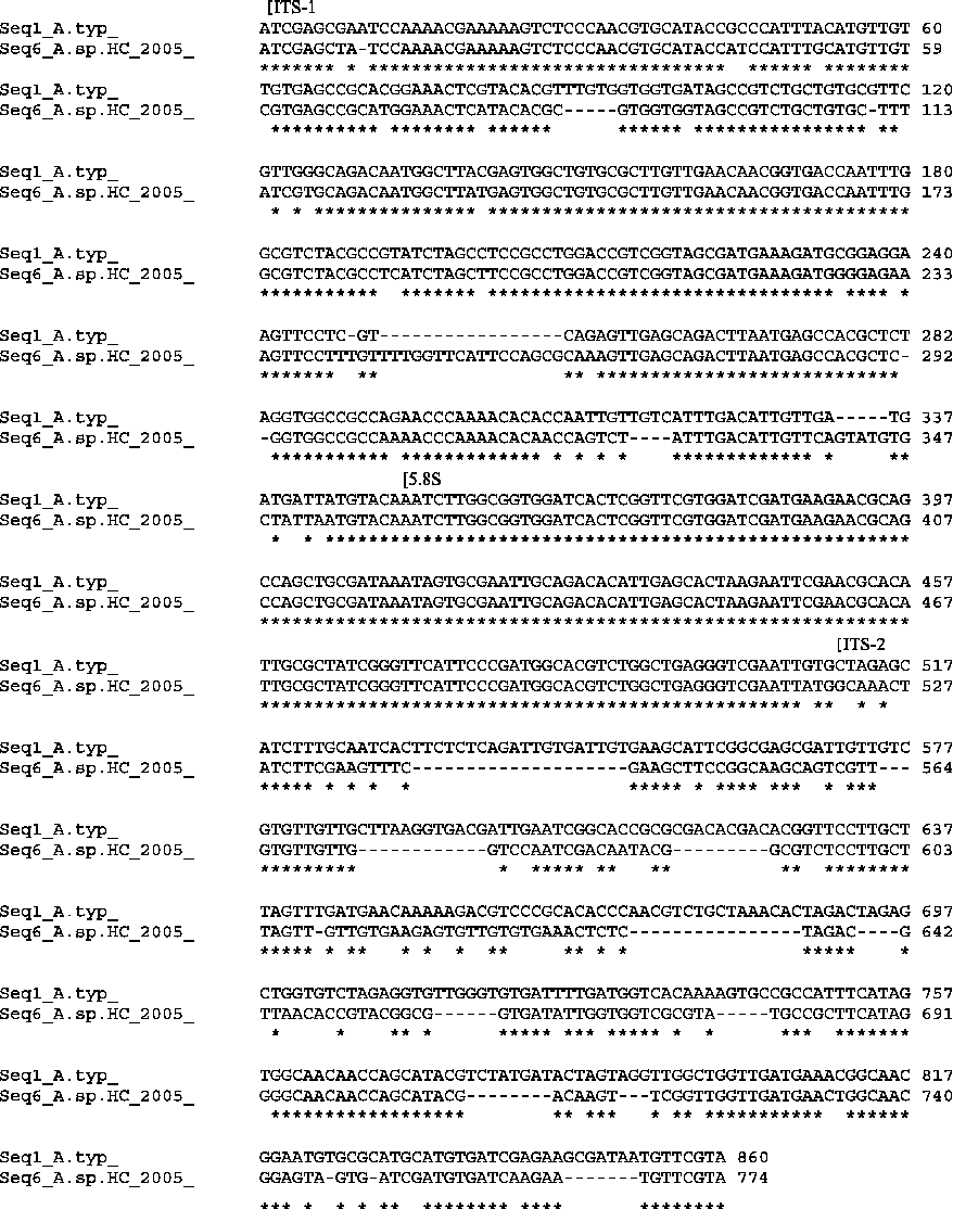
Molecular identification of 20 individual Anisakis larvae revealed 19 A. typica. Multiple sequence alignment of the A. typica indicated no genetic diversity within the sequenced rDNA region. The ITS-1, 5.8S, ITS-2 rDNA was identical for all investigated specimens at both sampling sites, compared with the reference sequence AB432909. No difference was observed between our material and Anisakis (typica) sp. 2 genotype as given in Palm et al. (Reference Palm, Damriyasa, Linda and Oka2008) (EU346093). A single specimen differed from the 19 identified A. typica by amplicon size and sequence. This individual was identified as Anisakis sp. HC-2005 (EU718474). A sequence alignment of the A. typica sequences from Java and Bali (AB432909) with the genotype Anisakis sp. HC-2005 is given in fig. 4. The total lengths of the PCR product from Anisakis sp. HC-2005 was 774 bp (ITS-1 358 bp, ITS-2 259 bp) compared with 859–860 bp for A. typica. Anisakis sp. HC-2005 was isolated first from Mauritian waters, and the present finding is a new locality record for this yet unspecified genotype from Indonesian waters.
Alignment of the ITS-1, 5.8S and ITS-2 region from larval Anisakis typica (Seq1_A.typ) and larval Anisakis sp. HC-2005 (Seq6_A.sp.HC_2005) of Epinephelus areolatus from the coastline of Segara Anakan.
Discussion
The Indonesian Archipelago is known to harbour an extremely high marine biodiversity on a global scale (Gray, Reference Gray1997; Myers et al., Reference Myers, Mittermeier, Mittermeier, da Fronesca and Kent2000; Allen & Werner, Reference Allen and Werner2002; Roberts et al., Reference Roberts, McClean, Veron, Hawkins, Allen, McAllister, Mittermeier, Schueler, Spalding, Wells, Vynne and Werner2002; Palm, Reference Palm2004), based on its geographical position and geological history (Rückert et al., Reference Rückert, Klimpel and Palm2010). Earlier studies proved high parasite diversity for Indonesian waters, although the number of investigations is relatively low. According to Jakob & Palm (Reference Jakob and Palm2006), up to 400 different parasite species have been recorded from Indonesian marine waters in 242 investigated fish species. Palm (Reference Palm2004) recorded 23% of the then known trypanorhynch cestode fauna from Indonesian waters.
The parasite fauna of some commercially important epinephelids from Indonesian waters, such as E. coioides and E. fuscoguttatus, has been studied in recent years (see Yuniar et al., Reference Yuniar, Palm and Walter2007; Palm & Rückert, Reference Palm and Rückert2009; Rückert et al., Reference Rückert, Hagen, Yuniar and Palm2009a, Reference Rückert, Klimpel, Mehlhorn and Palmb, Reference Rückert, Klimpel and Palm2010). This contrasts with E. areolatus, a species with only few parasite records worldwide (19 records, Hafeezullah & Siddiqi, Reference Hafeezullah and Siddiqi1970; Parukhin, Reference Parukhin1970, Reference Parukhin1976; Sey et al., Reference Sey, Nahhas, Uch and Vang2003; Moravec & Justine, Reference Moravec and Justine2008; Purivirojkul & Areechon, Reference Purivirojkul and Areechon2008; 5 of them from Indonesian waters) (Bu et al., Reference Bu, Leong, Wong, Woo and Foo1999, Palm, Reference Palm2004) (see table 2). So far, ten studies on the parasite fauna of E. areolatus revealed 19 parasite species/taxa, belonging to the Digenea (9 species), Monogenea (3 species), Cestoda (4 species), Nematoda (2 species) and a single crustacean (see table 2). Within the present study, 14 new host records could be established.
Different parasite species were applied within the stargraph system (see Palm & Rückert, Reference Palm and Rückert2009). The digenean trematode Didymodiclinus sp. has already been recorded from Segara Anakan lagoon for E. coioides (Rückert et al., Reference Rückert, Hagen, Yuniar and Palm2009a), indicating lower host specificity and probably a wide zoogeographical distribution in Indonesia. Groupers represent final hosts in the life cycle of this parasite, becoming infected via smaller fishes, possibly Johnius coitor and/or Nemipterus japonicus (Rückert et al., Reference Rückert, Klimpel, Mehlhorn and Palm2009b), both abundant hosts for that digenean at least in the Segara Anakan region (Yuniar, Reference Yuniar2005; Rückert, Reference Rückert2006; Rückert et al., Reference Rückert, Klimpel, Mehlhorn and Palm2009b; Theisen, Reference Theisen2009). According to Palm (Reference Palm2004) Parotobothrium balli is widely distributed in Indonesian waters. The genus Callitetrarhynchus has been isolated from N. japonicus off Segara Anakan by Theisen (Reference Theisen2009). Raphidascaris sp. has been already recorded for E. coioides at both sampling sites with a similar prevalence of infection (2.9–8.6% in Segara Anakan region versus 20.0–28.6% in Bali) (Kleinertz, Reference Kleinertz2010). The recorded Serrasentis sagittifer (Acanthocephalan) had already been found in the Segara Anakan region by Theisen (Reference Theisen2009) in three different fish species (J. coitor, N. japonicus and Platycephalus arenarius; see also Verweyen et al., Reference Verweyen, Klimpel and Palm2011). Rückert (Reference Rückert2006) and Rückert et al. (Reference Rückert, Klimpel, Mehlhorn and Palm2009b, Reference Rückert, Klimpel and Palm2010) suggested the epinephelids E. coioides and E. fuscoguttatus as paratenic and/or transport hosts for these species.
Because of its zoonotic importance, the first Indonesian research on anisakids in finfish started in Jakarta Bay around the 1980s (Burhanuddin & Djamali, Reference Burhanuddin and Djamali1978, Reference Burhanuddin and Djamali1983; Hadidjaja et al., Reference Hadidjaja, Ilahude, Mahfudin and Malikusworo1978; Hutomo et al., Reference Hutomo, Burhanuddin and Hadidjaja1978; Ilahude, Reference Ilahude1980; Petersen et al., Reference Petersen, Palm, Möller and Cuzi1993; Jakob & Palm, Reference Jakob and Palm2006; Palm et al., Reference Palm, Damriyasa, Linda and Oka2008). Hadidjaja et al. (Reference Hadidjaja, Ilahude, Mahfudin and Malikusworo1978), Hutomo et al. (Reference Hutomo, Burhanuddin and Hadidjaja1978) and Ilahude et al. (Reference Ilahude, Hadidjaja and Mahfudin1978) studied anisakid nematodes from East Sumatra and the North Java coast, and Burhanuddin & Djamali (Reference Burhanuddin and Djamali1978) used anisakid nematodes for stock separation in the roundscad Decapterus russelli in the Java Sea. Ilahude et al. (Reference Ilahude, Hadidjaja and Mahfudin1978) and Burhanuddin & Djamali (Reference Burhanuddin and Djamali1983) recorded 23 different fish species from the northern Java coast that were infected with anisakid nematodes, including four different Epinephelus species. However, no infection was recorded from the fish musculature, most probably due to the preservation of the fish in 10% formalin. Other Anisakis sp. records originate from Indonesian mariculture (e.g. Asmanelli et al., Reference Asmanelli, Yuliansyah and Muchari1993). Palm et al. (Reference Palm, Damriyasa, Linda and Oka2008) identified A. typica and Anisakis sp. 1 and 2 from Auxis rochei rochei, Caesio cuning, Coryphaena hippurus and E. areolatus by using molecular methods. They stated that larval A. typica or closely related siblings infest a wide range of clupeiform, perciform and also gadiform fish in warmer waters, and seem to be the most common anisakid nematodes in the Indonesian region. Anisakis typica is a common parasite of various dolphins of warmer temperate and tropical waters belonging to the families of Delphinidae, Phocoenidae and Pontoporidae (see Mattiucci et al., Reference Mattiucci, Paggi, Nascetti, Portes Santos, Costa, Di Beneditto, Ramos, Argyrou, Cianchi and Bullini2002). A total of 21 fish species were known to harbour Anisakis spp. or A. typica in Indonesia, with 34 fish species known to be infected with anisakid nematodes (Palm et al., Reference Palm, Damriyasa, Linda and Oka2008). The present study adds a new locality record from off Segara Anakan lagoon. Anisakis sp. HC-2005 is herewith identified from Indonesian waters for the first time. This genotype requires further attention, especially in order to estimate the potential risk for the fish consumer caused by fishery products from this geographical region that might include this nematode.
Fish parasites have been demonstrated to be useful as biological indicators to monitor environmental change (Vidal-Martínez et al., Reference Vidal-Martínez, Pech, Sures, Purucker and Poulin2010). They have been used to indicate, for example, bacterial biomass (Palm & Dobberstein, Reference Palm and Dobberstein1999; Palm & Rückert, Reference Palm and Rückert2009), heavy metals (Sures & Siddall, Reference Sures and Siddall2003) or environmental stress (Landsberg et al., Reference Landsberg, Blakesley, Reese, McRae and Forstchen1998) (for reviews on parasitological bioindicators, see Lafferty, Reference Lafferty1997; Marcogliese & Cone, Reference Marcogliese and Cone1997; Overstreet, Reference Overstreet1997; Williams & MacKenzie, Reference Williams and MacKenzie2003; Marcogliese, Reference Marcogliese2005; Palm, Reference Palm and Mehlhorn2011). Sasal et al. (Reference Sasal, Mouillot, Fichez, Chifflet and Kulbicki2007) utilized fish parasites to detect anthropogenic influences (urban and industrial pollution) in coral reefs, and Lafferty et al. (Reference Lafferty, Shaw and Kuris2008b) proposed them as a convenient method for assessing spatial variation in their final host distribution. Because their occurrence is dependent on the composition of the food web, a low biodiversity could impair parasite transmission by reducing the availability of hosts required by parasites with complex life cycles (Marcogliese, Reference Marcogliese2005; Lafferty et al., Reference Lafferty, Shaw and Kuris2008b). Consequently, heteroxenous fish parasites (multiple hosts) with complex life cycles are generally useful tools to indicate food web relationships in unaffected marine habitats (e.g. Palm, Reference Palm1999; Klimpel et al., Reference Klimpel, Rückert, Piatkowski, Palm and Hanel2006; Palm et al., Reference Palm, Klimpel and Walter2007; Lafferty et al., Reference Lafferty, Allesina, Arim, Briggs, De Leo, Dobson, Dunne, Johnson, Kuris, Marcogliese, Martinez, Memmott, Marquet, McLaughlin, Mordecai, Pascual, Poulin and Thieltges2008a). Thus, they reflect the status of the marine food web that might be affected through environmental change. The occurrence of endoparasites decreases in polluted waters (Nematoda; Kiceniuk & Khan, Reference Kiceniuk, Khan and Nraigu1983), whereas ectoparasitic parasites such as monogeneans are increasing (Monogenea, Khan & Kiceniuk, Reference Khan and Kiceniuk1988; Trichodina, Khan, Reference Khan1990; Palm & Dobberstein, Reference Palm and Dobberstein1999; Ogut & Palm, Reference Ogut and Palm2005). These effects can be used in order to analyse and monitor environmental conditions, especially where other information, such as water quality parameters, pollution or anthropogenic influences, are scarce.
Ten different bioindicators were chosen and visualized in a single stargraph, according to Bell & Morse (Reference Bell and Morse2003) and Palm & Rückert (Reference Palm and Rückert2009), demonstrating regional differences in the parasite infection of E. areolatus at both sampling sites (fig. 3). We applied: (1) the ratio of ecto- versus endoparasites, which has been suggested to indicate the common parasite infection of free-living groupers in two Indonesian coastal ecosystems (Vidal-Martínez et al., Reference Vidal-Martínez, Aguirre-Macedo, Vivas-Rodríguez and Moravec1998; Dzikowski et al., Reference Dzikowski, Paperna and Diamant2003; Jakob & Palm, Reference Jakob and Palm2006; Rückert et al., Reference Rückert, Hagen, Yuniar and Palm2009a); (2) the endohelminth parasite diversity, to indicate successful parasite transmission and host biodiversity (Rückert et al., Reference Rückert, Hagen, Yuniar and Palm2009a; Palm et al., Reference Palm, Kleinertz and Rückert2011); (3) the Evenness of endohelminths, to assess the availability of potential host organisms in the surroundings and/or other external factors resulting in low diversity and high dominance levels (Kleinertz, Reference Kleinertz2010); (4) the Simpson diversity index for both ecto- and endoparasite species (see 2 and 3 above); (5) the Hepatosomatic index to indicate high enzymatic activity in relation to stress (e.g. caused by pollution) (Munkittrik et al., Reference Munkittrik, Van der Kraak, McMaster, Portt, Van den Heuval and Servos1994). According to Lafferty et al. (Reference Lafferty, Shaw and Kuris2008b), using tetraphyllidean cestodes of elasmobranchs as biological indicators, and Dzikowski et al. (Reference Dzikowski, Paperna and Diamant2003) on the transmission of parasites with complex life cycles through a chain of different host species, we applied endoparasite prevalences to indicate regional differences at the chosen sampling sites. Moreover, we applied: (6) the prevalence of the digenean Didymodiclinus sp. (Hechinger et al., Reference Hechinger, Lafferty, Huspeni, Andrew and Armand2007); (7 and 8) the prevalence of trypanorhynch cestodes, C. gracilis and P. balli, that utilize elasmobranchs as final hosts (Palm, Reference Palm2004; Lafferty et al., Reference Lafferty, Shaw and Kuris2008b); (9) the prevalence of the nematode Raphidascaris sp. (Palm & Rückert, Reference Palm and Rückert2009, Palm et al., Reference Palm, Kleinertz and Rückert2011); and (10) the prevalence of the acanthocephalan S. sagittifer (Dzikowski et al., Reference Dzikowski, Paperna and Diamant2003; Marcogliese, Reference Marcogliese2003, Reference Marcogliese2005; Hechinger et al., Reference Hechinger, Lafferty, Huspeni, Andrew and Armand2007; Lafferty et al., Reference Lafferty, Shaw and Kuris2008b; Kleinertz, Reference Kleinertz2010; Palm, Reference Palm and Mehlhorn2011). Four of the chosen bioindicators demonstrated significant regional differences (Hepatosomatic index, prevalences of Didymodiclinus sp., Raphidascaris sp. and S. sagittifer). According to the chosen parasite bioindicators, the environmental conditions at both studied sampling sites differed significantly, reflecting either different abiotic conditions, such as regular salinity changes or anthropogenic pollutants (in Segara Anakan, see below), and/or biotic conditions resulting in different food webs and availability of prey organisms (off the Balinese coast).
Palm et al. (Reference Palm, Kleinertz and Rückert2011) applied the same methodology to monitor the parasite community of groupers from a mariculture facility in the Thousand Islands, Indonesia. By using six different parasite metrics from E. fuscoguttatus presented in a single figure, a significant change in parasite composition and abundance was recorded over six consecutive years. Their study was the first to use fish parasites to assess long-term changes in holding conditions within a commercially run tropical finfish mariculture farm. Their results suggested that groupers can also be used as biomarkers to monitor environmental change in the wild, requiring more detailed information on the parasite systematics and especially on taxonomy. Being directly linked to the surrounding invertebrate and vertebrate communities, fish parasites with multiple-host life cycles (Hechinger et al., Reference Hechinger, Lafferty, Huspeni, Andrew and Armand2007) are sensitive bioindicators of aquatic ecosystem health (Overstreet, Reference Overstreet1997; Dzikowski et al., Reference Dzikowski, Paperna and Diamant2003). They require unaffected environmental conditions to get access to the full range of potential parasite intermediate hosts, whereas monoxenous parasite species (single-host) may persist in highly perturbed, extreme environments (Dzikowski et al., Reference Dzikowski, Paperna and Diamant2003; Hechinger et al., Reference Hechinger, Lafferty, Huspeni, Andrew and Armand2007). Consequently, we can conclude that the observed parasite fauna in E. areolatus from Balinese waters reflects merely unaffected environmental conditions for this species in Indonesian coastal waters, while the parasite fauna off Segara Anakan lagoon is widely influenced by the specific environmental conditions at that sampling site (see below). Other available methods such as multi-dimensional scaling (MDS) for community analyses (see fig. 2) are likewise useful tools to demonstrate significant differences in parasite composition at different sampling sites. However, they fall short of providing any possible reasons for the observed differences.
According to the stargraphs, groupers from both sampling sites originate from two different habitats, and the differences in the metazoan parasite fauna are either a result of altered feeding behaviour or of food composition. For decades, Segara Anakan lagoon has been facing a number of environmental problems due to extensive resource exploitation (Jennerjahn et al., Reference Jennerjahn, Nasir and Pohlenga2009). Most important are overfishing, logging of mangrove wood, high sediment input through the Citanduy River because of poor upland agricultural practices, agricultural runoff, potential pesticide, oil pollution and organic contaminants in water, sediment and macrobenthic invertebrates (White et al., Reference White, Marosubroto and Sadorra1989; Jennerjahn et al., Reference Jennerjahn, Nasir and Pohlenga2009; Dsikowitzky et al., Reference Dsikowitzky, Nordhaus, Jennerjahn, Khrycheva, Sivatharshan, Yuwono and Schwarzbauer2011). Because of the hydrological impact of the nearby mangrove-fringed Segara Anakan lagoon with high water mass influx, freshwater input mainly from Citanduy River (Holtermann et al., Reference Holtermann, Burchard and Jennerjahn2009), tidal variability (Jennerjahn et al., Reference Jennerjahn, Nasir and Pohlenga2009) and two water exchange channels with direct connection to the ocean (Holtermann et al., Reference Holtermann, Burchard and Jennerjahn2009), the coastal zone of Segara Anakan is more heavily influenced compared to the sampled Balinese waters. We are aware that it is difficult to correlate directly the observed parasite communities, without replication, to specific environmental or anthropogenic factors at both sampling sites. However, both localities that likewise offer suitable living conditions for E. areolatus, are distinctly different, a situation that is reflected by the recorded parasite fauna.
Acknowledgements
We are thankful to Professor Dr E. Yuwono from Universitas Jenderal Soedirman (UNSOED), Purwokerto, Java, for providing laboratory space and institutional support. Special thanks to Mr A. Rinto Suncoko and Mr E. Hermawaran from UNSOED for their personal initiative and organizational support during fieldwork. We are thankful to Professor Dr S. Klimpel (Senckenberg Biodiversity and Climate Research Centre (BIK-F), Frankfurt, Germany) for supporting the molecular nematode identification. Financial support was provided by the German Academic Exchange Service (DAAD), the German Ministry for Education and Research (BMBF Grant No. 03F0471) within the framework of the joint Indonesian–German research programme SPICE (Science for the Protection of Indonesian Coastal Marine Ecosystems, RISTEK research permit No. Surat Izin: 0037/FRP/SM/II/09) (S.K., H.W.P.), and the German Research Council (DFG PA 664/4-1 and 2, 664/6-1) for the molecular study (H.W.P.). This is publication No. 2 under the Memorandum of Understanding between the Faculty of Veterinary Medicine, Udayana University, Bali and Aquaculture and Sea-Ranching, University of Rostock, Germany, in order to promote research on fish parasites and biodiversity data collection in Indonesia.