





Introduction
Microbial pigments are natural compounds synthesised by a wide variety of microorganisms, including bacteria, fungi and algae (Maoka, Reference Maoka2020; Pailliè-Jiménez et al., Reference Pailliè-Jiménez, Stincone and Brandelli2020). Beyond their role in colouration, these compounds are critical for maintaining cell integrity and metabolic continuity under extreme environmental stresses such as high radiation, oxidative stress and nutrient limitation (Barreto et al., Reference Barreto, Casanova, Junior, Reis-Mansur and Vermelho2023; Dieser et al., Reference Dieser, Greenwood and Foreman2010; Maoka, Reference Maoka2020). Carotenoids are particularly notable for protecting against reactive oxygen species by acting as antioxidants (Maoka, Reference Maoka2020). This phenomenon has been documented in Deinococcus species, where carotenoid production contributes to their resistance to ionising radiation alongside efficient DNA repair mechanisms (Leuko et al., Reference Leuko, Bohmeier, Hanke, Böttger, Rabbow, Parpart, Rettberg and de Vera2017; Wang et al., Reference Wang, Liu, Yi, Zhu, Liu, Zhang, Xie and Jiang2024).
Microbial pigments are increasingly considered as potential biosignatures, chemical markers of life that could, in principle, be detected from a distance (Kumar et al., Reference Kumar, Kampe, Rösch and Popp2015; Leuko et al., Reference Leuko, Bohmeier, Hanke, Böttger, Rabbow, Parpart, Rettberg and de Vera2017; Vítek et al., Reference Vítek, Jehlička, Edwards, Hutchinson, Ascaso and Wierzchos2014). The incorporation of deep-UV and visible Raman spectroscopy into current Mars missions underscores the scientific community’s recognition of carotenoids as significant targets in the search for extraterrestrial life. This phenomenon is largely due to carotenoids exhibiting highly specific molecular vibrations that produce intense, identifiable signals even at very low concentrations, making them ideal candidates for remote detection via non-destructive laser analysis (Baqué et al., Reference Baqué, Backhaus, Meeßen, Hanke, Böttger, Ramkissoon, Olsson-Francis, Baumgärtner, Billi, Cassaro, de la Torre Noetzel, Ré, Edwards, Ehrenfreund, Elsaesser, Foing, Fédéric, Börn, Joshi, Kozyrovska, Lasch, Lee, Leuko, Onofri, Ott, Pacelli, Rabbow, Rothschild, Schulze-Makuch, Selbmann, Serrano, Szewzyk, Verseux, Wagner, Westall, Zucconi and de Vera2022; Vítek et al., Reference Vítek, Jehlička, Edwards, Hutchinson, Ascaso and Wierzchos2014). Subterranean ecosystems provide distinctive natural settings for investigating these adaptive processes (Zhu et al., Reference Zhu, Jiang and Liu2022). The complete darkness, extreme nutrient scarcity and enduring physicochemical stability inherent to cave environments necessitate that resident microorganisms evolve specialised survival strategies, including improved cellular stress responses and alternative energy pathways (Çolak and Güngör, Reference Çolak and Güngör2022; Wu et al., Reference Wu, Ke, Hernandez, Wang, Dumont, Jia and Conrad2015).
Consequently, these shared characteristics make cave systems useful models on Earth for simulating the potential for life in the subsurface environments of other planets, particularly Mars (Çolak and Güngör, Reference Çolak and Güngör2022). Although the Martian surface is inhospitable to life as we know it, protected underground areas might offer more suitable conditions. The European Space Agency has taken this parallel seriously enough to embed it in astronaut training; its CAVES programme uses cave environments specifically because the isolation, resource constraints and operational risks they impose are among the closest things on Earth to the demands of space (Sauro et al., Reference Sauro, De Waele, Payler, Vattano, Sauro, Turchi and Bessone2021).
Morca Cave, located on the Taşeli Plateau in the central Taurus Mountains of southern Türkiye, is an example of an extreme underground environment that makes cave microbiology vital to astrobiology. One of Türkiye’s deepest cave systems, it has been the subject of ongoing research since 2013 (Ahamada Rachid and Doğruöz Güngör, Reference Ahamada Rachid and Doğruöz Güngör2023; Özbek and Aydın, Reference Özbek and Aydın2023). Its near-complete isolation from surface light and nutrient inputs, combined with the long-term stability of its physicochemical parameters, makes it a particularly informative setting for studying microbial persistence under prolonged darkness and oligotrophy.
A critical yet under-researched aspect of cave microbiology is the vertical depth gradient. In deep cave systems, increasing distance from the surface brings about a progressive transformation in geochemical conditions: nutrient availability decreases sharply, water chemistry varies with depth-dependent mineral dissolution, and the residual effect of surface-derived organic carbon becomes negligible at extreme depths (Turrini et al., Reference Turrini, Chebbi, Riggio and Visca2024). These gradients impose increasingly intense selective pressures on resident microbial communities, triggering depth-specific adaptations in metabolic strategies, pigment biosynthesis and cellular defence mechanisms (Bogdan et al., Reference Bogdan, Baricz, Chiciudean, Bulzu, Cristea, Năstase-Bucur, Levei, Cadar, Sitar, Banciu and Moldovan2023; Faitová, Reference Faitová2021).
From an astrobiological perspective, this vertical dimension offers a direct analogy, given the geochemical heterogeneity of the Martian subsurface environment. Current Mars habitability models acknowledge that depth-dependent variations in radiation flux, water activity and redox chemistry create distinct ecological niches at different stratigraphic levels (Stamenković et al., Reference Stamenković, Lynch, Boston, Tarnas, Edwards, Sherwood-Lollar, Atreya, Templeton, Freeman, Fischer, Spohn, Webster, Fairén, Mustard, Mischna, Onstott, Osburn, Kieft, Grimm, Brinckerhoff, Johnson, Beegle, Head, Haldemann, Cockell, Hernlund, Wilcox, Paige, Etiope, Glavin, Zorzano M-P, Sekine, Stalport, Kirschvink, Magnabosco, Orosei, Grott, Rummel, Kobayashi, Inagaki, Bishop, Chevrier, Bell, Orcutt, McIntosh, Miljkovic, Breuer, Usui, Zacny, Heggy, Rivera-Valentin, Barba, Woolley, Warr, Malaska, Blank, Ruffatto, Sapers, Matthies, Ward, Shkolyar, Schmelzbach, Gabriel, Parker, Bolivar-Torres, Pál, Schulze-Makuch, Torres Celis, Kereszturi, Spry, Uckert, Hesse, Harris, Plesa, Hu, Agha-mohammadi, Wade, Chatterjee, McGarey, Graham, Suzuki, Schrenk, Sherrill, Howe, Manthena, Burgin, Carpenter, Giersch, Cormarkovic, Smith, McDonnell, Michalski, Jha, Cable, Gloesener, Paul, Gault, Kedar, Marteau, Temel, Krieger and Timoney2021; Tarnas et al., Reference Tarnas, Mustard, Lollar, Stamenković, Cannon, Lorand, Onstott, Michalski, Warr, Palumbo and Plesa2021). In this context, characterising the responses of pigment-producing bacteria in deep terrestrial caves to depth gradients provides a structured, analogue-based framework for predicting at which depths and in what form biosignatures might be preserved in extraterrestrial subsurface environments, including, most immediately, the Martian subsurface (Merino et al., Reference Merino, Aronson, Bojanova, Feyhl-Buska, Wong, Zhang and Giovannelli2019; Onstott et al., Reference Onstott, Ehlmann, Sapers, Coleman, Ivarsson, Marlow and Nealson2019).
Relatively few studies have combined direct pigment characterisation with molecular identification of cave bacteria across significant vertical depth gradients, and Morca Cave, one of Türkiye’s deepest and least-disturbed subterranean environments, offered a compelling opportunity to do exactly that. We studied three sampling depths and used 16S rRNA gene sequencing, TLC and HPLC-based pigment analysis to describe the carotenoid profiles of pigment-producing bacteria from their aphotic zones. The functional roles of carotenoids outlined above, including antioxidant activity, radiation resistance, membrane stabilisation and biosignature potential, provide the ecological rationale for characterising cave-derived carotenoid producers, but they were not experimentally evaluated in the present study. Functional assays fall outside the scope of the present work and are identified as priorities for follow-up research. Beyond the characterisation itself, we wanted to understand what these pigments might mean more broadly: whether they reflect biosynthetic features compatible with persistence under prolonged darkness and nutrient limitation and whether the same biochemical signatures could one day help us recognise life in the subsurface of another planet. These hypotheses are formulated to respond to these two questions: do heterotrophic bacteria cultivable from aphotic zones of Morca Cave retain carotenoid biosynthetic capacity across a vertical depth gradient extending to −680 m, and what taxonomic groups and pigment profiles characterise the producing isolates?
Materials and methods
Cave background and sampling site
Morca Cave (36° 18′ 34.37″ N, 32° 39′ 37.10″ E) is located in the southern part of the Taşeli Plateau, within the central Taurus Mountains of southern Türkiye (Figures 1 and 2). The plateau, situated along the Alpine-Himalayan orogenic belt, is generally flat and has been shaped by northwest-trending faults. Its limestone layers vary in thickness and age, reaching up to 1600 m in thickness (Usuloğlu, Reference Usuloğlu2020). Neighbouring systems on the same plateau, including EGMA Peynirlikönü (−1429 m) and Kuzgun Düdeni (−1400 m), share this geological structure and have entrances located approximately 200 m above Morca Cave. A 2020 siphon dive confirmed that the main branch does not extend further than −1210 m, although exploration of side branches is ongoing (Usuloğlu, Reference Usuloğlu2020).
During the 2019 exploratory expedition organised by the Anatolian Speleology Group Association (ASPEG), which included sampling, Morca Cave was surveyed to a depth of 1210 m and a total length of approximately 4 km, placing it among Türkiye’s deepest cave systems. Its near-complete isolation from surface light and nutrient input, combined with the long-term stability of its physicochemical parameters, makes it a compelling site for microbiological sampling across significant depth gradients. Ambient conditions at the sampling depths were recorded during sampling as approximately 5°C and 95% relative humidity, consistent with the thermally and hygrometrically buffered regime characteristic of deep karst systems.
Location of Morca Cave. (on the left) Satellite image of the Taşeli Plateau showing the cave entrance area (red outline). (on the right) Aerial view of the cave entrance and base camp, with colour-coded exploration routes mapped between 2013 and 2020.

Mapping studies of the Morca Cave between 2013 and 2020.

Sample collection and isolation of microorganisms
In the summer of 2019, samples were collected from five locations within Morca Cave. Three surface samples were collected (MY1 at −620 m, MY2 at −680 m and MY3 at −400 m) along with two composite cave-sediment samples (MT1 at −620 m and MT2 at −400 m), all positioned away from the cavers’ route to minimise contamination (Figure 3). Pigmented isolates from these sediment samples are listed in Table 2. After collection under aseptic conditions, samples were transported to the microbiology laboratory via the cold chain as quickly as possible.
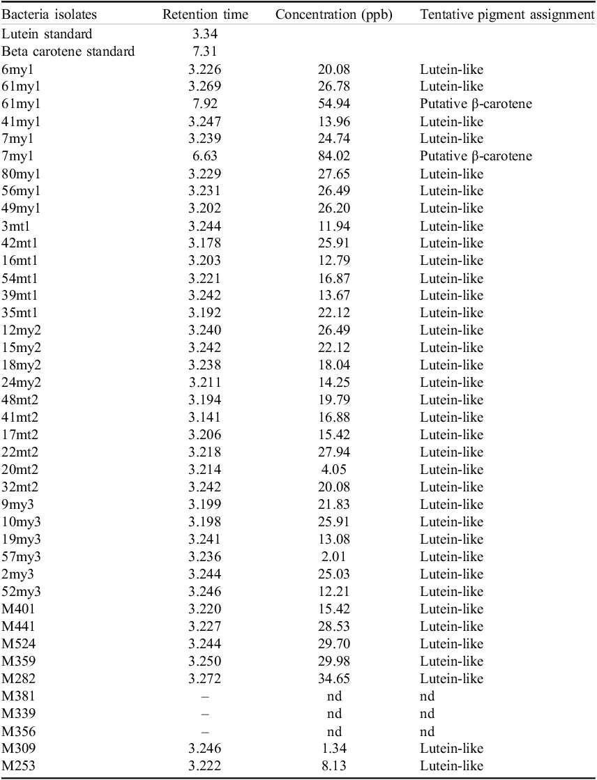
Retention times and pigment concentration and nature

Table 1. Long description
A table with 40 rows and 4 columns. The columns are labeled Bacteria isolates, Retention time, Concentration (ppb), and Tentative pigment assignment. The table lists various bacteria isolates along with their retention times, concentrations in parts per billion, and tentative pigment assignments. Notable trends include multiple isolates with lutein-like pigments and a few with putative beta-carotene. Some isolates have ‘nd’ indicating no data.
Note: Peaks reported as “Lutein-like” in this table are tentative assignments based on co-elution near the authentic Sigma–Aldrich lutein standard (RT 3.34 min); observed retention times systematically eluted earlier (3.14–3.27 min), and the dominant compound is therefore best regarded as a lutein-like xanthophyll pending LC-MS confirmation. Peaks reported as “Putative β-carotene” deviated from the authentic β-carotene standard (RT 7.31 min) at 6.63 min and 7.92 min, respectively, and may correspond to co-eluting apolar carotenoids (e.g., γ-carotene, cis-β-carotene isomers) rather than all-trans-β-carotene. See Discussion HPLC quantification for details.
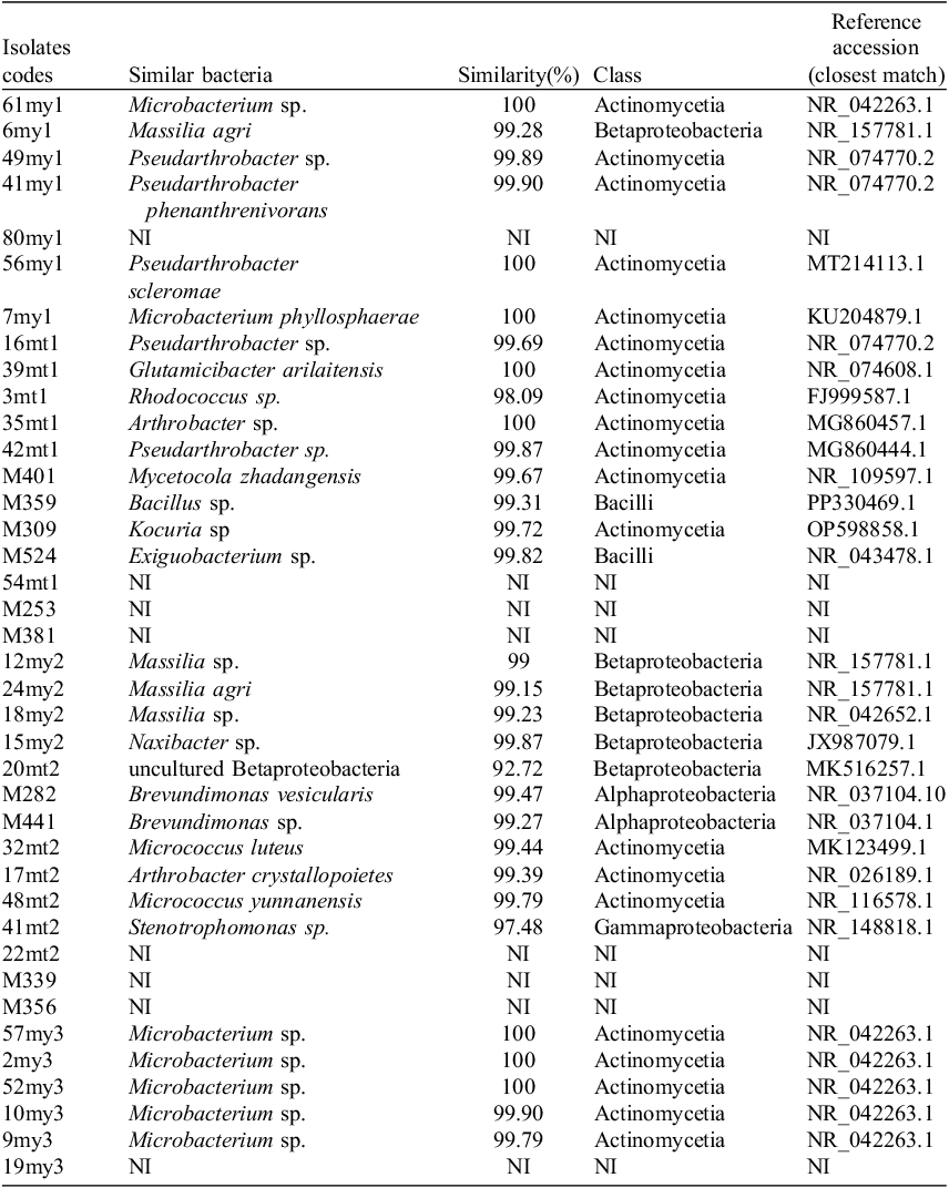
Genera of identified isolates and their classes

Table 2. Long description
A table with 38 rows and 5 columns. The columns are labeled Isolates codes, Similar bacteria, Similarity(%), Class, and Reference accession (closest match). The table lists various bacterial isolates identified from samples, their closest similar bacteria, the percentage of similarity, the class they belong to, and the reference accession numbers for the closest match. Each row provides specific details for different isolates. Notable entries include Microbacterium sp. with 100 percent similarity in multiple instances, and various other bacteria with different similarity percentages and classes. The table provides a comprehensive overview of the bacterial diversity identified in the samples.
NI: non identified.
One gram of each soil sample was weighed under sterile conditions and suspended in a phosphate buffer. Both the buffered soil suspensions and the cave surface samples collected from 1 cm2 areas using sterile swabs were serially diluted at ratios of 10−1 to 10−5. The diluted samples were inoculated onto R2A medium (Reasoner and Geldreich, Reference Reasoner and Geldreich1985) in triplicate and incubated at 28°C for 7 days. Fresh 24-hour cultures of the resulting isolates were collected from the medium using a phosphate buffer and stored at −86°C.
Sampling locations within Morca Cave: A (MY3, M441, M356, M339, M282 and MT2 at −400 m), B (MY1, M524, M401, M381, M359, M309, M253 and MT1 at −620 m), C (MY2 at −680 m).

Extraction of carotenoid pigments
Pigmented isolates were subcultured onto nutrient agar (NA) prior to pigment extraction to obtain sufficient biomass for downstream HPLC quantification. Orange- and yellow-pigmented colonies were then harvested by adding 5 ml of methanol directly to the plate and transferring the suspension to 15 ml Falcon tubes. The tubes were centrifuged at 6000 rpm for 15 minutes, vortexed, and the extraction was repeated until the pellet became pale. The pooled supernatant was distributed into 1.5 ml microcentrifuge tubes, and methanol was evaporated at 60°C. To prevent pigment degradation, all steps were carried out under low-light conditions (Craft and Soares, Reference Craft and Soares1992; Sobin and Stahly, Reference Sobin and Stahly1942; Thirkell and Strang, Reference Thirkell and Strang1967). Methanol was selected as the sole extraction solvent for compatibility with downstream HPLC injection; the implications of this choice for the recovery of nonpolar carotenoids are addressed in the Discussion.
Thin-Layer Chromatography (TLC) analysis
Thin-layer chromatography was used to characterise the carotenoid content of the pigmented isolates. A silica gel 60 plate (10 cm × 10 cm, Supelco, 1.05721) was used as the stationary phase. Among the solvent systems tested, including acetone:hexane (6:4), methanol:acetone:water (4:4:2), chloroform: methanol (6:4) and ethanol:water:chloroform (4:2:4), a chloroform: methanol mixture at a ratio of 9:1 (v/v) yielded the best separation and was used as the mobile phase throughout. Pigment extracts dissolved in methanol were spotted onto the plates and air-dried. The plates were developed for 15 minutes and then left to dry at room temperature. Given the pigments’ susceptibility to rapid colour loss under visible light, visualisation was performed under short-wavelength UV light (254 nm), where carotenoid spots appeared as distinct dark bands against a fluorescent background. Spots were marked, and their retention factor (Rf) values were calculated using the following formula (Bele and Khale, Reference Bele and Khale2011):
 $${\text{Rf Distance traveled by the solute/Distance traveled by the solvent front}}$$
HPLC analysis of β-carotene and lutein
HPLC analysis was carried out by EGE MATAL ANALIZ as a service. The system used was an Agilent 1260 Infinity HPLC-DAD equipped with an Agilent Eclipse Plus C18 column (5 µm, 4.6 × 250 mm). High-purity β-carotene and lutein standards (Sigma–Aldrich) were used as external standards for pigment identification and quantification, with calibration curves constructed at multiple concentrations spanning the working range and linearity confirmed by R 2 > 0.99. The mobile phase was a mixture of methanol, acetonitrile and tert-butyl methyl ether at a 50:40:10 (v/v/v) ratio, with a flow rate of 0.8 ml/min, a column temperature of 25°C, a fixed detection wavelength of 450 nm and an injection volume of 100 µL (Hagaggi and Abdul-Raouf, Reference Hagaggi and Abdul-Raouf2023). Methanol extracts were collected into 1.5 ml microcentrifuge tubes; a single aliquot per isolate was injected for HPLC analysis.
Genomic DNA isolation
DNA was extracted from pigment-producing isolates using a commercial kit (GeneAll Exgene™ Cell SV mini, Cat. No. 106-101, GeneAll Biotechnology Co., Ltd., Seoul, Korea) according to the manufacturer’s protocol. DNA concentrations were measured with a NanoDrop spectrophotometer (Thermo Scientific), and the quality of the extracted DNA was checked using A260/A280 and A260/A230 absorbance ratios; only samples with A260/A280 values between 1.8 and 2.0 were used for downstream PCR amplification. Samples were stored at −20°C until PCR amplification.
PCR amplification and sequencing
The universal primers 27F (5′-AGAGTTTGATCCTGGCTCAG-3’) and 1492R (5′-GGTTACCTTGTTACGACTT-3’) were used to amplify 16S rRNA gene regions (Lane, Reference Lane, Stackebrandt and Goodfellow1991). DNA concentrations were adjusted to 10 ng/µl prior to amplification. PCR was carried out in a BIORAD T100 Thermal Cycler using EmeraldAmp MAX HS PCR Master Mix (Takara) under the following conditions: initial denaturation at 94°C for 30 s (1 cycle); denaturation at 94°C for 30 s, annealing at 58°C for 30 s and extension at 72°C for 60 s (30 cycles each); and a final extension at 72°C for 10 min. PCR products (1500 bp) were verified by electrophoresis on a 1% agarose gel stained with Gel Red (Fisher Biotec) at 100 V for 30 min and visualised under UV transillumination.
Amplicons were sent to BM Laboratuvar Sistemleri (Ankara) for Sanger sequencing using an Applied Biosystems ABI3730XL DNA Analyser. The resulting chromatograms were inspected in FinchTV software, with low-quality regions at the 5′ and 3′ ends manually trimmed before downstream analysis. The trimmed sequences were converted to FASTA format and compared against sequences in the NCBI BLAST database (http://www.ncbi.nlm.nih.gov/BLAST) to identify the closest-matching species. Sequences generated in this study have been deposited in the NCBI GenBank database under accession numbers PQ363299–PQ363304, PV446154–PV446157, ON184090–ON184109 and ON310864 (Supplementary Table S1).
Phylogenetic analysis
The 16S rRNA gene sequences of the pigmented isolates, together with the closest reference sequences retrieved from GenBank (accession numbers listed in Table 2), were aligned in MEGA12 (Kumar et al., Reference Kumar, Stecher, Suleski, Sanderford, Sharma and Tamura2024) using the MUSCLE algorithm. A phylogenetic tree was reconstructed using the Neighbour-Joining method (Saitou and Nei, Reference Saitou and Nei1987), with evolutionary distances computed by the Maximum Composite Likelihood model (Tamura et al., Reference Tamura, Nei and Kumar2004) and expressed as the number of base substitutions per site. All ambiguous positions were removed for each sequence pair using the pairwise deletion option, yielding a final analysed dataset of 1527 positions. The robustness of the inferred topology was evaluated by bootstrap resampling with 1000 replicates (Felsenstein, Reference Felsenstein1985), and only bootstrap support values ≥50% are displayed at the corresponding nodes. Aquifex aeolicus (GenBank accession AJ309733.1) was included as the outgroup to root the tree. All phylogenetic analyses were performed in MEGA12 (Kumar et al., Reference Kumar, Stecher, Suleski, Sanderford, Sharma and Tamura2024).
Statistical analysis
All statistical analyses were performed in R (version 4.5.2) within the RStudio environment. Differences in lutein-like xanthophyll concentration among sampling depths (−400, −620, −680 m) and among bacterial classes were assessed using non-parametric Kruskal–Wallis tests, given the small and unequal group sizes and the non-normal distribution of the pigment concentration data. Associations between Gram morphology and sampling site were evaluated by chi-square tests. Significance was set at α = 0.05 throughout, and continuous data are reported as mean ± standard deviation. Because of the limited number of isolates per depth–class combination, statistical results are reported as exploratory and pairwise post-hoc analyses were not pursued.
Results
Bacterial isolates distribution
Bacterial colony counts according to the isolation zones (depths) and Gram morphology: A) colony count per sampling depth; B) Proportion of Gram identity through sampling depth.

A total of 265 bacterial isolates were recovered from the five sampling sites distributed across the three depths of Morca Cave (Figure 4). The highest colony count was obtained from the surface sample collected at −620 m (MY1, n = 83), followed by the surface sample at −400 m (MY3, n = 59), the soil sample at −620 m (MT1, n = 55), the surface sample at −680 m (MY2, n = 35) and the soil sample at −400 m (MT2, n = 33). Gram-positive bacteria predominated at four of the five sampling sites, accounting for between 64.4% and 86.7% of isolates, with the highest proportion at the −620 m surface site. The only exception was the −680 m surface sample (MY2), where Gram-negative bacteria constituted most isolates (51.4%). Among all 265 isolates, 39 displayed visible pigmentation, predominantly yellow or orange colonies, and were selected for further characterisation by TLC, HPLC and 16S rRNA gene sequencing.
Thin-layer chromatography analysis
Figure 5 illustrates the typical carotenoid TLC profile of cave bacteria from Morca Cave: a dominant nonpolar fraction migrating near the solvent front (spot E, Rf 0.91–0.95, within the β-carotene migration range) is present in most isolates, while polar xanthophyll fractions (spots A–D) are detected at variable intensities in the mid-plate region. The isolate 49my1 (Pseudarthrobacter sp.) stands out as the most carotenoid-rich in this particular plate, displaying the full complement of five spots, a pattern consistent with the multi-pigment profile described in Figure 6. The figure also visually confirms why M356 was recorded as negative: while a faint high-Rf signal is visible under UV, it was not reproducible or intense enough to be reliably recorded as a confirmed spot.
The appearance of the spots under UV light (A) and marking the spots on the silica plate (B).

Thin-layer chromatography resolved up to five distinct pigment spots (A through E) in the 39 pigmented isolates (Figure 6). Spots A through D, with Rf values ranging from 0.06 to 0.58, are consistent with the mobility range of xanthophyll-type carotenoids under the chloroform:methanol (9:1) solvent system used. Spot E, with Rf values between 0.91 and 0.95, falls within the Rf range reported for β-carotene under this solvent system but may also include other apolar carotenoids of similar polarity. Bands were detected in 35 of the 39 isolates, with spot C the most consistently observed (present in 34 isolates, ∼87%), indicating that a single dominant xanthophyll, likely lutein or a structurally related xanthophyll, is ubiquitous across depths and sample types. Spot E, in the β-carotene Rf range, was detected in 22 isolates. Two isolates, 49my1 and 41my1, displayed all five spots simultaneously. Isolate 17mt2 produced no detectable spots under UV, and isolates M381 isolated from −620 m, M339 and M356 isolated from −400 m likewise yielded no visible bands, indicating either pigment concentrations below TLC detection thresholds or the presence of non-carotenoid chromophores not visible at 254 nm.
The Rf values of different pigments for each sample.

HPLC analysis results of beta carotene and lutein compounds
HPLC analysis provisionally identified a lutein-like xanthophyll in 36 of the 39 isolates, with concentrations ranging from 1.34 ppb (M309) to 34.65 ppb (M282), and a mean of approximately 19.5 ppb (methanol extract) (Table 1). Isolate 17mt2 showed no detectable bands on TLC but yielded a quantifiable lutein-like xanthophyll signal by HPLC (15.41 ppb); the basis for this discrepancy is discussed further below. Three isolates, M381, M339 and M356, yielded no detectable signal for either compound, consistent with their absence of TLC spots. HPLC peaks eluting near the retention time of the β-carotene standard were detected in only two isolates: 7my1 (RT 6.63 min, 84.02 ppb) and 61my1 (RT 7.92 min, 54.94 ppb); both retention times deviate from the β-carotene standard (RT 7.31 min), and these signals are therefore reported here as putative β-carotene pending confirmation by additional standards or mass spectrometry. These two isolates were the only strains in which both a high-Rf TLC band and a high-RT HPLC peak in the β-carotene region were observed.
The isolate M282, identified as Brevundimonas vesicularis (99.47% similarity), yielded the highest lutein-like xanthophyll concentration in the entire dataset (34.65 ppb), while M309 (Kocuria sp.) produced the lowest detectable amount (1.34 ppb).
Mean lutein-like xanthophyll concentrations did not show a consistent trend with sampling depth, ranging from 17.02 ppb at −400 m to 20.71 ppb at −620 m and 20.23 ppb at −680 m, suggesting that lutein-like xanthophyll biosynthesis is maintained at broadly comparable levels regardless of depth (Kruskal–Wallis, p > 0.05; Figure 7). However, the two isolates producing putative β-carotene peaks by HPLC, 7my1 and 61my1, were both recovered from the −620 m surface site, and the two isolates displaying the most complex TLC profiles (five spots each) were also from this depth. Whether this reflects a site-specific microbiological feature or sampling variation cannot be determined from the present dataset and warrants verification through replicated sampling.
Pigments concentrations: A) Lutein-like xanthophyll concentration by zone; B) pigment concentration per isolate.

Sequencing of the isolates’ DNA and identification of the isolates
16S rRNA gene sequencing identified 30 of the 39 pigmented isolates to the genus or species level, with one additional isolate (20mt2) resolved only to the class level (Betaproteobacteria); the remaining 8 isolates (80my1, 54mt1, 22mt2, 19my3, M253, M381, M339 and M356) could not be assigned to any known taxon and are designated as not identified (NI) (Table 2). The dominant class was Actinomycetia, comprising 20 isolates (51.3% of all pigmented sequenced strains), represented by the genera Microbacterium, Pseudarthrobacter, Arthrobacter, Glutamicibacter, Micrococcus, Kocuria, Rhodococcus and Mycetocola. Betaproteobacteria accounted for 6 isolates (15.4%); five were assignable to the genus Massilia or the related genus Naxibacter, while isolate 20mt2 (92.72% similarity to its closest match) was resolvable only at the class level. Alphaproteobacteria were represented by two isolates of Brevundimonas (M282 and M441, 5.1%), Bacilli by two isolates (M524 identified as Exiguobacterium sp. and M359 as Bacillus sp., 5.1%) and Gammaproteobacteria by a single isolate of Stenotrophomonas sp. (41mt2, 2.6%) (Figures 8 and 9). The −620 m sampling sites (MY1 and MT1) yielded the greatest taxonomic diversity among the identified strains, including Microbacterium, Pseudarthrobacter, Massilia, Glutamicibacter, Arthrobacter, Rhodococcus, Mycetocola, Kocuria, Bacillus and Exiguobacterium. The −400 m surface sample (MY3) was dominated by Microbacterium species (five out of six identified isolates), while the −400 m sediment sample (MT2) contributed the most phylogenetically varied community, spanning Actinomycetia, Alphaproteobacteria, Betaproteobacteria and Gammaproteobacteria (Figures 8 and 9).
Bacterial class composition by zone.

Neighbour-joining phylogenetic tree based on 16S rRNA gene sequences of 31 pigment-producing bacterial isolates from Morca Cave, with Aquifex aeolicus (GenBank AJ309733.1) as the outgroup. Tip labels include the GenBank accession, taxonomic assignment, isolate code and sampling depth/substrate. Bootstrap support values ≥50% (1000 replicates) are shown in black at the nodes; branch lengths, expressed as substitutions per site, are shown in red and the scale bar represents 0.05 substitutions per site. Analyses were performed in MEGA12 with the Maximum Composite Likelihood distance model and pairwise deletion of ambiguous positions. Isolate 20mt2 (PQ363301.1) is labelled at the class level (Betaproteobacterium) because its closest BLAST match shared only 92.7% 16S rRNA gene sequence similarity, below the threshold for reliable genus-level assignment.

Lutein-like xanthophyll concentration differed significantly among bacterial classes (Kruskal–Wallis, p < 0.05; Figure 10). While most of the lutein-like xanthophyll-producing bacteria belong to Actinomycetia, the highest concentrations of the produced lutein-like xanthophyll have been observed from members of Alphaproteobacteria (Figure 10).
Lutein-like xanthophyll concentration across bacterial classes.

Discussion
Bacterial diversity and pigmentation in Morca Cave
The isolation of 265 bacterial strains from five sampling sites across three depths points to a microbial community shaped by the particular physicochemical conditions of subterranean life. The dominance of Gram-positive bacteria at four of the five sites, ranging from 64.4% to 86.7%, is consistent with what has been reported for other karst cave systems, where Actinomycetes and related Gram-positive lineages tend to be more abundant, owing to their capacity for spore formation, secondary metabolite production and tolerance of nutrient-poor conditions (Barton and Northup, Reference Barton and Northup2007; Tomova et al., Reference Tomova, Lazarkevich, Tomova, Kambourova and Vasileva-Tonkova2013). The exception was the −680 m surface sample, where Gram-negative bacteria reached 51.4%, a reversal that may reflect localised conditions at this site rather than a general depth-dependent trend, given that this pattern is based on a single sampling point.
The highest isolate counts came from the −620 m surface sample (MY1, n = 83), which also yielded the greatest proportion of Gram-positive organisms (86.7%). Pigment production in cave bacteria is often seen as a way to protect against light or oxidative damage, but in permanently dark environments, carotenoids likely play additional roles, potentially helping to stabilise membranes, tolerate osmotic stress and protect against reactive oxygen species produced through non-light-related processes, although these functional roles remain to be experimentally verified for the specific isolates characterised here (Tian and Hua, Reference Tian and Hua2010). From an astrobiological standpoint, the recovery of pigment-producing isolates from a permanently dark environment is a notable observation: it indicates that carotenoid biosynthesis is maintained under heterotrophic, light-independent cultivation in these strains, which is relevant to the question of whether pigmented chemotrophic life could persist in the permanently dark subsurface environments of Mars or the icy moons of the outer solar system. This depth-dependent ecological structuring within Morca Cave finds a conceptual parallel in the stratified habitability gradients proposed for the Martian crust (Michalski et al., Reference Michalski, Cuadros, Niles, Parnell, Rogers and Wright2013; Stamenković et al., Reference Stamenković, Lynch, Boston, Tarnas, Edwards, Sherwood-Lollar, Atreya, Templeton, Freeman, Fischer, Spohn, Webster, Fairén, Mustard, Mischna, Onstott, Osburn, Kieft, Grimm, Brinckerhoff, Johnson, Beegle, Head, Haldemann, Cockell, Hernlund, Wilcox, Paige, Etiope, Glavin, Zorzano M-P, Sekine, Stalport, Kirschvink, Magnabosco, Orosei, Grott, Rummel, Kobayashi, Inagaki, Bishop, Chevrier, Bell, Orcutt, McIntosh, Miljkovic, Breuer, Usui, Zacny, Heggy, Rivera-Valentin, Barba, Woolley, Warr, Malaska, Blank, Ruffatto, Sapers, Matthies, Ward, Shkolyar, Schmelzbach, Gabriel, Parker, Bolivar-Torres, Pál, Schulze-Makuch, Torres Celis, Kereszturi, Spry, Uckert, Hesse, Harris, Plesa, Hu, Agha-mohammadi, Wade, Chatterjee, McGarey, Graham, Suzuki, Schrenk, Sherrill, Howe, Manthena, Burgin, Carpenter, Giersch, Cormarkovic, Smith, McDonnell, Michalski, Jha, Cable, Gloesener, Paul, Gault, Kedar, Marteau, Temel, Krieger and Timoney2021).
In a broader context, deep karst caves studied for their microbial communities – including Krubera-Voronja and Veryovkina Caves in Georgia, both exceeding 2 km depth, Lechuguilla Cave (USA, −489 m), and Movile Cave (Romania, isolated chemoautotrophic system) – have each contributed distinct insights to subterranean microbiology (Itcus et al., Reference Itcus, Pascu, Lavin, Brad and Purcarea2018; Northup et al., Reference Northup, Barns, Yu, Spilde, Schelble, Dano, Crossey, Connolly, Boston, Natvig and Dahm2003; Sarbu et al., Reference Sarbu, Kane and Kinkle1996). Morca Cave occupies a complementary niche within this comparative framework: it offers a deep, oligotrophic and minimally disturbed limestone environment and is among Türkiye’s deepest cave systems characterised microbiologically to date. The combined depth, geological isolation and pristine state make it a particularly informative site for understanding microbial pigment biosynthesis under the extended physicochemical isolation that defines candidate planetary analogue habitats.
TLC analysis and pigment identification
Thin-layer chromatography resolved up to five distinct pigment spots (A through E) across the selected isolates, with spots A–D falling within the xanthophyll Rf range and spot E within the β-carotene range, consistent with published reference values (Dieser et al., Reference Dieser, Greenwood and Foreman2010). The prevalence of spot C in approximately 87% of isolates points to a single dominant xanthophyll type across all sampling depths and substrate types, most probably the lutein-like xanthophyll characterised below. This cross-site consistency suggests that lutein-like xanthophyll production is a broadly shared metabolic feature in the Morca Cave community rather than a depth- or substrate-specific response.
The two isolates displaying all five spots simultaneously, 49my1 and 41my1, both identified as Pseudarthrobacter species, stand out as the most carotenoid-rich strains in the collection. Producing both multiple xanthophyll types and a high-Rf TLC band consistent with β-carotene at the same time is consistent with the capacity of certain actinobacterial strains to run branched isoprenoid biosynthetic pathways, generating cyclised and hydroxylated carotenoid end-products in parallel (Takaichi, Reference Takaichi2011). Pseudarthrobacter has been isolated from various extreme and oligotrophic environments and is metabolically versatile, though detailed carotenoid characterisation of cave-dwelling strains at the biochemical level remains scarce (Margesin and Schinner, Reference Margesin and Schinner2001). Of the four isolates that yielded no TLC signal, 17mt2, M381, M339 and M356, despite their yellow or orange colony colour, absence of detectable bands under UV has been also noted. Isolate 17mt2 showed no detectable bands on TLC but yielded a quantifiable lutein-like xanthophyll signal by HPLC (15.41 ppb). While TLC sensitivity is generally lower than HPLC, the absence of a TLC signal in this isolate at a concentration detectable by HPLC likely reflects strain-specific differences in extraction efficiency or pigment-matrix interactions rather than a simple detection threshold effect, as several other isolates produced visible TLC spots at comparably low concentrations. The remaining three isolates, M381, M339 and M356, yielded no signal by either method.
It should be noted that pigment assignment in this study relied on Rf-based classification supported by HPLC retention-time matching against authentic lutein and β-carotene standards. While diode-array detection (400–500 nm) was used to verify spectral consistency with reference compounds, definitive structural identification of all xanthophyll spots – particularly the four spots assigned to the broader xanthophyll Rf range – would require complementary techniques such as LC-MS, NMR or comparison with a wider panel of carotenoid standards. The pigment assignments reported here should therefore be regarded as tentative for compounds beyond the lutein-like xanthophyll and putative β-carotene reported above, and future work using mass spectrometry-based identification will allow more precise characterisation of the carotenoid diversity in this community.
HPLC quantification: lutein-like xanthophyll prevalence and the β-carotene paradox
HPLC provisionally identified a lutein-like xanthophyll as the dominant carotenoid across the Morca Cave isolates, detected in 36 of 39 strains at concentrations ranging from 1.34 to 34.65 ppb, with a mean of approximately 19.5 ppb. The highest-yielding strain, M282, was identified as Brevundimonas vesicularis (34.65 ppb), a genus previously documented as a producer of ketocarotenoids including astaxanthin and adonixanthin (Asker et al., Reference Asker, Awad, Beppu and Ueda2018). That the highest-yielding strain in the dataset belongs to the numerically minor Alphaproteobacteria is a useful reminder that carotenoid output does not simply track taxonomic abundance.
The discrepancy between 22 isolates with TLC bands at β-carotene Rf and only two with HPLC peaks matching the β-carotene standard most likely indicates that the high-Rf TLC bands include other apolar carotenoids (e.g., lycopene, γ-carotene, cis-β-carotene isomers) that co-migrate with β-carotene under the chloroform:methanol (9:1) system but resolve at different retention times by HPLC. An additional contributing factor may be the relatively low solubility of β-carotene in pure methanol compared with apolar solvents such as hexane or methyl tert-butyl ether (Craft and Soares, Reference Craft and Soares1992), which could systematically reduce the recovery of nonpolar carotenoids relative to xanthophylls during the extraction step. Thermal degradation during methanol evaporation at 60°C may contribute additional losses in low-titre samples (Aman et al., Reference Aman, Schieber and Carle2005; Henry et al., Reference Henry, Catignani and Schwartz1998), but cannot by itself account for the magnitude of the TLC–HPLC discrepancy. This interpretation highlights the complementary value of TLC and HPLC: TLC provides qualitative breadth for the broader carotenoid pool, while HPLC delivers quantitative precision but is constrained by the matching of authentic standards. Future work using apolar extraction solvents (e.g., hexane or methyl tert-butyl ether), low-temperature or nitrogen-stream evaporation and an extended panel of carotenoid standards or LC-MS confirmation would allow more comprehensive resolution of the apolar carotenoid fraction in these isolates. It is also worth noting that both strains retaining quantifiable putative β-carotene belong to Microbacterium, which may reflect a particularly high initial putative β-carotene titre in these strains rather than any intrinsic biochemical resilience; this hypothesis warrants direct testing in future work. Both putative β-carotene peaks (7my1: 6.63 min; 61my1: 7.92 min) deviated more substantially from the authentic standard (7.31 min) than did the lutein-like xanthophyll peaks, consistent with the possibility that one or both signals correspond to a co-eluting apolar carotenoid (e.g., γ-carotene, cis-β-carotene isomer) rather than all-trans-β-carotene. As with the lutein assignment above, LC-MS confirmation will be needed to resolve these candidates definitively.
A second source of uncertainty applies specifically to the lutein assignment. All HPLC peaks reported here as lutein eluted between 3.14 and 3.27 min, systematically earlier than the authentic lutein standard (3.34 min). Although such retention-time offsets can arise from matrix effects, column equilibration or sample-buffer differences, the consistency of the early shift across 36 independent extracts suggests that the eluting compound may not be lutein itself but a structurally related xanthophyll of comparable polarity. This consideration carries additional weight because lutein biosynthesis in bacteria is uncommon: the canonical lutein pathway depends on lycopene ϵ-cyclase, an enzyme well documented in plants, algae and cyanobacteria but rare in heterotrophic actinobacteria and proteobacteria, where pigmented strains more typically accumulate decaprenoxanthin, sarcinaxanthin, zeaxanthin, β-cryptoxanthin or other hydroxylated C40/C50 xanthophylls (Osawa et al., Reference Osawa, Ishii, Sasamura, Morita, Kasahara, Maoka and Ojima2010; Takaichi, Reference Takaichi2011; Trutko et al., Reference Trutko, Dorofeeva, Evtushenko, Ostrovskii, Hintz, Wiesner and Akimenko2005). The genera identified in this collection, Microbacterium, Pseudarthrobacter, Arthrobacter, Brevundimonas, are consistent with this latter group (Takaichi, Reference Takaichi2011). Definitive identification of the dominant xanthophyll in Morca Cave isolates will therefore require LC-MS or LC-MS/MS confirmation against an expanded panel of bacterial carotenoid standards; until then, the principal xanthophyll reported here is best regarded as a lutein-like compound co-eluting near, but not identical to, authentic lutein, rather than lutein sensu stricto.
Taxonomic diversity and ecological significance
16S rRNA gene sequencing placed Actinomycetia as the dominant class among pigmented isolates, accounting for 51.3% of identified strains across the genera Microbacterium, Pseudarthrobacter, Arthrobacter, Glutamicibacter, Micrococcus, Kocuria, Rhodococcus and Mycetocola. This finding is in line with the broader cave microbiology literature, in which Actinomycetia consistently emerge as the predominant cultivable carotenoid producers in aphotic subterranean habitats (Riquelme et al., Reference Riquelme, Marshall Hathaway, Enes Dapkevicius, Miller, Kooser, Northup, Jurado, Fernandez, Saiz-Jimenez and Cheeptham2015; Tomova et al., Reference Tomova, Lazarkevich, Tomova, Kambourova and Vasileva-Tonkova2013). Within this class, Microbacterium was the most numerically prominent genus, with multiple isolates recovered from all three depths and both substrate types. Its high relative abundance at the −400 m surface site (MY3), where five of the six identified isolates belong to this genus, is noteworthy, although this observation comes from a single sampling point and would require replicated sampling to establish as a depth- or substrate-specific pattern.
Isolate 48mt2, identified as Micrococcus yunnanensis (99.79% similarity), is of particular interest. A rare 50-carbon tricyclic carotenoid with strong antioxidant properties has been isolated from this species (Osawa et al., Reference Osawa, Ishii, Sasamura, Morita, Kasahara, Maoka and Ojima2010), and its detection here suggests that unusual carotenoid structures beyond the common C40 compounds may be present in the Morca Cave community. Raman spectroscopy-based investigations of cave pigments have attributed coloured bacterial signals to exactly this suite of genera, Micrococcus, Deinococcus and Kocuria, reinforcing the view that these lineages are consistent carotenoid contributors in mineral-rich subsurface habitats (Kumar et al., Reference Kumar, Kampe, Rösch and Popp2015). It is also worth noting that a significant proportion of Microbacteriaceae produce yellow, red or orange colonies with colouration attributable to isoprenoids whose absorption spectra closely resemble those of C40 carotenoids (Leiva et al., Reference Leiva, Alvarado, Huang, Wang and Garrido2015; Trutko et al., Reference Trutko, Dorofeeva, Evtushenko, Ostrovskii, Hintz, Wiesner and Akimenko2005).
Betaproteobacteria, represented exclusively by Massilia and Naxibacter isolates, constituted 15.4% of identified strains. Members of Massilia have been reported from cave environments and soil ecosystems, with some strains known to produce bioactive pigments (Lou et al., Reference Lou, Gu, Wang, An and Xu2016). Their concentration at the −680 m surface site (MY2), the deepest sampled location and the only one where Gram-negative bacteria were in the majority, is consistent with depth-related shifts in community composition reported for other karst cave systems, although in the absence of in situ geochemical characterisation (water chemistry, pH, redox), the underlying environmental drivers cannot be identified from the present data. The single Stenotrophomonas sp. isolate (41mt2, Gammaproteobacteria, 97.48% similarity) further illustrates the phylogenetic breadth of pigmented bacteria here, showing that carotenoid biosynthesis is not limited to any single lineage but is a widely used metabolic strategy across the cave bacterial community. Both Brevundimonas isolates (M282 and M441, Alphaproteobacteria) were recovered exclusively from the −400 m soil sample (MT2); however, with only two isolates this distribution should not be over-interpreted and requires expanded sampling to assess whether the genus is genuinely restricted to this depth-substrate combination. Notably, M282, identified as Brevundimonas vesicularis, had the highest lutein-like xanthophyll concentration in the entire dataset (34.65 ppb), showing that this depth–substrate combination supports particularly active carotenoid biosynthesis even though the genus is numerically minor at the community level. Among the Bacilli, Exiguobacterium sp. (M524, 29.70 ppb lutein-like xanthophyll) and Bacillus sp. (M359, 29.98 ppb lutein-like xanthophyll) were both recovered from the −620 m soil sample and were among the higher lutein-like xanthophyll producers despite representing only 5.1% of identified isolates, another reminder that carotenoid yield and community abundance do not go hand in hand. The eight unidentified isolates (NI), some of which showed visible pigmentation but yielded no HPLC signal, likely represent undescribed or poorly characterised lineages, and resolving them through metagenomic or whole-genome sequencing would fill an important gap in the picture of this cave’s pigmented community.
Astrobiological implications and future perspectives
Taken together, the results of this study carry several implications worth spelling out. The first concerns the physical envelope: pigmented heterotrophic bacteria were recovered across all three sampled depths and from both surface and soil substrates within a system defined by permanent darkness, severe nutrient limitation and the stable physicochemical conditions typical of deep karst environments down to −680 m. These conditions share several abiotic features, permanent darkness, oligotrophy and long-term physicochemical stability, with subsurface habitats proposed for Mars, including the hypothesised brine pockets beneath the Martian regolith, deep crustal fracture systems inferred from geomorphological data, and ancient lake sediments now buried beneath basaltic layers (Michalski et al., Reference Michalski, Cuadros, Niles, Parnell, Rogers and Wright2013; Onstott et al., Reference Onstott, Ehlmann, Sapers, Coleman, Ivarsson, Marlow and Nealson2019; Pavlov et al., Reference Pavlov, Shelegedin, Vdovina and Pavlov2010). The recurrence of carotenoid-producing Actinomycetia across this gradient suggests that organisms of this kind, or their functional analogues, could in principle persist in comparable planetary subsurface settings.
Carotenoid production was assayed under standard heterotrophic cultivation on R2A and nutrient agar. Our data therefore demonstrate that these isolates retain the genetic and enzymatic capacity for carotenoid biosynthesis, but do not directly establish that pigment biosynthesis remains active at comparable rates in situ within the cave. Confirming constitutive carotenoid expression under cave-analogue conditions (oligotrophic media, low temperature, no light) would require dedicated growth-phase pigment-quantification experiments, which we identify as a priority for follow-up work.
Direct one-to-one comparison between Morca Cave and the Martian subsurface is not possible. Mars’s surface and shallow subsurface temperatures are well below the ∼5°C recorded at the Morca sampling depths, and Martian lithology is dominated by basaltic rather than carbonate parent material. The combination of low but non-freezing temperature, near-saturated humidity (∼95% RH) and long-term physicochemical stability that characterises Morca is nonetheless closer to the conditions hypothesised for Martian subsurface brine pockets and rock-hosted aqueous habitats (Stamenković et al., Reference Stamenković, Lynch, Boston, Tarnas, Edwards, Sherwood-Lollar, Atreya, Templeton, Freeman, Fischer, Spohn, Webster, Fairén, Mustard, Mischna, Onstott, Osburn, Kieft, Grimm, Brinckerhoff, Johnson, Beegle, Head, Haldemann, Cockell, Hernlund, Wilcox, Paige, Etiope, Glavin, Zorzano M-P, Sekine, Stalport, Kirschvink, Magnabosco, Orosei, Grott, Rummel, Kobayashi, Inagaki, Bishop, Chevrier, Bell, Orcutt, McIntosh, Miljkovic, Breuer, Usui, Zacny, Heggy, Rivera-Valentin, Barba, Woolley, Warr, Malaska, Blank, Ruffatto, Sapers, Matthies, Ward, Shkolyar, Schmelzbach, Gabriel, Parker, Bolivar-Torres, Pál, Schulze-Makuch, Torres Celis, Kereszturi, Spry, Uckert, Hesse, Harris, Plesa, Hu, Agha-mohammadi, Wade, Chatterjee, McGarey, Graham, Suzuki, Schrenk, Sherrill, Howe, Manthena, Burgin, Carpenter, Giersch, Cormarkovic, Smith, McDonnell, Michalski, Jha, Cable, Gloesener, Paul, Gault, Kedar, Marteau, Temel, Krieger and Timoney2021; Tarnas et al., Reference Tarnas, Mustard, Lollar, Stamenković, Cannon, Lorand, Onstott, Michalski, Warr, Palumbo and Plesa2021) than to surface conditions, supporting its analogue value for those specific niches rather than the Martian regolith surface.
It is also worth noting that, despite the broader basaltic character of the Martian crust, carbonate-bearing sedimentary deposits have been identified on Mars by orbital and rover-based observations (Horgan et al., Reference Horgan, Anderson, Dromart, Amador and Rice2020; Wray et al., Reference Wray, Murchie, Bishop, Ehlmann, Milliken, Wilhelm, Seelos and Chojnacki2016), which makes the carbonate-dominated lithology of the Taşeli Plateau a relevant, though partial, point of geological comparison. Detailed elemental and mineralogical characterisation of the cave substrate was beyond the scope of this microbiological study, but is identified as a priority for future astrobiological refinement of the site.
The second implication is more specific. Lutein and β-carotene are among the carotenoids currently prioritised as biosignature targets for Mars exploration instruments, particularly the deep-UV and visible Raman spectrometers deployed on recent rovers (Baqué et al., Reference Baqué, Backhaus, Meeßen, Hanke, Böttger, Ramkissoon, Olsson-Francis, Baumgärtner, Billi, Cassaro, de la Torre Noetzel, Ré, Edwards, Ehrenfreund, Elsaesser, Foing, Fédéric, Börn, Joshi, Kozyrovska, Lasch, Lee, Leuko, Onofri, Ott, Pacelli, Rabbow, Rothschild, Schulze-Makuch, Selbmann, Serrano, Szewzyk, Verseux, Wagner, Westall, Zucconi and de Vera2022; Marshall and Olcott Marshall, Reference Marshall and Olcott Marshall2010; Vítek et al., Reference Vítek, Jehlička, Edwards, Hutchinson, Ascaso and Wierzchos2014). Carotenoids produce intense, highly characteristic Raman signals at very low concentrations, making them detectable in microbial samples even at low biomass and potentially preservable in mineral matrices over diagenetic timescales (Jehlička et al., Reference Jehlička, Edwards, Osterrothová, Novotná, Nedbalová, Kopecký and Oren2014; Marshall and Olcott Marshall, Reference Marshall and Olcott Marshall2010). The present study provides direct empirical evidence that a lutein-like xanthophyll, and in two strains a peak in the β-carotene retention-time range, are produced by chemotrophic bacteria isolated from a permanently dark, deep-cave environment, at concentrations ranging from 1.34 to 84.02 ppb depending on strain and compound. Crucially, because pigment biosynthesis in these organisms is decoupled from photosynthesis, the detection of carotenoid biosignatures on another planetary body cannot be treated as evidence of photosynthetic life, but neither can it be dismissed as a by-product of light-driven metabolism.
A third point concerns the genera identified here. Several, notably Arthrobacter, Rhodococcus, Micrococcus and Bacillus, have been extensively studied as model extremophiles in astrobiology for their resistance to ionising radiation, UV, desiccation and vacuum (Horneck et al., Reference Horneck, Klaus and Mancinelli2010; Nicholson et al., Reference Nicholson, Schuerger and Setlow2005; Pavlov et al., Reference Pavlov, Shelegedin, Vdovina and Pavlov2010). Finding their close relatives among the pigmented community of Morca Cave, simultaneously enduring extreme oligotrophy and complete darkness, reinforces the plausibility that such organisms, or their biosignature residues, could survive over geological timescales in planetary subsurface settings. Compared with DNA or proteins, carotenoids show relatively greater preservation potential in mineralised and sedimentary matrices, and their diagenetic transformation products can still retain Raman-detectable polyene backbones (Marshall and Olcott Marshall, Reference Marshall and Olcott Marshall2010). Their detection has accordingly been proposed as a target for life-detection instruments on Mars rovers and future Europa lander missions (Hand et al., Reference Hand, Murray, Garvin, Brinckerhoff, Christner, Edgett, Ehlmann, Hayes, Hoehler, Horst, Lunine, Nealson, Paranicas, Schmidt, Smith, Rhoden, Russell, Templeton, Willis, Yingst, Phillips, Cable, Craft, Hofmann, Nordheim and Pappalardo2017; Rothschild and Mancinelli, Reference Rothschild and Mancinelli2001; Vago et al., Reference Vago, Westall, Coates and Jaumann2017). The Morca Cave data thus contribute an additional terrestrial reference point relevant to the expected carotenoid signatures of chemotrophic subsurface life.
Several of the genera identified here (Microbacterium, Arthrobacter, Bacillus, Brevundimonas) are commonly pigmented in surface soil and aquatic environments. The present study therefore does not distinguish between de novo adaptive pigmentation in the cave lineage and phylogenetically conserved carotenoid production inherited from surface ancestors. Comparative pangenomic and biosynthetic gene cluster (BGC) analysis of cave and surface isolates from these genera offers a natural avenue for resolving this question.
For M381, M339 and M356 which yielded no signal by either method, their pigmentation warrants further investigation using more sensitive approaches such as HPLC-DAD or mass spectrometry applied directly to crude cell extracts without prior evaporation.
Limitations
The interpretations presented here are constrained by three methodological features that should be made explicit. First, sampling at each depth was carried out at a single time point and from a single field replicate, reflecting the operational realities of deep-cave work; depth-related comparisons should accordingly be regarded as exploratory. Second, HPLC measurements were based on a single injection per isolate, so between-strain rankings should be interpreted as indicative rather than statistically calibrated. Third, the culture-dependent approach captures only a fraction of the resident community, and the carotenoid-producing taxa identified here are best read as representative examples of biosynthetic potential at this site rather than a complete inventory.
Conclusion
Morca Cave supports a diverse bacterial community capable of producing carotenoid pigments down to the deepest sampled depth of −680 m, where light is absent and nutrient inputs are negligible. A lutein-like xanthophyll was detected consistently across all three sampling depths and from both surface and sediment substrates where each was collected, showing that carotenoid biosynthesis in these isolates does not require light induction under standard cultivation and is not confined to a single taxonomic group. The dominance of Actinomycetia, with secondary contributions from Betaproteobacteria, Alphaproteobacteria, Gammaproteobacteria and Bacilli, reflects a broadly distributed metabolic capacity rather than the signature of a single well-adapted lineage. These results provide an initial baseline characterisation of pigment-producing bacterial communities in Morca Cave, supporting its use as a terrestrial analogue for extraterrestrial subsurface environments, offering an empirical reference point for carotenoid-based biosignature research and connecting naturally to comparative pangenomic and in situ Raman spectroscopic approaches now central to planetary subsurface astrobiology.
Supplementary material
The supplementary material for this article can be found at https://doi.org/10.1017/S147355042610038X.
Acknowledgements
This study was funded by the Scientific Research Projects Coordination Unit of Istanbul University. Project numbers: 37545.
Competing interests
The authors declare none.




 Open access

