Introduction
Paragonimiasis, whether human or veterinary, has never been considered an endemic disease in South Africa. Despite this, many parasitology textbooks have included South Africa in the global distribution of human paragonimiasis, as has the most recent review of African paragonimiasis (Aka et al., Reference Aka, Adoubryn, Rondelaud and Dreyfuss2008). Inclusion in this latter review rests on a single report. The present contribution therefore reviews the available evidence for paragonimiasis transmission in South Africa in order to justify, or otherwise, the listing of the country as endemic for the disease.
Suggestions that paragonimiasis might occur in the province of KwaZulu-Natal date back to the early 1920s. In 1923 Cawston drew attention to the presence of several unidentified types of xiphidiocercariae developing in freshwater snails around Durban, and to the abundance of the snail Melanoides tuberculata in the same area. He argued that these might indicate the presence of paragonimiasis, which might account, in turn, for the high prevalence of chronic coughs in people living along the KwaZulu-Natal coast. Melanoides tuberculata has long been reported to serve as a first intermediate host of Paragonimus westermani in China (World Health Organization, 1995; Blair et al., Reference Blair, Xu and Agatsuma1999).
Paragonimus spp. typically produce microcercous cercariae but Cawston (Reference Cawston1923) did not find any snails infected with trematodes having microcercous cercariae. Similarly, Porter (Reference Porter1938) did not record any microcercous cercariae in her monograph on South African freshwater trematode cercariae, and neither did Nadasan & Appleton (Reference Nadasan, Appleton, Madsen and Appleton2006) who surveyed cercariae in the Durban area. Microcercous cercariae characteristically have a reduced tail, which means that they crawl rather than swim after being shed from their snail host and, because of this, are not as easy to see as the swimming ‘tailed’ xiphidiocercariae when snails are screened for patent trematode infections in test tubes.
The cases of paragonimiasis from KwaZulu-Natal are separated by some 4000 km from the nearest endemic area, in Cameroon, West Africa, where the disease is caused by two endemic flukes, P. africanus and P. uterobilateralis. These West African flukes use snails of the genus Potadoma (family Pachychilidae) as first intermediate hosts and crabs of the genera Liberonautes and Sudanonautes (family Potamonautidae) as second intermediate hosts (World Health Organization, 1995).
Methods
All available published and unpublished reports on paragomimiasis in South Africa were accessed and reviewed, as was the literature (published and unpublished) on trematode infections in freshwater snails. Enquiries were made at relevant institutions about any other unpublished records and whether or not specimens of local Paragonimus had been lodged in their collections.
The evidence
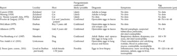
Table 1 lists a total of ten cases of paragonimiasis – six confirmed, three considered likely and one possible case – reported from South Africa since 1928. These cases include 4 cats, 2 dogs and 3 children, with 1 possible case from an adult female patient. Nine of the ten cases were from KwaZulu-Natal but with only four identifiable localities (Mandeni, Stanger, Durban and Umkomaas). The tenth case was an adult who had previously lived in Durban.
Details of the ten records of Paragonimus infection from South Africa. No precise localities are available for the three reports from ‘Zululand’ or for the adult patient. Zululand is usually taken as being that part of the province of KwaZulu-Natal north of the Thukela River, i.e. north of 29o15′S (see fig. 2).

Feline cases
Unfortunately few details are available for the three earliest feline cases (Curson, Reference Curson1928; Mönnig, Reference Mönnig1928; A.Verster, unpubl. data, 1972). Curson (Reference Curson1928) tentatively identified the adult flukes recovered from the lungs of a domestic cat in Zululand as the Asian species Paragonimus (?) westermani. Mönnig (Reference Mönnig1928) recovered his specimens, presumably adults, also from ‘the lungs of a cat in Zululand’ but referred them to Paragonimus kellicotti, a parasite of North American carnivores. Proctor & Gregory (Reference Proctor and Gregory1974) compared sections of unembryonated Paragonimus eggs from cat and human faeces under the transmission electron microscope in an attempt to use eggshell morphology and structure to identify the parasite(s). They did not find any morphological or size differences between eggs from the two hosts (table 1) and were unable to identify the species involved.
Canine cases
A necropsy on an infected dog by van Rensburg et al. (Reference Van Rensburg, Verster and Hiza1987) presents the most detailed account of the clinical symptoms of paragonimiasis in South Africa. The cause of death was given as parasitic pneumonia following a spell of severe respiratory distress. The lungs showed emphysema with large, air-filled bullae, and approximately 20 thin-walled, granulomatous lesions were found embedded in the lung parenchyma. These lesions measured about 15 mm in diameter and each contained two light-brown, conical flukes about 10 mm long. The flukes lay in a brown exudate, possibly haematin of parasitic origin. Numerous eggs were present in the granulomatous layer surrounding the flukes. Faeces were not examined.
Van Andel et al. (Reference Van Andel, Howell, McKenna, Seddon, Julian and Appleton2011) gave similar details for the most recent case, a dog imported into New Zealand from Umkomaas, KwaZulu-Natal, in 2010. The animal was depressed and was coughing two or three times a week with sputum containing flecks of blood. Clinical examination was consistent with a large dog in good condition. Two weeks later, however, the owner reported that the frequency of coughing had increased and the sputum was tinged with blood. Radiographic and other observations reported by van Andel et al. (Reference Van Andel, Howell, McKenna, Seddon, Julian and Appleton2011) conformed to those described previously for an infected dog by van Rensburg et al. (Reference Van Rensburg, Verster and Hiza1987).
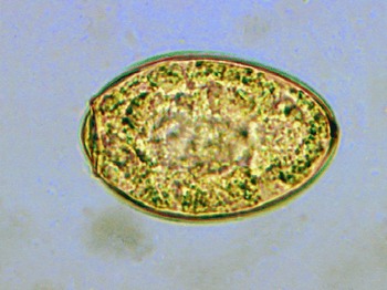
Blood and mucus were noted on the endotracheal tube on removal after the radiographic procedure. Microscopy identified eosinophils, neutrophils and macrophages in the mucus and eggs were scattered throughout the smear. These eggs were oval, operculate and measured approximately 75 × 45 μm and were identified as a species of Paragonimus, a parasite not known in New Zealand (fig. 1). A circulating eosinophilia was noted and Paragonimus eggs were also present in the dog's faeces. The dog was treated with praziquantel (Biltricide® 600 mg) at 50 mg kg− 1 for 3 days and retested at 2-week intervals for circulating eosinophilia and faecal egg counts. All testing undertaken after the administration of praziquantel was negative.
(colour online) Unembryonated egg of Paragonimus sp. (75 × 45 μm) from pulmonary blood/mucus from the infected dog from Umkomaas (photo F. Hill).

Human cases
Proctor & Gregory (Reference Proctor and Gregory1974) compared eggs recovered from the faeces of a child (described as a ‘paediatric patient’) and a cat from Durban (see above) but could not distinguish between them. McCallum (Reference McCallum1975) recovered eggs from a cyst filled with a greenish fluid in the region of the lower ribs on the back of a 9-year-old boy, an ectopic site for a Paragonimus cyst. Samples of the fluid, stained with Papanicolaou stain and counterstained with OG6 and EA50, showed many golden-brown eggs in a field of neutrophils, histiocytes and cellular debris, but without eosinophilia. Eggs were also found in the patient's faeces. The eggs were oval-shaped and operculate with, as also noted by Proctor & Gregory (Reference Proctor and Gregory1974), a slightly thickened ‘shoulder’ at the opercular margins. They were identified as P. kellicotti.
The only adult patient recorded (J. Frean, pers. comm., 2011) presented with eosinophilia and an inflammatory mass involving the liver, pancreas and spleen. A liver biopsy showed eggs that were considered referable to Paragonimus, leading to a diagnosis of possible paragonimiasis.
Discussion
These reports cover a period of approximately 90 years, from 1923 to 2010. The first reports were by Curson (Reference Curson1928) and Mönnig (Reference Mönnig1928), although Curson's material had been collected in Zululand several years earlier, between 1921 and 1923. Mönnig (Reference Mönnig1928) gave no collection date for his record. These two reports may refer to the same case, which may then have been perpetuated as from ‘South Africa’ in Mönnig's Veterinary helminthology and entomology (Reference Mönnig1934) and its various revisions.
Mönnig (Reference Mönnig1928) and McCallum (Reference McCallum1975) attributed the infections they saw to the North American P. kellicotti. Curson (Reference Curson1928) suggested, however, that his material conformed to P. (?) westermani and van Rensburg et al. (Reference Van Rensburg, Verster and Hiza1987) noted that the adult flukes they recovered had bifid spines on the tegument, resembling P. westermani. Blair et al. (Reference Blair, Xu and Agatsuma1999) have pointed out, however, that the tegumental spination of the adult fluke is not a useful character for identification. Unfortunately no specimens were kept for further examination.
The identity of the species involved thus remains uncertain. Several authors have pointed to the fact that infections have been diagnosed in different vertebrate hosts, viz. cats, dogs and humans, and suggested that this apparently low host specificity is indicative of the Asian P. westermani. But Blair et al. (Reference Blair, Xu and Agatsuma1999) have shown that a variety of mammalian hosts have been reported for many species of Paragonimus.
Human paragonimiasis is a zoonosis, and several authors have speculated on the existence of a focus of paragonimiasis in South Africa which could be the source of the human infections reported from the country. Allanson (Reference Allanson1975) thought that the human cases recorded by 1975 were unlikely to have been isolated, and he, McCallum (Reference McCallum1975) and van Rensburg et al. (Reference Van Rensburg, Verster and Hiza1987) all suggested that a reservoir of infection may be present in KwaZulu-Natal. D.M. Blair suggested (in litt., 02.06.2011) that the South African cases may represent an undescribed African species of Paragonimus. Alternatively, it is possible that since other platyhelminth parasites, both trematodes and cestodes, have become invasive in South Africa (Picker & Griffiths, Reference Picker and Griffiths2011), the Paragonimus discussed here might have been introduced.
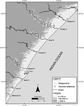
The present review has shown that, with the possible exception of the single adult patient, all the cases reported from South Africa came from the province of KwaZulu-Natal. The four identifiable localities all lie in a narrow strip ± 130 km long and ± 11 km wide (area approximately 1450 km2) below 100 m altitude on the province's coastal plain (fig. 2).
Map of KwaZulu-Natal, South Africa, showing the known distributions of paragonimiasis (squares) and the snail Tomichia natalensis (crosses).

It is arguable that the infection reported by van Andel et al. (Reference Van Andel, Howell, McKenna, Seddon, Julian and Appleton2011) could have been contracted en route from South Africa to New Zealand from food eaten during a stopover in South-East Asia, an endemic area for paragonimiasis. To comply with quarantine regulations, however, pets are always flown on the most direct route, in this case from Durban (an endemic area) to Auckland via Johannesburg and Sydney, all non-endemic areas.
Diagnoses of paragonimiasis in the South African cases have been made on the recovery of adult flukes from the lungs; symptoms such as respiratory difficulties (coughing) comparable to tuberculosis; eosinophilia and neutrophilia and the presence of granulomatous lesions (cysts) in the lungs and extrapulmonary sites. None of the recovered flukes was authoritatively identified, but in most cases oval, golden-brown, operculate eggs conforming to Paragonimus eggs were recovered from cysts, pulmonary blood/mucus and/or faeces. The eggs were unembryonated, with measurements ranging from 68–84 × 40–50 μm (Proctor & Gregory, Reference Proctor and Gregory1974; Allanson, Reference Allanson1975; van Andel et al., Reference Van Andel, Howell, McKenna, Seddon, Julian and Appleton2011) to 90–120 × 46–60 μm (van Rensburg et al., Reference Van Rensburg, Verster and Hiza1987) (see table 1). McCallum (Reference McCallum1975) gave no measurements for his eggs, but following Allanson (Reference Allanson1975), the eggs studied by McCallum (Reference McCallum1975) and Proctor & Gregory (Reference Proctor and Gregory1974) may have come from the same patient, in which case they should have the same measurements, 68–84 × 40–50 μm.
The only adult patient had lived in Durban previously but had since travelled to the Central America/Caribbean area where human paragonimiasis is endemic, and admitted to being ‘an adventurous eater’ (J. Frean, pers. comm., 2011). Eggs seen in the liver biopsy taken from this patient were similar in colour, shape and measurements to those reported in the other cases, but no opercula were seen in the sections examined. As with McCallum's (Reference McCallum1975) case, the affected organs are ectopic locations for paragonimiasis. Extrapulmonary locations have been reported for several Paragonimus species (Blair et al., Reference Blair, Xu and Agatsuma1999).
Final and intermediate hosts
No local mammalian host is known, but the Cape clawless otter, Aonyx capensis, which feeds extensively on crabs, is an obvious candidate. However, attempts by C.H.J. Schutte (South African Medical Research Council, Durban) to find Paragonimus eggs in A. capensis scats and metacercarial cysts in freshwater crabs from rivers in KwaZulu-Natal in the 1980s were unsuccessful.
All known snail intermediate hosts for Paragonimus spp. are prosobranchs (World Health Organization, 1995; Blair et al., Reference Blair, Xu and Agatsuma1999). Two indigenous prosobranch species occur in the area of KwaZulu-Natal from which paragonimiasis has been reported: M. tuberculata (family Thiaridae) and Tomichia natalensis (family Pomatiopsidae). The review by Blair et al. (Reference Blair, Xu and Agatsuma1999) shows that members of both families serve as first intermediate hosts for Paragonimus spp. elsewhere in the world.
Melanoides tuberculata is listed as a first intermediate host of P. westermani in China by the World Health Organization (1995) although Blair et al. (Reference Blair, Xu and Agatsuma1999) note that this has not been proven. Melanoides tuberculata is widespread over the lowlands of KwaZulu-Natal, mostly below 500 m altitude (de Kock & Wolmarans, Reference De Kock and Wolmarans2009), which suggests that if it is indeed susceptible to the local Paragonimus, infections should have been reported over a wider area than is the case. Tomichia natalensis is a poorly known species and has only been collected from a narrow area of the KwaZulu-Natal lowlands, with 11 of the 13 records below 100 m, from Mtunzini in the north to Umkomaas in the south (Porter, Reference Porter1938; Connolly, Reference Connolly1939; Davis, Reference Davis1981; Brown, Reference Brown1994; K.N. de Kock, unpubl. data). It occurs on mud and fallen leaves in streams and rivers shaded by coastal forest. The International Union for Conservation of Nature (IUCN) recognizes T. natalensis as endemic to KwaZulu-Natal and has categorized it as ‘critically endangered’ based on its very restricted distribution (Darwall et al., Reference Darwall, Smith, Allen, Holland, Harrison and Brooks2011).
Only one species of river crab, Potamonautes sidneyi, occurs in the lowlands of KwaZulu-Natal where paragonimiasis has been reported, viz. between the 100 m altitudinal contour and the coast (Hart et al., Reference Hart, Stewart, Bickerton, Day, Stewart, de Moor and Louw2001; Cumberlidge & Daniels, Reference Cumberlidge, Daniels, Darwall, Smith, Tweddle and Skelton2009). This species is frequently found away from water and often in suburban gardens. The dog from Umkomaas diagnosed with paragonimiasis by van Andel et al. (Reference Van Andel, Howell, McKenna, Seddon, Julian and Appleton2011) repeatedly ate crabs. Anecdotal evidence (M. van Andel, pers. comm., 2011) indicates that other dogs that lived on the same property had to be euthanized after lung X-rays showed changes consistent with paragonimiasis.
A comment by Hart et al. (2001) that river crabs are eaten as a delicacy by people in KwaZulu-Natal was supported by S. Kasavan (University of KwaZulu-Natal) who told the author that members of the Indian community often collect crabs, particularly river crabs, and cook them to make a spicy soup – especially to alleviate respiratory symptoms such as congested or ‘tight’ chests and asthma. Cooking will, of course, kill any metacercarial cysts present in the crabs, which might explain the small number of paragonimiasis infections seen in people.
A complicating factor may be the recent invasion of KwaZulu-Natal by the South-East Asian thiarid snail Tarebia granifera (Appleton et al., Reference Appleton, Forbes and Demetriades2009). Although the literature contains many reports that T. granifera is susceptible to P. westermani and serves as its first intermediate host in Asia, Michelson (Reference Michelson1992) contends that there is no evidence for these claims. Tarebia granifera is, however, spreading in eastern South Africa and, since it has been shown to compete with indigenous gastropods in a lake in the same area of KwaZulu-Natal (Miranda & Perissinotto, Reference Miranda and Perissinotto2012), anecdotal evidence that it is displacing M. tuberculata in the province may be correct. Tarebia granifera has also invaded several of the rivers where T. natalensis occurs, but there is no information on its impact on this species.
Conclusions
Nine of the ten cases of paragonimiasis reported from South Africa involved cats, dogs and children in the province of KwaZulu-Natal. The tenth, involving the only adult person, was tentatively diagnosed and could have been contracted in the province or outside the country. Four of the infections reported from cats and dogs (Curson, Reference Curson1928; Mönnig, Reference Mönnig1928; van Rensburg et al., Reference Van Rensburg, Verster and Hiza1987; van Andel et al., Reference Van Andel, Howell, McKenna, Seddon, Julian and Appleton2011) had lung involvement, while the two human cases for which adequate details are available (McCallum, Reference McCallum1975; J. Frean, pers. comm., 2011) were extrapulmonary infections.
The diagnosis of paragonimiasis in a dog from KwaZulu-Natal in 2010 (van Andel et al., Reference Van Andel, Howell, McKenna, Seddon, Julian and Appleton2011), 25 years after the next most recent autochthonous case in 1985 (van Rensburg et al., Reference Van Rensburg, Verster and Hiza1987), suggests that transmission is active in KwaZulu-Natal although not commonly reported. It seems probable that a zoonotic focus of paragonimiasis exists in the province but is confined to a narrow belt below 100 m altitude in the coastal lowlands. No specimens of the adult fluke were kept, so it cannot be identified. The definitive host is likely to be the otter A. capensis. Both thiarid and pomatiopsid snails have been shown to serve as first intermediate hosts for Paragonimus spp. elsewhere in the world (Blair et al., Reference Blair, Xu and Agatsuma1999). In the present situation, therefore, the snail host could be either M. tuberculata or T. natalensis. Melanoides tuberculata is widely distributed, but the range of T. natalensis is narrow, with almost all records lying below 100 m. It thus matches that of the reported localities for the disease (fig. 2). The second intermediate host is probably the widely distributed river crab P. sidneyi.
The similarity between the geographic ranges of the South African paragonimiasis cases and the endemic pomatiopsid snail T. natalensis (fig. 2) suggests that T. natalensis may be its first intermediate host and that the parasite is an indigenous and undescribed species of Paragonimus. If the invasive T. granifera does displace, or even compete with, the snail intermediate host of the Paragonimus species under review, either M. tuberculata or T. natalensis, it could bring transmission to an end.
Future research should confirm transmission in the area by demonstrating the parasite's life cycle, i.e. determining whether M. tuberculata or T. natalensis is the first intermediate host and whether P. sidneyi is the second intermediate host in the area identified in fig. 2. A faecal survey of dogs and cats across KwaZulu-Natal could establish the extent of transmission, as could a questionnaire survey of veterinarians in the province, asking whether or not they have observed the pulmonary masses characteristic of paragonimiasis in cats and dogs on X-ray.
Acknowledgements
I am grateful to several colleagues for assistance. In particular I acknowledge Mary van Andel (MAF Biosecurity, New Zealand) for providing details on the latest canine case (ex. Umkomaas) and Dr Fraser Hill (Gribbles Veterinary Laboratories, Palmerston North, New Zealand) for supplying the photomicrograph of an egg from this dog (fig. 1). Professor J. Frean (National Institute for Communicable Diseases, Johannesburg, South Africa) provided details on the only recorded adult paragonimiasis patient and Professor D. Blair (James Cook University, Australia) helped with the literature. Professors J. Boomker and L. Prozesky (University of Pretoria, South Africa) confirmed that no local Paragonimus material was lodged in the collections at Onderstepoort, and Professor K.N. de Kock (Northwest University, South Africa) made available the KwaZulu-Natal locality records for Tomichia natalensis from the National Freshwater Snail Collection.