Glucocorticoids are beneficial when secreted after acute stress, but prolonged glucorticoid elevations, due to chronic stress or illness, produce adverse affective, cognitive, and physical consequences, including mood symptoms and memory impairments in adults (Charney, Reference Charney2004; de Kloet, Joels, & Holsboer, Reference de Kloet, Joels and Holsboer2005; McEwen, Reference McEwen1998). These deleterious effects are thought to result from abnormal modulation of glucorticoid receptors in the medial temporal lobe (MTL), encompassing the amygdala and hippocampus (Charney, Reference Charney2004; de Kloet et al., Reference de Kloet, Joels and Holsboer2005; Lupien & Lepage, Reference Lupien and Lepage2001; Nemeroff, Reference Nemeroff2004; Roozendaal, Reference Roozendaal2003).
We recently demonstrated that adolescents with major depressive disorder (MDD) exhibit abnormally high MTL responses when encoding affective stimuli into memory (Roberson-Nay et al., Reference Roberson-Nay, McClure, Monk, Nelson, Guyer and Froom2006). Such findings may relate to prior research noting associations among glucocorticoid hypersecretion, MDD, and perturbed MTL function. However, elevated cortisol levels in pediatric MDD is reported less consistently than in adult MDD (see reviews by de Kloet et al., Reference de Kloet, Joels and Holsboer2005; Goodyer, Park, Netherton, & Herbert, Reference Goodyer, Park, Netherton and Herbert2001). Such data have led to the suggestion that glucocorticoids show minimal relationships with MTL function in children and adolescents.
This relationship, however, has not been thoroughly examined in youths because of the difficulty in conducting such studies. Manipulating glucocorticoid levels in pediatric populations, pharmacologically or through stress challenges, poses ethical problems. Here, we investigate the association between glucocorticoids and MTL function in adolescents who suffer from chronic endogenous hypersecretion of cortisol due to Cushing syndrome. This condition provides a unique naturalistic model for studying the cerebral consequences of excess cortisol. Similar to lesion studies, the examination of pathological development can shed light on normal development (Cicchetti, Reference Cicchetti2003; O'Connor, Reference O'Connor2003). Adults with Cushing syndrome present reduced hippocampal volume, declarative memory impairments, and depressive symptoms (Bourdeau et al., Reference Bourdeau, Bard, Forget, Boulanger, Cohen and Lacroix2005; Forget, Lacroix, Somma, & Cohen, Reference Forget, Lacroix, Somma and Cohen2000). Surprisingly, adolescents with Cushing syndrome seem to fail to manifest depressive symptoms (Magiakou & Chrousos, Reference Magiakou and Chrousos2002; Merke et al., Reference Merke, Giedd, Keil, Mehlinger, Wiggs and Holzer2005). This would suggest that the combination of elevated cortisol and MTL alteration is not sufficient to trigger the emergence of mood symptoms in these adolescents, implying the presence of protective factors that are lacking in adults with depression.
Although no animal or human studies have yet examined the effects of chronically elevated corticosteroid levels on emotional memory, acute elevations in cortisol levels have been shown to enhance memory for emotional stimuli (Abercrombie, Kalin, Thurow, Rosenkranz, & Davidson, Reference Abercrombie, Kalin, Thurow, Rosenkranz and Davidson2003; Buchanan & Lovallo, Reference Buchanan and Lovallo2001; Cahill, Gorski, & Le, Reference Cahill, Gorski and Le2003; Roozendaal, Reference Roozendaal2003). Recent animal studies suggest that acute, as well as chronic, stress stimulates the amygdala (Mitra, Jadhav, McEwen, Vyas, & Chattarji, Reference Mitra, Jadhav, McEwen, Vyas and Chattarji2005; Poeggel et al., Reference Poeggel, Helmeke, Abraham, Schwabe, Friedrich and Braun2003; Roozendaal, Reference Roozendaal2003; Vyas, Mitra, Shankaranarayana Rao, & Chattarji, Reference Vyas, Mitra, Shankaranarayana Rao and Chattarji2002), which is implicated in emotional memory (Phelps & LeDoux, Reference Phelps and LeDoux2005).
In line with these results, we expect chronically elevated cortisol levels to predict enhanced MTL activation during the encoding of emotional information. To this aim, we compared adolescents with Cushing syndrome and healthy adolescents on MTL reactivity using the exact same paradigm and analysis employed in our previous work on adolescent MDD (Roberson-Nay et al., Reference Roberson-Nay, McClure, Monk, Nelson, Guyer and Froom2006).
Methods
Participants
Twelve adolescents with endogenous Cushing syndrome were compared to 22 healthy adolescents. Endogenous Cushing syndrome is rare, and almost always due to one of two conditions, pituitary adenoma or adrenal gland tumors (Magiakou & Chrousos, Reference Magiakou and Chrousos2002). Patients with Cushing syndrome were being followed at the NIH Clinical Center and enrolled in the study on a voluntary basis. Healthy subjects were recruited in the community. The study was approved by the institutional review boards of the National Institute of Mental Health and National Institute of Child Health and Human Development. Prior to participation in the study, parents and adolescents gave written consent and assent, respectively. All subjects were compensated for participation in the study following the guidelines provided by the National Institute of Mental Health.
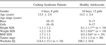
All subjects underwent physical and psychiatric examinations. Exclusion criteria included chronic medical condition (except for Cushing syndrome) and pharmacological treatment unrelated to Cushing syndrome. All participants were free from any present or lifetime history of psychiatric disorders, as assessed by the Kiddie Schedule for Affective Disorders and Schizophrenia—Present and Lifetime Version (KSADS-PL; Kaufman et al., Reference Kaufman, Birmaher, Brent, Rao, Flynn and Moreci1997) with parents and adolescents separately. Tanner stage in all patients was determined by a pediatrician and a self-administered questionnaire in control subjects (Duke, Litt, & Gross, Reference Duke, Litt and Gross1980). Patients with Cushing syndrome had hormones measured as part of their clinical evaluation. All participants scored within the average/above average on the Wechsler Abbreviated Scale of Intelligence for Children (Wechsler, Reference Wechsler1999). Demographic and clinical characteristics are presented in Table 1.
Demographic and clinical characteristics of patients with Cushing syndrome and healthy adolescents

Note: Values are reported as means ± SD. Reduced n indicates missing data.
*p < .05. **p < .01.
Face memory task
The memory paradigm consisted of two performance phases, encoding during functional magnetic resonance imaging (fMRI) scanning and recognition outside the scanner. This memory paradigm has previously been used successfully in our laboratory for behavioral and fMRI studies of adolescents (Nelson et al., Reference Nelson, McClure, Monk, Zarahn, Leibenluft and Pine2003; Pine et al., Reference Pine, Lissek, Klein, Mannuzza, Moulton and Guardino2004; Roberson-Nay et al., Reference Roberson-Nay, McClure, Monk, Nelson, Guyer and Froom2006). The paradigm uses a variant of the frequently employed “subsequent memory” paradigm from cognitive neuroscience.
During the encoding phase, participants viewed 32 portraits of adult actors selected randomly from a larger pool of 56 actors. These photographs were selected from three standardized sets of gray-scale photographs depicting different facial expressions constructed by Ekman and Friesen (Reference Ekman and Friesen1976), Gur (www.uphs.upenn.edu/bbl/pubs/downloads/nptasks.shtml), and Tottenham and Nelson (www.macbrain.org/faces/index.htm). Each actor presented one of four emotional expressions (happy, fear, angry, neutral) throughout the entire encoding paradigm, and eight different actors were viewed for each expression (8 × 4 = 32). Although facial expressions for a given actor were held constant within each participant's task, expressions varied randomly across different participants' tasks. Thus, one participant may have viewed a given actor displaying a consistently happy expression, whereas a subsequent participant may have viewed the same actor displaying a consistently angry expression. This feature controlled for variability in nonemotional aspects of the actors (e.g., ethnicity, gender, hair texture) that influences memory formation. While in the scanner, participants viewed the series of 32 adult faces (8 happy, 8 angry, 8 fearful, 8 neutral) under four conditions. Three conditions required participants to attend to different aspects of the face stimuli: (a) How afraid did the stimulus make you feel? (b) How hostile does the face appear? (c) How large is the nose? These questions were rated on a five-button box (1 = not very to 5 = extremely). In the fourth condition, participants' attention was unconstrained, such that the faces were viewed passively without participants making any ratings.
As described in Roberson-Nay et al. (Reference Roberson-Nay, McClure, Monk, Nelson, Guyer and Froom2006), the use of these different attention manipulations facilitates encoding success by reducing boredom associated with repeated viewing of the same pictures. Moreover, as in all of our prior studies of memory encoding, fMRI analyses reported here contrast “successfully” relative to “unsuccessfully” encoded pictures by collapsing data acquired in each of the four conditions. This approach maximizes statistical power by allowing a more precise estimation of encoding-related brain activation. Moreover, as delineated in Roberson-Nay et al. (Reference Roberson-Nay, McClure, Monk, Nelson, Guyer and Froom2006), the inclusion of 32 “null-event” trials, consisting of fixation crosses, occurred randomly within each condition to facilitate interpretation of brain activations.
The four conditions were presented as a 14-min single run comprising 160 trials (8 actors × 5 stimuli [4 emotions and fixation] × 4 conditions). Each facial expression was presented a total of four times, once during each of the four conditions. Trials within a given condition were blocked together, and presentation order of condition and facial expressions was randomized across participants. Rating instructions appeared for 3 s before each condition block. Facial expression and fixation trials were shown for 4 s each. Each face and fixation trial was followed by an intertrial interval showing a blank screen that varied randomly from 750 to 1250 ms.
Stimuli were displayed using Avotec Silent Vision Glasses (Stuart, FL), and responses were recorded by a five-key button box developed by MRI Devices (Waukesha, WI). Participants were trained in an MRI simulator prior to entering the scanner to become familiar with the actual MRI environment and response device. Participants were also administered a practice version of the task to ensure understanding of the task. The practice version contained pictures of only neutral facial expressions that were not shown in the MRI scanner.
After completing the fMRI scanning session, participants performed the surprise recognition memory test. As indicated previously (Nelson et al., Reference Nelson, McClure, Monk, Zarahn, Leibenluft and Pine2003; Pine et al., Reference Pine, Lissek, Klein, Mannuzza, Moulton and Guardino2004; Roberson-Nay et al., Reference Roberson-Nay, McClure, Monk, Nelson, Guyer and Froom2006), the use of this incidental memory paradigm, with a surprise postscan recognition test, was designed to address two issues. First, these procedures mirror memory-encoding processes elicited when dealing with emotionally salient stimuli of everyday life. Second, the procedures minimize potential effects on memory encoding related to underlying between-group differences in effort, strategic approaches to memory encoding, or compliance with encoding instructions.
The recognition memory test consisted of 48 photographs, including 24 previously seen and 24 new actors, presented on a computer screen. Previous work showed that the inclusion of neutral faces at encoding and recall interfered with memory formation (see Nelson et al., Reference Nelson, McClure, Monk, Zarahn, Leibenluft and Pine2003; Pine et al., Reference Pine, Lissek, Klein, Mannuzza, Moulton and Guardino2004). Thus, the actors portraying the neutral faces during encoding were not used at recall. In addition, the pictures included at recall comprised only actors portraying neutral faces, that is, different expressions from the emotional expressions seen during encoding. Actors were randomly selected for each research participants to serve as “targets” that were presented both at encoding and recall, or new actors (“lures”) that were only presented at recall. This procedure minimized the possibility that actor-specific characteristics, apart from the portrayed emotion, would influence memory. During the recognition memory test, which occurred approximately 30–40 min after presentation of the faces in the scanner, participants were asked to rate the actors as old or new. The selective use of neutral expressions at recall significantly increased the difficulty of recognition. Subjects were required to encode features of individuals, who displayed a given emotion, and then to use these representations, when attempting to recognize individuals depicting no emotion. At recall, each face was presented for 4 s, during which time subjects indicated “old” or “new” by button press.
Memory scores were based on the signal detection theory. Specifically, a measure of signal detection threshold (d′) was calculated for each participant for each emotion by subtracting the z score for hits for each emotion from the overall z score for false alarms (Snodgrass & Corwin, Reference Snodgrass and Corwin1988). This generated d′ estimates for each face type: “happy,” “fearful,” and “angry.” A d′ value for total hits (z score for false alarms – z score for sum of hits for all emotional face types) was also calculated for each participant. Conceptually, d′ represents the degree to which a subject is capable of differentiating signals or “true targets” from noise. Higher d′ values indicate increasing ability to correctly pick out a specific target type from a series of distracters, and positive d′ values indicate higher true signal reporting than false alarm reporting. The postscan memory test allowed data acquired at encoding in the scanner to be “binned” based on the postscan recognition (remembered vs. forgotten).
MRI data acquisition
Whole-brain blood oxygen level dependent (BOLD) fMRI data were acquired on a General Electric Signa 3-tesla magnet (Waukesha, WI). Following sagittal localization and a manual shim procedure, functional T2*-weighted images were gathered using an echo planar single-shot gradient echo pulse sequence with a matrix size of 64 × 64 mm, repetition time (TR) of 2000 ms, echo time (TE) of 40 ms, field of view (FOV) of 240 mm, and voxels of 3.75 × 3.75 × 5 mm, providing whole-brain coverage. Echo planar images (EPI) were acquired in 23 contiguous 5-mm axial slices per brain volume positioned parallel to the anterior commissure and posterior commissure (AC–PC) line. All functional data were obtained in a single 14-min run per subject. Following EPI data collection, a high-resolution T1-weighted anatomical image was acquired for each subject using a standardized magnetization-prepared gradient echo sequence (180 1-mm sagittal slices, FOV = 256, number of excitations [NEX] = 1, TR = 11.4 ms, TE = 4.4 ms, matrix size = 256 × 256, time to inversion [TI] = 300 ms, bandwidth = 130 Hz/pixel, 33 kHz/256 pixels) to facilitate spatial normalization.
Data analysis
Demographic and clinical characteristics
Demographic and clinical characteristics were compared between patients with Cushing syndrome and healthy adolescents using two-sample Student t tests for continuous measures or chi-square tests for nominal measures. Body mass index (BMI), height and weight standard deviation score (SDS) were determined using anthropometric reference data for US children (Center for Disease Control, Epi InfoTM, version 3.3.2).
Behavioral data
Memory recall d′ values were evaluated for assumptions of normality and sphericity. These variables were distributed normally, and Greenhouse–Geisser (Reference Greenhouse and Geisser1959) corrections were applied when the sphericity criterion was not met. A mixed repeated-measures analysis of variance (ANOVA) with diagnosis (Cushing syndrome vs. healthy) as the between-subjects factor and emotional faces (happy vs. fearful vs. angry) as the within-subjects factor was conducted to assess effects of diagnosis on performance and its potential interaction with emotional faces.
Imaging data
Preprocessing data procedures and fMRI data analyses were performed using the Statistical Parametric Mapping (SPM99, Wellcome Department of Cognitive Neurology, London) software and supplemental routines written in Matlab 6 (Mathworks, Natick, MA). Imaging data for participants moving more than 2.0 mm in any plane as assessed with MedX software (Medical Numerics, Sterling, VA) were excluded. Preprocessing procedures included corrections for slice timing and motion, coregistration to the anatomical data, and spatial normalization to a Montreal Neurologic Institute (MNI) T1-weighted template image supplied with SPM99b.
Individual subject level event-related response amplitudes were estimated using a general linear model for each event type. Event-types were specified based on the emotion of the faces, and whether faces viewed at encoding were remembered or forgotten. Fixation trials served as an implicit baseline. The waveform used to model event-related responses was a rectangular pulse (4-s duration) convolved with the hemodynamic response function specified in SPM99. Contrast images were created for each subject using pairwise comparisons of the event-related BOLD response amplitudes across conditions. Before performing group-level analyses, each contrast image was divided by the subject-specific voxel time series mean, generating values proportional to percentage fMRI signal change (Zarahn, Aguire, & D'Esposito, Reference Zarahn, Aguirre and D'Esposito1997). These normalized contrast images were then smoothed with an isotropic Gaussian kernel (full width at half-maximum = 11.4) to reduce nonstationarity in the spatial autocorrelation structure produced by the previous step (Friston, Mechelli, Turner, & Price, Reference Friston, Mechelli, Turner and Price2000).
A priori hypotheses motivated a region of interest (ROI)-based analysis of the amygdala, and anterior hippocampus. These ROIs were ascertained from standard anatomical criteria on a single MNI template and applied to all normalized brains at the group level (Szeszko et al., Reference Szeszko, Robinson, Alvir, Bilder, Lencz and Ashtari1999, Reference Szeszko, Strous, Goldman, Ashtari, Knuth and Lieberman2002). Voxelwise tests were conducted in these anatomically defined volumes of interest. Consistent with the current standard (Hariri, Tessitore, Mattay, Fera, & Weinberger, Reference Hariri, Tessitore, Mattay, Fera and Weinberger2002; Winston, Strange, O'Doherty, & Dolan, Reference Winston, Strange, O'Doherty and Dolan2002), we utilized the Gaussian random field threshold (α = .05, corrected) with small volume correction (SVC) implemented in SPM99 (Worsley, Marrett, Neelin, Vandal, Friston, & Evans, Reference Worsley, Marrett, Neelin, Vandal, Friston and Evans1996). Statistical significance of activation in regions of interest was set to corrected p < .05.
Similar to our previous work in depressed adolescents (Roberson-Nay et al., Reference Roberson-Nay, McClure, Monk, Nelson, Guyer and Froom2006), we tested between-group differences in the contrast of BOLD signal changes during the viewing of the faces that were remembered on later recall versus during the viewing of the faces that were forgotten on later recall (remembered faces vs. forgotten faces). Again, consistent with our prior study, and because we had no specific a priori hypotheses regarding effects of emotion type, we pooled emotions together in this analysis. This strategy permitted to optimize statistical power.
Finally, to better understand the nature of significant activations, we evaluated separately significant loci of activation for the contrasts remembered versus baseline and forgotten versus baseline in each group. These analyses were conducted in SPSS-14.0 on the mean extracted BOLD signal changes of each significant locus. In addition, correlations were performed among these extracted mean values, memory performance in the whole sample, and cortisol levels in the Cushing syndrome group (Figure 1).
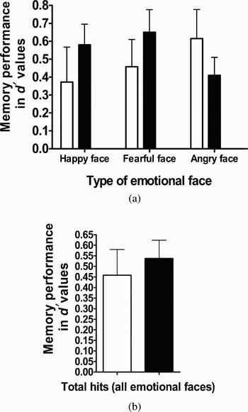
A comparison of memory performance between groups. Mean memory d′ values between patients with Cushing syndrome (open bars) and healthy adolescents (filled bars) for (a) each emotional face and (b) total hits (collapsed across all emotional faces). There were no group differences regarding memory performance (all ps > .10). Error bars represent standard error of the mean.

Two control regions, the orbitofrontal cortex (OFC) and fusiform gyrus, were used to examine the regional specificity of the findings. These regions were chosen because of the absence of reported functional abnormalities in these areas in Cushing syndrome, and because of their role in emotion processing (OFC; Phillips, Drevets, Rauch, & Lane, Reference Phillips, Drevets, Rauch and Lane2003) and stimulus identification (Adolphs, Reference Adolphs2003), rather than in memory encoding.
Results
Demographic and clinical characteristics
Patients with Cushing syndrome and healthy adolescents did not differ on age (Cushing syndrome patients: 13.5, SD = 2.9; Healthy adolescents: 14.2, SD = 2.4), gender distribution (Cushing syndrome patients: 4 boys, 8 girls; healthy adolescents: 10 boys, 12 girls), puberty level, or Wechsler IQ (Cushing syndrome patients: 114.4, SD = 13.1; healthy adolescents: 108.1, SD = 16.0; see Table 1). As expected, patients with Cushing syndrome had significantly higher BMI, t (29) = 4.3, p < .001, and weight, t (29) = 2.5, p < .03, and shorter stature, t (29) = 2.9, p < .009, than age-matched controls. All patients with Cushing syndrome had biochemically confirmed hypercortisolism (mean ± SD, urinary free cortisol corrected for body surface area: Cushing syndrome group: 132.66 ± 55.77 µg/m2/24 hr; normal range in healthy adolescents of the same Tanner puberty stage: <68 µg/m2/24 hr) and lacked diurnal variation in plasma cortisol concentrations (mean ± SD, morning cortisol, 21.5 ± 9.7 µg/dl; midnight cortisol, 19.1 ± 8.5 µg/dl). The average duration of Cushing syndrome based on onset of decreased growth velocity was 2.6 years (range = 1–4.5 years). None of the adolescents with Cushing syndrome were receiving treatment at the time of testing, and all patients were evaluated before surgery for removal of the pituitary tumor (10 patients) or bilateral adrenalectomy (2 patients with primary pigmented neoplastic adrenal disease).
Behavioral data
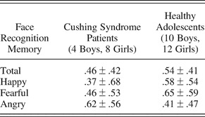
The d′ values used to measure memory for happy and fearful and angry faces are displayed in Table 2. The d′ values for total hits (i.e., hits for the three types of emotional faces combined) were significantly different from zero in both the Cushing syndrome patients, t (11) = 3.8, p < .004, and healthy adolescents, t (21) = 6.2, p < .001, indicating that overall hit rates were significantly higher than the false alarm rates in both the patient and healthy groups. The mixed repeated-measures ANOVA measuring the influence of diagnosis on memory for happy, fearful and angry faces revealed no main effects of emotional faces, F (2, 64) = 0.23, p > .10, or of diagnosis, F (1, 32) = .19, p > .10, and no Emotional Faces × Diagnosis interaction, F (2, 64) = 2.02, p > .10. Therefore, both the patient and the healthy adolescents performed similarly on memory recall of each emotional face types. In addition, the absence of effect of Emotional faces on performance supported the pooling of emotions in the fMRI analysis.
Predictors of memory performance (d′ values) in patients with Cushing syndrome and healthy adolescents

Note: Values are means ± SD. significant difference between both groups on memory recall for total hits and types of emotional faces (ps > .10).
Imaging data
Amygdala ROI analysis
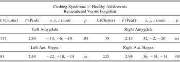
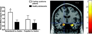
Amygdala response to remembered versus forgotten faces revealed greater activation in the left amygdala in Cushing syndrome patients compared to healthy adolescents, t = 2.84 (MNI x, y, z = −14, −6, −10 mm), p < .04 SVC corrected. No group differences emerged in the right amygdala (see Table 3).
Regional activations in the group comparison of remembered versus forgotten

Note: The regions of interest (ROI) analyses for the left amygdala and right anterior hippocampus. The statistical threshold is set at p < .05 after a small volume correction of the ROI.
Next, a repeated-measures ANOVA was conducted in SPSS-14.0 on the extracted BOLD signal percent changes of the left amygdala ROI in the contrasts remembered versus fixation and forgotten versus fixation. This analysis revealed that the significant group difference in left amygdala for the contrast remembered versus forgotten reflected greater activation for remembered versus fixation in patients with Cushing syndrome than in healthy adolescents, F (1, 32) = 8.9, p < .007. No significant group differences emerged for forgotten versus fixation, F (1, 32) = .54, p > .10 (Figure 2).
Mean BOLD signal differences in the left amygdala (MNI −14, −6, −10 mm) for contrasts remembered versus fixation and forgotten versus fixation in Cushing syndrome patients and healthy adolescents. Adolescents with Cushing syndrome showed significantly higher activation when compared to healthy adolescents (p < .05). Error bars represent standard error of the mean.

Left amygdala activations in the contrasts of remembered versus forgotten, remembered versus fixation, or forgotten versus fixation were not correlated with d′ performance scores in the Cushing syndrome and healthy adolescents groups, combined or separately. Moreover, measures of 24-hr urinary cortisol levels in the Cushing syndrome patients did not correlate with left amygdala activation (remembered vs. forgotten; p > .10).
Anterior hippocampal ROI analysis
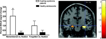
Anterior hippocampal response to remembered versus forgotten faces revealed greater activation in the right anterior hippocampus, t = 2.98 (MNI x, y, z = 36, −14, −14 mm), p < .03, SVC corrected, in Cushing syndrome patients compared to healthy adolescents (see Table 3). No group differences emerged in the left anterior hippocampus.
Next, a repeated-measures ANOVA was conducted in SPSS-14.0 on the extracted BOLD signal percent changes of the right anterior hippocampus ROI in the contrasts remembered versus fixation and forgotten versus fixation (Figure 3). Similarly to the findings in the left amygdala, the significant group difference in right anterior hippocampus for the contrast remembered versus forgotten reflected greater activation for remembered versus fixation in patients with Cushing syndrome than in healthy control, F (1, 32) = 9.8, p < .005. No significant group differences emerged for forgotten versus fixation, F (1, 32) = 2.21, p < .15.
Mean BOLD signal differences in the right anterior hippocampus (MNI 36, −14, 14 mm) for contrasts remembered versus fixation and forgotten versus fixation in Cushing syndrome patients and healthy adolescents. Adolescents with Cushing syndrome showed significantly higher activation when compared to healthy adolescents (p < .05). Error bars represent standard error of the mean.

Right anterior hippocampal activations in the contrasts of remembered versus forgotten, remembered versus fixation, or forgotten versus fixation were not correlated with d′ performance scores in the Cushing syndrome and healthy adolescents groups, combined or separately. Moreover, measures of 24-hr urinary cortisol levels in the Cushing syndrome patients did not correlate with right anterior hippocampal activation (remembered vs. forgotten contrast; p > .10).
Control ROIs: OFC and fusiform gyrus
ROI analyses of left and right OFC, and fusiform gyrus showed no significant group differences in the main contrast (remembered vs. forgotten; ps > .10), supporting a regional specificity of the significant group differences reported above.
Discussion
This study is the first to demonstrate associations between endogenous chronic hypersecretion of cortisol levels and perturbed engagement of brain substrates previously associated with depression and emotional memory. This work uses a naturalistic approach that provides a unique opportunity to address potential mechanisms of developmental psychopathology. Adolescents with Cushing syndrome suffer from chronic endogeneous hypersecretion of cortisol by pituitary adenoma or adrenal gland tumors (Magiakou & Chrousos, Reference Magiakou and Chrousos2002), and represent a natural model of chronic exposure to cortisol during development. This approach is based on the tenet held in the field of developmental psychopathology, advocating for the notion that the study of atypicality is an important avenue to inform normal development and reciprocity (Cicchetti, Reference Cicchetti2003).
We show here that the study of adolescents with Cushing syndrome reproduces imaging findings previously obtained with an independent sample of depressed adolescents, using an identical paradigm and analysis (Roberson-Nay et al., Reference Roberson-Nay, McClure, Monk, Nelson, Guyer and Froom2006). Left amygdala activation in response to successful encoding of evocative faces was significantly higher in adolescents with Cushing syndrome than in healthy controls. In addition, activation of the right anterior hippocampus, a region involved in emotional processing (Bannerman et al., Reference Bannerman, Rawlins, McHugh, Deacon, Yee and Bast2004; Dolcos, LaBar, & Cabeza, Reference Dolcos, LaBar and Cabeza2004), was also abnormally elevated in these adolescents.
None of the adolescents with Cushing syndrome suffered from a psychiatric disorder, including mood disorder. This is important, as it suggests that exaggerated amygdala reactivity combined with chronic elevation of glucocorticoids, a marker of depression in adults (Charney, Reference Charney2004; de Kloet et al., Reference de Kloet, Joels and Holsboer2005; Nemeroff, Reference Nemeroff2004), is not sufficient for mediating depression in adolescents. This observation further suggests that protective factors may be at play. A number of neurotransmitters, neurohormones, brain circuits, and genes have been linked to the protective mechanisms underlying resiliency (see Curtis & Cicchetti, Reference Curtis and Cicchetti2003; Charney, Reference Charney2004). Adolescents with Cushing syndrome may activate such protective neural mechanisms, which may account for their resiliency to depression. These protective mechanisms may be specific to the endocrine disease or to the potentially more plastic, still maturing neural networks in adolescents. The high prevalence of depression in adults with Cushing syndrome (see Bourdeau et al., Reference Bourdeau, Bard, Forget, Boulanger, Cohen and Lacroix2005; Forget et al., Reference Forget, Lacroix, Somma and Cohen2000) relative to children and adolescents with Cushing syndrome (Magiakou & Chrousos, Reference Magiakou and Chrousos2002; Merke et al., Reference Merke, Giedd, Keil, Mehlinger, Wiggs and Holzer2005) suggests that the latter proposition (plasticity) or a combination of both (plasticity interacting with disease specificity) could explain this age difference in the psychiatric symptoms associated with Cushing syndrome. Clearly, further research investigating possible protective neurobiological factors conferring resiliency to psychopathology is critical.
Age-related differences in the biological correlates of depression have also been suggested within the corticosteroid system. In a significant percentage of adults with MDD, cortisol levels were reported to be elevated throughout the day (de Kloet et al., Reference de Kloet, Joels and Holsboer2005; Pariante, Reference Pariante2006; Pariante and Miller, Reference Pariante and Miller2001). In children and adolescents, however, most studies have failed to detect elevated circadian levels of cortisol (Birmaher et al., Reference Birmaher, Dahl, Perel, Williamson, Nelson and Stull1996; Dahl et al., Reference Dahl, Puig-Antich, Ryan, Nelson, Novacenko and Twomey1989; Gipsen-de Wied, Jansen, Duyx, Thijssen, & van Engeland, Reference Gispen-de Wied, Jansen, Duyx, Thijssen and van Engeland2000; Puig-Antich et al., Reference Puig-Antich, Dahl, Ryan, Novacenko, Goetz and Goetz1989; see Kutcher & Sokolov, Reference Kutcher, Sokolov and Goodyer2001). Subtle developmental differences in the pattern of cortisol secretion in depression have been proposed with respect to the typical diurnal variation of this hormone (Forbes et al., Reference Forbes, Williamson, Ryan, Birmaher, Axelson and Dahl2006). Hence, cortisol hypersecretion has been found around sleep onset in depressed adolescents (Dahl et al., Reference Dahl, Ryan, Puig-Antich, Nguyen, al-Shabbout and Meyer1991; Goodyer, Park, & Herberg, Reference Goodyer, Park and Herbert2001). The density threshold (temporal and magnitude characteristics) of cortisol levels at which central effects occur is not known. Therefore, although circadian cortisol patterns in depressed adolescents seem to less perturbed than in depressed adults, brain substrates in adolescents might be exquisitely vulnerable to potentially deleterious effects of elevated corticosteroids around sleep onset, which could mediate amygdala hyperreactivity in depressed teenagers.
Pharmacological manipulations of corticosteroid levels in animals have been performed in the adult and perinatal periods, but not in the adolescent period (see review by Romeo & McEwen, Reference Romeo and McEwen2006). This limits our ability to infer from animal work the cerebral consequences of chronic hypercortisolemia of late childhood onset, a time of unique developmental changes (e.g., Gogtay et al., Reference Gogtay, Giedd, Lusk, Hayashi, Greenstein and Vaituzis2004). The distinct neural plasticity of the adolescent period is clearly evidenced in the maturation of reward systems. Indeed, the juvenile animal has been shown to harbor vulnerabilities to addictive drugs that differ quantitatively and qualitatively from those reported neonatally or in adulthood (see reviews by Ernst, Pine, & Hardin, Reference Ernst, Pine and Hardin2006; Spear, Reference Spear2000).
Animal studies of the neural and behavioral consequences of chronic stress accompanied with hypercorticosteroidism have been examined when stressors were applied at different periods of development (perinatal, juvenile, or adult). Findings include amygdala cellular (Mitra et al., Reference Mitra, Jadhav, McEwen, Vyas and Chattarji2005; Poeggel et al., Reference Poeggel, Helmeke, Abraham, Schwabe, Friedrich and Braun2003; Vyas et al., Reference Vyas, Mitra, Shankaranarayana Rao and Chattarji2002) and hippocampal structural alterations (de Kloet et al., Reference de Kloet, Joels and Holsboer2005; Isgor et al., Reference Isgor, Kabbaj, Akil and Watson2004; Kim & Diamond, Reference Kim and Diamond2002), which were associated with memory deficits and enhanced anxiety. The timing of these neural and behavioral changes, as well as their potential for normalization once the stress was removed, appear to differ as a function of when the chronic stress was applied (see review by Romeo & McEwen, 2006).
In humans, similar findings of hippocampal atrophy associated with memory and anxiety problems have been reported in adults who suffered a chronic stress (Bourdeau et al., Reference Bourdeau, Bard, Forget, Boulanger, Cohen and Lacroix2005; de Kloet et al., Reference de Kloet, Joels and Holsboer2005; Lupien et al., Reference Lupien, Fiocco, Wan, Maheu, Lord and Schramek2005). The only study that examined the behavioral and brain structural consequences of chronically elevated corticosteroids in 11 children and adolescents with Cushing syndrome reported reduced amygdala volume, no hippocampal size alterations, and, similar to the present findings, no affective symptoms or memory deficits (Merke et al., Reference Merke, Giedd, Keil, Mehlinger, Wiggs and Holzer2005). Here, we report for the first time the presence of functional neural abnormalities in these structures associated with hypercortisolemia.
A number of limitations need to be mentioned. First, the sample size of the Cushing syndrome patients is relatively small for the detection of memory deficits. However, this sample size is quite acceptable for an fMRI study, particularly considering the rarity of this condition and the clear-cut underlying neurobiology. We are planning to examine these same subjects after corrective surgery, and test whether the functional cerebral alterations noted here revert to normal patterns with normalized cortisol levels. Second, the healthy group, although similar on all demographic characteristics to the patient group, does not permit to control for the effects of a chronic illness on brain development. A comparison group with a chronic illness that does not directly affect brain function would be important to include in the next generation of studies.
This first study of cerebral function in adolescents with chronic endogeneous hypercortisolemia due to Cushing syndrome demonstrates the presence of functional alterations in amygdala and hippocampus, that are not associated with affective or memory symptoms. Such findings need to be followed by work examining the role of age and related brain maturational stage on these effects, as well as the identification of possible protective factors conferring resilience to affective and cognitive consequences in this disease and/or during this stage of cerebral development.