I. INTRODUCTION
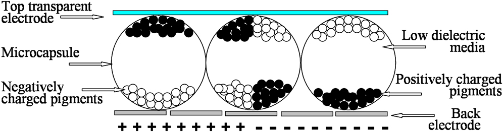
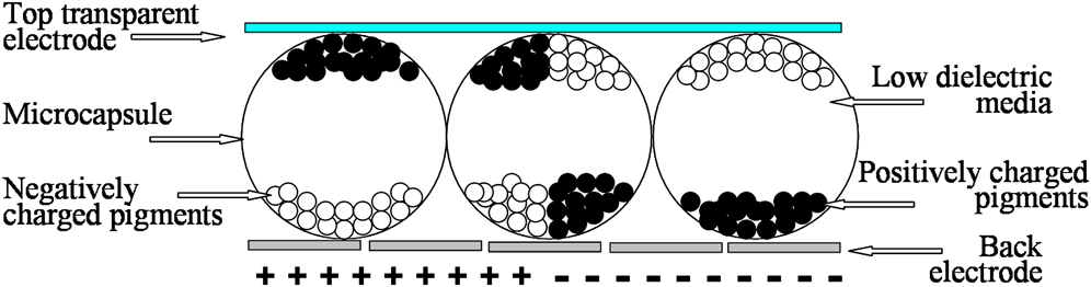
Electrophoretic displays (EPDs), the most efficient solution for the reflective digital display, combine the advantages of electronic displays and conventional paper. EPD technology is based on the movement of charged pigments encapsulated in a liquid media of low dielectric constant when a voltage is applied.Reference Jang, Sung, Choi and Chin1–Reference Jang, Sung, Choi and Chin5 Charged pigment particles with low density, high mobility, and suspension stability are the basis of EPD. Though the first EPD device was introduced as early as 1969,Reference Evans, Lees, Maltz and Dailey6 it did not develop during the next 20 years until Joseph Jacobson first microencapsulated the electrophoretic suspension into individual microcapsules by in situ polymerization,Reference Comiskey, Albert, Yoshizawa and Jacobson7 which reduced undesirable particle clustering and agglomeration to a scale less than the microcapsule size.
Although research on EPD has made rapid progress in the last decade, the efforts on improving the contrast and the response speed never stopped. For the white and black display, titanium dioxide (TiO2) is commonly selected to act as the white pigment due to its excellent whiteness. To avoid the sedimentation of the TiO2 in the suspension resulting from its high density (3.9–4.1 g/cm3), TiO2/polymer composite particles and hollow particles were prepared to decrease the density of TiO2 to match the media.Reference Meng, Tang, Peng and Ren8–Reference Li, Qin, Yang and Liu13 Carbon black (CB) is applied as the black pigment. Because of its ultra-large surface area, CB easily aggregates. Thus, surface modification is needed to prevent the aggregation of the CB particles.Reference Kim, Garoff, Anderson and Schlangen14, Reference Yin, Wu and Chen15 Also, modification of other black pigments were under research to meet the specific needs of EPD applications.Reference Meng, Wen, Sun, Zheng, Ren and Tang16–Reference Yu, An, Bae, Ahn, Kang and Suh18
Microencapsulation refers to the techniques using natural or synthetic polymer materials to enwrap solid, liquid, and/or gas materials. Since the development of the first carbon paper prepared by microcapsule technology in the early 1950s, the technique of microcapsules has attracted increasing academic and commercial interest and has been applied to cosmetics, pesticides, foods, biology, catalysis, pharmaceutical, medical applications, etc.Reference Chan, Craig, Schrock and Cohen19–Reference Sung, Lee, Jang, Choi and Jhon24 Both physical and chemical methods can be applied to prepare microcapsules. Commonly, the microcapsules prepared by physical methods are not suitable to the application of EPD because the capsule wall is not compact enough to inhibit the release of the core material. Up to now, only limited chemical methods have been successfully applied in the preparation of microcapsules, including interfacial polymerization,Reference Hong and Park25, Reference Hong and Park26 in situ polymerization,Reference Yu, An, Bae, Jung, Kim, Ahn, Kang and Suh3, Reference Wang, Zhao, Guo and Zheng27, Reference Guo, Zhao and Wang28 and complex coacervation.Reference Mayya, Bhattacharyya and Argillier29, Reference Kruif, Weinbreck and Vries30
Complex coacervation is one approach to realize microencapsulation, during which two countercharged polyelectrolytes interact and coacervate droplets are formed and accumulated to be wall membrane of microcapsules. The complex coacervation method is attractive because the gelatin (GE)-based microcapsules prepared by this method are more transparent and flexible than microcapsules synthesized by other methods. G. Sun and Z. ZhangReference Sun and Zhang31 studied the deformation of microcapsules prepared from urea–formaldehyde resin and GE–arabic gum under excess pressure. It was reported that the urea–formaldehyde microcapsule ruptures at 35% deformation, while the GE–arabic gum capsules do not. Though the GE–arabic gum system has become the most common material in the study of the capsules prepared by complex coacervation,Reference Chang, Leung and Lin32, Reference Dong, Xia, Hua, Hayat, Zhang and Xu33 searching and developing new wall materials with better properties is still important.
In our research, black and white electrophoretic particles with good suspension stability were prepared by surface modification of TiO2, CB, and Cu2Cr2O3 through chemical or physical methods. GE-based microcapsules with a compact wall were prepared by complex coacervation methods. EPD prototype devices based on the obtained electrophoretic particles and microcapsules were prepared and driven at a low voltage.
II. EXPERIMENTAL PROCEDURES
TiO2 (R960; rutile coated with silicon oxide, 3.9 g/cm3) was produced by Dupont Co., New Johnsonville, TN. CB (MA-100, ρ = 1.7 g/cm3) was supplied by Mitsubishi Chemical Corporation, Tokyo, Japan. Polyisobutylene succinimide (T151), 3-(meth-acryloyloxy) propyltrimethoxysilane (MPS), and lauryl methacrylate (LMA) were commercially available.
N-(p-vinyl benzyl) phthalimide (VBP) and TiO2/PVBP composite particles were prepared according to the reported procedures.Reference Dai, Wu and Chen34 Four grams (4 g) of TiO2 was added into a mixture of 40 mL ethanol, 4 mL water, and 2 g MPS and was rapidly stirred. The resultant suspension was purified to obtain TiO2/MPS particles without free MPS by three cycles of centrifugation and redispersion in ethanol. Four grams (4 g) of TiO2/MPS particles and 4 g VBP were added into 8 mL toluene with mechanical agitation and was slowly heated to 358 K under nitrogen protection. The toluene solution (8 mL) of azobisisobutyronitrile (AIBN, 0.04 g) was dropped slowly into the reaction system. Free radical polymerization of VBP on the TiO2 surface was performed at 358 K in the nitrogen atmosphere for 24 h. The resultant suspension was refined from the poly(N-vinylbenzyl phthalimide) by three cycles of centrifugation and redispersion in toluene.
Cu2Cr2O3 (type 30C965 supplied by the Shepherd Color Company, Cincinnati, OH) was first encapsulated by SiO2 through sol-gel method before compositing with PLMA. Ten grams (10 g) of Cu2Cr2O3 particles, 4 mL tetraethyl orthosilicate, and 12 g span85 were added into 40 mL deionized water, and the mixture was ultrasonicated to become a stable emulsion. The emulsion was added into a mixture of 50 mL ethanol and 12 mL NH3·H2O. The reaction solution was stirred at a rate of 400 rpm at room temperature for 4 h. Cu2Cr2O3 coated with SiO2 was separated by centrifugation.
PLMA was anchored onto the TiO2 and Cu2Cr2O3/SiO2 particle surface via free radical polymerization by one-step method. Five grams (5 g) of TiO2 (Cu2Cr2O3/SiO2) and 2.5 g LMA were added into 15 mL toluene with mechanical agitation and was slowly heated to 358 K under nitrogen protection. The toluene solution (10 mL) of AIBN (0.05 g) was dropped slowly into the reaction system. Free radical polymerization of LMA on the TiO2 surface was performed at 358 K in the nitrogen atmosphere for 24 h. The resultant suspension was refined from the free PLMA by three cycles of centrifugation and redispersion in toluene.
The CB/T151 composite particles were prepared by adsorption on the CB surface. The mixture of CB (0.5 g), T151 (0.2 g), and toluene (30 mL) was treated by ultrasonication for 5 h. The polymer-treated inorganic white/black particles acting as electrophoretic particles were dispersed in tetrachloroethylene (TCE) by ultrasonication to form electrophoretic liquid.
GE (type B) is the primary wall material. Styrene–maleic anhydride (SMA) copolymers , sodium alginate (SA), and sodium carboxymethylcellulose (NaCMC) act as polymeric anionic polyelectrolyte (PAP). Sodium dodecyl sulfate (SDS), sodium lauryl sulfate (SLS), dioctyl sulfosuccinate sodium (DSS), and perfluoro-nonene oxy benzene sulfonate (OBS) serve as surfactants. Glutaraldehyde (25% aqueous solution) is a cross-link agent. TCE is the electrophoretic media. Solvent blue 35 offers background color. All the chemicals above are commercially available.
Microcapsules were prepared via complex coacervation at 318 K. The electrophoretic liquid, TCE containing electrophoretic particles inside, as core materials were dispersed in the aqueous solution of GE/PAP/surfactant blend. Then, the pH of the system was adjusted by the addition of acetic acid (10 wt% aqueous). When the pH is below 4.5, GE and PAP/surfactant of opposite charges react through complex coacervation process due to charge neutralization. Most of complex coacervates in the colloid phase gathered on the oil/water (O/W) interface to form complex coacervation layer, which formed compact microcapsules after being cross-linked by glutaraldehyde. The matrix character display prototype was fabricated by coating the obtained microcapsules on indium tin oxide/polyethylene terephthalate (ITO/PET) film.
The yield is defined as the mass ratio of the obtained microcapsules to the all initial ingredients used during microcapsule formation. The morphology of electrophoretic particles and microcapsules were characterized by scanning electron microscopy (SEM, JSM-5510, JEOL CO., Tokyo, Japan) and transmission electron microscopy (TEM, JEM-1200EX, Tokyo, Japan). Particle size distribution was measured by phase analysis light scattering (Brookhaven Instruments Co., Holtsville, NY). Thermal stability was measured on a Pyris θ Thermogravimetic Analyzer (PekinElmer Inc., Waltham, MA) at 10 K/min ramp rate in a stream of nitrogen. The barrier property of the capsule was measured by annealing the microcapsule at high temperature for different times. Surface tension (σ) of the aqueous NaCMC solution with different surfactants was tested by drop weight method at 298 K.Reference Dai, Wu, Li, Zhou, Li and Chen35, Reference Chatterjee, Moulik, Sanyal, Mishra and Puri36 The optical photos of EPD prototype were taken on Power Shot A2000 IS (Canon China, Inc., Beijing, China). The reflectance of color display was measured on Portable Reflectance JFB-1, Chemical Machinery Co., Ltd, Shanghai PuShen, China.
III. EXPERIMENTAL RESULTS
A. Preparation of the black and white electrophoretic particles
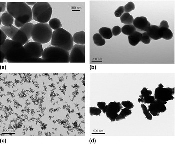
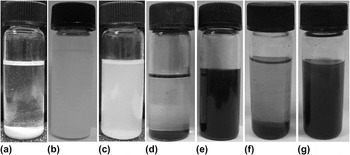
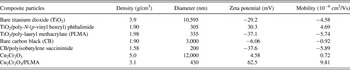
Electrophoretic particles of low density and high mobility are required by the EPD application. In this study, TiO2 was selected as white pigment and modified by polymers such as PVBP and PLMA through radical polymerization. CB and Cu2Cr2O3 were chosen to act as black pigments, in which CB was modified by T151 through physical absorption and Cu2Cr2O3 was modified by PLMA by radical polymerization. The TEM images shown in Fig. 1 give the morphology of TiO2/PVBP, TiO2/PLMA, CB/T151, and Cu2Cr2O3/PLMA composite particles. A thin layer of polymer can be found around the particles. It is seen in Fig. 2 that the suspension stability of the composite particles in the solvent was greatly enhanced due to the decrease of the particle density as well as the steric effect induced by the soluble polymer chains around the pigments. The polymer chains around the bare pigment can extend into solvent, providing enough steric repulsion over electrostatic attraction among particles. Table I gives the characteristics of the bare white/black pigments and the composite particles including diameter, mobility, zeta potential, etc. For the reason that the polymer modification hinders the aggregation of the bare pigment, the diameter of the composite particles is much smaller than that of the bare one. The polymer can also charge the composite particles in the low dielectric media such as TCE because of the functional group in the polymer. Blending positive and negative electrophoretic particles together in the low dielectric media can result in a dual-particle electrophoretic suspension which can be driven under direct current voltage to display black/white image.
Transmission electron microscopic images of (a) titanium dioxide (TiO2)/poly-N-(p-vinyl benzyl) phthalimide (PVBP), (b) TiO2/poly-lauryl methacrylate (PLMA), (c) carbon black (CB)/polyisobutylene succinimide (T151), and (d) Cu2Cr2O3/PLMA composite particles.

Suspension stability of the particles in tetrachloroethylene: (a) bare TiO2, (b) TiO2/PLMA, (c) TiO2/PVBP, (d) bare CB, (e) CB/T151, (f) bare Cu2Cr2O3, (g) Cu2Cr2O3/PLMA.

Properties of the pigments and the composite particles in tetrachloroethylene.

B. Microcapsules prepared by complex coacervation method
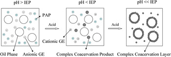
GE, a natural, nontoxic, water-soluble polyampholyte obtained from partial hydrolysis of insoluble collagen, has carboxyl, amino, and hydroxyl functional groups as well as good film-forming property,Reference Emregiil, Sungur and Akbulut37 all of which makes GE-based microcapsule a very important capsule in large areas. As shown in Scheme I, GE is PAP in aqueous solution when the pH is higher than its isoelectric point (IEP). When pH is lower than IEP, GE is positively charged and can react with another PAP through complex coacervate process.
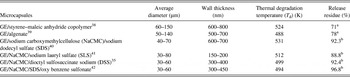
Except for the natural polymer of arabic gum, there are rarely reports about the PAP which can coacervate with GE to form microcapsules appropriate to EPD application. Here, natural polymer (SA), synthetic polymer (SMA), and semi-natural polymer (NaCMC) were applied separately as PAP to react with GE and the corresponding microcapsules were prepared by complex coacervation method. The characteristics of six kinds of GE-based microcapsules including thermal degradation temperature (T d), release residue (the residual wt% of microcapsules after releasing), the range of the average diameter, and wall thickness were summarized in Table II. In general, the average diameter of the capsule is no more than 200 μm, the wall thickness is below 1 μm, and the T d is much higher than the boiling temperature of TCE. Higher release residue means higher barrier property.
Characteristics of the gelatin (GE)-based microcapsules prepared by complex coacervation method.

a The capsules were annealed at 363 K for 70 h.
b The capsules were annealed at 395 K for 120 min.
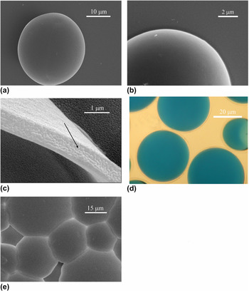
Figure 3 gives the morphology of the GE/NaCMC/DSS microcapsule as an example. Good microcapsules are commonly spherical as shown in Fig. 3(a). The wall of the microcapsule should be compact and defect-free just like the image given in Fig. 3(b), which is beneficial to enhance the barrier property of the microcapsules. That is to say, the core material cannot penetrate outside the compact capsule wall. Compared with GE/arabic gum microcapsules formed in the presence of SDS which has a porous capsule wall microstructure,Reference Mayya, Bhattacharyya and Argillier43 the capsule wall of GE/NaCMC/DSS microcapsule is compact and smooth. It is believed that coacervate droplets of GE/NaCMC/DSS have more affinity and wettability than those of GE/arabic gum/SDS. As shown in Fig. 3(c), the thickness of the capsule is no more than 1 μm to ensure transparency [Fig. 3(d)]. Domains in size of about 100 nm can be found in Fig. 3(c), which suggests that the wall membranes of the microcapsules are formed from nanosized coacervate droplets. When microcapsules are put on the flat plate, as shown in Fig. 3(e), they will deform next to each other and the appearance changes from sphericity to pentagon or hexagon due to the elasticity of the capsule wall. Since the microcapsules contact with each other compactly, the area for display is enlarged, which is advantageous to the application of microcapsules in EPD.
Images of the gelatin (GE)/sodium carboxymethylcellulose (NaCMC)/dioctyl sulfosuccinate sodium (DSS) microcapsules. (a, d, e) Complete microcapsules, (b) wall of the capsule, and (c) cross section of the capsule (domains of about 100 nm are pointed by the arrow).

C. Microcapsules of GE/NaCMC prepared by complex coacervation method
NaCMC is one of the most important nontoxic anionic water-soluble linear polymers. NaCMC is produced by partial substitution of the 2, 3, and 6 hydroxymethyl groups of cellulose. At concentrations below 7 g/L, NaCMC has no surface activity and can hardly be adsorbed at O/W interface.Reference Guillot, Delsanti, Desert and Langevin44 It is known that the amount of polyeletrolyte adsorbed on the oil droplet surfaces before and during coacervation is the important factor determining the microencapsulability of the core material. Since there are not enough NaCMC adsorbed on the O/W interface without assistance, GE/NaCMC microcapsules are impossible to form through the interaction between GE and NaCMC. For reason that microencapsulability depends also on the affinity and wettability between oil droplets and coacervate droplets,Reference Luzzi and Gerraughty45 some surfactants were used to increase the encapsulation yield.Reference Mayya, Bhattacharyya and Argillier46 In the GE/NaCMC system, SDS, SLS, and DSS are such kind of surfactants.
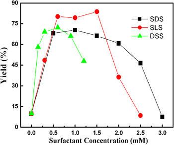
Figure 4 gives the yield of the complex coacervation as a function of the concentration of the surfactants such as SDS, SLS, and DSS. Obviously, the yield of the GE/NaCMC system without excess addition of surfactant is very low. Unstable droplets can be found in water without addition of surfactants. The low yield of the microcapsules could be attributed to the weak interaction between GE and NaCMC. The existence of surfactant enhances the yield remarkably. It is reported that additional small molecular surfactant in the GE/NaCMC system could improve the amount of NaCMC adsorbed on the O/W interface because the hydrophobic interaction between NaCMC and the surfactant outweigh the Coulombic repulsion arising from similarly charged polymer sites and surfactant headgroup.Reference Dhara and Shah47 After the yield reaches the highest value, further increase of the surfactant concentration leads to a quick decrease of the yield. The reason for the yield decreasing is that the hydrophobic sites of the neighboring polymer chains are solubilized separately by the excess surfactant and the formed complexes are gradually dissolved.Reference Dickinson and Matsumura48
Effects of the concentration of different surfactants on yield of the GE/NaCMC-based microcapsules.

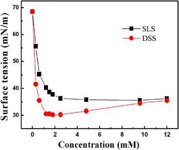
A different concentration sensitivity on the microcapsule yield for the three surfactants can also be found from Fig. 4. The surfactant concentration for the highest microcapsule yield is different for different surfactants, which is around 1, 1, and 0.6 mM for SDS, SLS, and DSS, respectively. This phenomenon can be partially explained according to the effect of surfactant concentration (SLS and DSS) on the surface tension of the aqueous NaCMC solution shown in Fig. 5. The surface tension of the aqueous NaCMC solution (68.52 mN/m) decreases sharply with the addition of SLS/DSS. The influence of DSS on the surface tension of the aqueous NaCMC solution is much more obvious than that of SLS, which coincides with the fact that the alkyl chain length of DSS is longer than that of SDS, because of which the hydrophobic interaction between DSS and NaCMC may be stronger than that between SLS and NaCMC. As a result, it is easier for DSS than SLS to promote the process of the complex coacervation reaction. This result is identical to the above-mentioned data that the concentration of DSS to form stable capsules in GE/NaCMC/DSS system is 0.6 mM, lower than that of SLS (1.0 mM).
Effect of surfactant sodium lauryl sulfate (SLS) and DSS in different concentrations on surface tension of NaCMC aqueous solution.

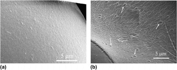
The wall structure of the microcapsules is influenced heavily by the amount of surfactant addition, similar to the yield of microcapsules. The morphology of the wall of GE/NaCMC/SLS microcapsule is given in Fig. 6 as an example. Figures 6(a) and 6(b) were SEM images from the samples prepared with 1.0 and 2.0 mM SLS, respectively. In Fig. 6(a), the capsule wall is very compact. While in Fig. 6(b), the pinholes spread all over the capsule wall. If the amount of SLS increases to 2.5 mM, the capsules fracture under high vacuum. It is suggested that with increasing SLS concentration, the amounts of the defects on the capsule wall increase remarkably and the strength of the capsule wall weakens simultaneously. In other words, excessive SLS would weaken the interaction of coacervate droplets by desorbing GE from the O/W surface.
Scanning electron microscopic images of the wall of GE/NaCMC/SLS microcapsules prepared with various SLS contents: (a) 0.1 mM and (b) 2.0 mM SLS (the pinholes on the wall are pointed by the arrows).

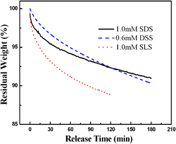
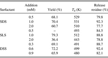
The thermal stability of the GE/NaCMC microcapsules with the aid of appropriate amount of surfactant is given in Fig. 7. The thermal degradation temperature (T d) of all capsules is around 500 K which is much higher than the boiling temperature of the solvent TCE (394 K). Figure 8 gives the releasing behavior of the capsules at 394 K. Both GE/NaCMC/DSS and GE/NaCMC/SDS capsules have similar good barrier properties, while GE/NaCMC/SLS system has relatively poor property. Detailed information, including T d and release residue (after 120 min thermal treatment at 395 K) of GE/NaCMC microcapsules prepared with surfactant in different amounts, is given in Table III. The T d of capsules is much higher than the boiling temperature of solvent TCE (394 K) except for the GE/NaCMC/SLS system with the SLS addition of 2.0 mM, the yield of which is only 36.4, and much lower than that of other systems. In other words, the microcapsules have good thermal stability if the yield of the system is high enough. Slight variation of the addition amount of the surfactant will not influence the degradation temperature greatly. While the release residue of the capsules treated at 395 K for 120 min is quite different with the change of the surfactant concentration. Over 10% change of the residue can be easily detected. As we know, the speed of the weight loss for the microcapsules under annealing can be used to judge the barrier property of the capsules, i.e., the ability of the capsule wall to protect core materials and prevent core materials from releasing outside. Commonly, at this extreme temperature, any defect in capsule walls can cause microcapsule rapture and weight loss would be found instantaneous. In Table III, after 120 min treatment, the highest release residue is about 90%, which indicates the good barrier property of the corresponding system and thus the long-term stability for microcapsules.
Thermogravimetric analysis of the microcapsules prepared with different surfactants in suitable amount.

Residual weights (%) of the microcapsules prepared with different surfactants in suitable amount annealing at 395K.

Characteristics of GE/NaCMC-based microcapsules with various surfactants in different concentrations.

D. Fabrication and properties of the matrix display EPD prototype devices
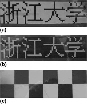
A matrix display prototype device was fabricated by coating the obtained microcapsules on flexible ITO/PET film applied as front electrode. The back electrode was patterned to offer addressing signals. Scheme II gives the structure of the EPD prototype device. Here, we give the properties of a typical EPD device based on GE/NaCMC/SDS microcapsule containing the white positively charged TiO2/PVBP particles and the black negatively charged CB/T151 particles. The matrix display prototype, driven under static mode at 9 V, can display the Chinese characters of “Zhejiang University” as shown in Figs. 9(a) and 9(b). The response time of the prototype is about 700–800 ms. The reflectance of white and black colors displayed in Fig. 9(c) are 36.5–37.4% and 3.6–4.2%, respectively. The contrast ratio is 8.9–10.4:1.
Images of the matrix display prototype devices based on the GE/NaCMC/sodium dodecyl sulfate (SDS) microcapsule (containing the TiO2/PVBP and CB/T151 as electrophoretic particles) driven at 9 V: (a, b) Chinese characters of “Zhejiang University” and (c) white and black diamonds.

The schematic diagram of the complex coacervation.

The schematic diagram of the electrophoretic display prototype device.
IV. CONCLUSIONS
The white electrophoretic particles of TiO2/PVBP and TiO2/PLMA composite particles and the black electrophoretic particles of CB/T151 and Cu2Cr2O3/PLMA composite particles with good suspension stability were prepared by anchoring suitable polymers onto the pigments’ surface. The stable dual-particle electrophoretic systems containing positively charged TiO2/PVBP and Cu2Cr2O3/PLMA particles and negatively charged TiO2/PLMA and CB/T151 particles were prepared. The elastic, optical transparent, and spherical GE-based microcapsules with good barrier property were prepared via complex coacervation. The species and the concentration of the anionic surfactants have great influence on the morphology and the property of the GE/NaCMC-based microcapsules. The matrix character EPD prototype devices were fabricated using the obtained electrophoretic particles and microcapsules. The response time of EPD prototype was 700–800 ms, and the maximum contrast ratio can be higher than 10:1 driven at 9 V.
ACKNOWLEDGMENTS
This work was financially supported by the National High-tech Research Development Program (863 Program) of China (Grant No. 2008AA03A331) and the Science & Research Program of Zhejiang Province (Grant No. 2009C21024).