The morphology of the common atrioventricular junction in patients with atrioventricular septal defect is complex. The valve guarding the common junction is usually committed equally to both ventricles through symmetrical bi-ventricular connections.Reference Bharati and Lev 1 – Reference Anderson, Ho, Falcao, Daliento and Rigby 3 In a small proportion of patients, however, the junction is unequally shared between the ventricles, and the arrangement is then described in terms of ventricular imbalance.Reference Mahle, Shirali and Anderson 4 Standard cross-sectional echocardiography has traditionally been thought sufficient to provide pre-operative information for clinical decision-making with regard to the suitability for the creation of bi-ventricular circulations.Reference Smallhorn 5 No uniform guidelines exist at present, however, to permit accurate decision-making in borderline cases. Real-time three-dimensional echocardiography, specifically its multiplanar formatting option, has now provided new insights into evaluation of the atrioventricular junctions, and can provide significant information in this challenging arena.Reference Bharucha, Roman, Anderson and Vettukattil 6 , Reference Bharucha, Sivaprakasam, Haw, Anderson and Vettukattil 7
The outcome of surgical repair in patients with atrioventricular septal defects depends on many factors, including left atrioventricular valve regurgitation,Reference Bharucha, Sivaprakasam, Haw, Anderson and Vettukattil 7 , Reference Ten Harkel, Cromme-Dijkhuis, Heinerman, Hop and Bogers 8 but most of all, the feasibility of performing a biventricular repair. In the normal heart, the atrial septum is aligned with the ventricular septum at the cardiac crux, resulting in equal distribution of blood flow to the ventricles, and thus appropriate biventricular development. We hypothesised that disruption of such atrioventricular septal alignment at the crux of the heart in patients with atrioventricular septal defect would lead to imbalance of venous return, and thus ventricular imbalance. The multiplanar re-formatting technique allows the operator to view the heart in any cross-sectional plane, and provides the option to move sequentially through the data set. During analysis using the multiplanar mode, the data set is cut in three planes simultaneously, providing three sets of dynamic cross-sectional images. Each plane can be moved and aligned in such a way that the individual planes are oriented to visualise the structure of interest.
Using the multiplanar re-formatting mode, therefore, we sought to identify the atrioventricular septal angulation in a group of patients with atrioventricular septal defect undergoing surgery, comparing the values obtained in those patients undergoing biventricular repair as opposed to those with ventricular imbalance and undergoing functionally univentricular repair.
Methods
We evaluated 31 patients with atrioventricular septal defect and common atrioventricular junction before surgery using standard cross-sectional and three-dimensional echocardiography. Ventricular imbalance was deemed present in nine patients following conventional cross-sectional assessment, one of the ventricles being considered hypoplastic, and therefore inadequate for independent function in biventricular circulations. The suggested decision was subsequently confirmed at surgical inspection when the surgeon chose to create a functionally univentricular circulation. The remaining 22 patients were all considered to have two adequately sized ventricles subsequent to standard cross-sectional echocardiographic assessment, and all underwent successful biventricular repair, except for one patient who died in the ICU.
Images were acquired using a 3–5 MHz matrix phased array transducer and a real-time three-dimensional imaging system (Philips Sonos 7500 or IE33; Philips Co., DA Best, the Netherlands). Stored full-volume three-dimensional echocardiographic data sets were assessed offline using Tom Tec software (image arena version 3.0 Build 3.04.48; Tom Tec imaging systems, Munich, Germany).
Offline analysis was carried out in the multiplanar re-formatting mode, allowing the operator to view three orthogonal cardiac planes simultaneously, and to move these planes throughout the images in order to examine the data set in its entirety. Using this multiplanar re-formatting mode, we identified the four-chamber plane at the crux of the heart. A line was then drawn through the plane of the apical muscular ventricular septum, with another line drawn along the plane of the atrial septum from its insertion on the atrial roof to its leading edge. We measured the angle formed by the intersection of these two lines, and recorded it as the atrioventricular septal angle (Fig 1).
Shows the three-dimensional data set being analysed in multiplanar reformatting mode in diastole. The crux of the heart was identified by positioning two planes perpendicular to each other through the long axis of the atrioventricular septum. Protractor was used to measure the angle of the atrial septum in relation to the ventricular septum. Panel ( a ) shows that there is no angle between the atrial and ventricular septum as they are aligned with each other. Panel ( b ) shows that the atrial septum is at an angle in relation to the ventricular septum, leading to a hypoplastic ventricle. LA=left atrium; LV=left ventricle; RA=right atrium; RV=right ventricle.

Statistical analysis was performed using SPSS version 16.0. Simple descriptive statistics were used to define demographic and echocardiographic data, including the atrioventricular septal angle. Non-parametric tests including Student’s t-test were used to compare continuous variables between the 22 patients in whom the surgeon had attempted biventricular repair and the nine who were treated in functionally univentricular manner. A value for p<0.05 was considered significant. Ethical approval was obtained according to local requirements.
Results
Entire group
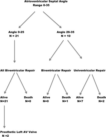
We reviewed the findings from 31 patients, of whom 17 were boys, with a mean age of 52.09 months, and an age range from 2 to 264 months (Table 1). Of these patients, 15 had trisomy 21. In 25 of these patients, the atrioventricular junction was guarded by a common valvar orifice, whereas the other six had separate valvar orifices for the right and left ventricles within the common junction, with the potential for shunting confined at the atrial level, so-called partial defects. Of the overall group, 22 patients were considered to have a balanced ventricular arrangement, and underwent biventricular surgical repair. The remaining nine patients were deemed unsuitable for biventricular repair because of ventricular imbalance and underwent functionally univentricular palliation (Fig 2).
Flow chart showing the outcome of the groups in relation to the atrioventricular (AV) septal angulation.

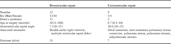
Demographics and outcome in patients undergoing biventricular as opposed to functionally univentricular repair.

Patients undergoing biventricular repair
Of the 22 patients undergoing biventricular repair, 14 had an atrioventricular septal angle of 0, the septal structures being in direct alignment. In the remaining eight patients, it ranged from 1 to 35°, with the mean atrioventricular septal angle being 7.4°. The mean age at biventricular repair was 20 months, with standard deviation of 56 months, and with a range from 2 to 200 months. Associated defects included coarctation of the aorta, double-outlet right ventricle, and pulmonary stenosis, each present in one patient. Trisomy 21 was present in 13 patients. All except one of the patients underwent successful biventricular surgical repair. The outstanding patient initially underwent biventricular repair at the age of 4 months. The right ventricle, however, proved to be of inadequate size, and the patient died due to low cardiac output at the age of 4 months, while still under intensive care.
Replacement of the left atrioventricular valve with a mechanical prosthesis was subsequently required in two patients. There have been no late deaths to date.
In two of the patients, the initial decision had been to proceed to functionally univentricular management, but when pre-operative assessment demonstrated adequately sized ventricles, they were put forward for biventricular repair. The first, with an atrioventricular septal angle of 24°, had a common valvar orifice and pulmonary stenosis. Initially, the left ventricle was thought to be hypoplastic, but re-evaluation suggested it would be adequate, and the patient underwent biventricular repair at the age of 6 months. Owing to severe post-operative left atrioventricular valvar regurgitation, the left atrioventricular valve was replaced with a 19 mm St Jude’s mechanical valve at 1 year of age. At follow-up of 6 years, the patient is well and has not required re-operation.
The second patient, with an atrioventricular septal angle of 22°, also had a common valvar orifice and a small left ventricle, initially thought to be the consequence of ventricular imbalance. The patient underwent pulmonary arterial banding at 2 months of age, followed by creation of a bidirectional cavopulmonary shunt and a Damus–Kaye–Stansel connection at the age of 9 months. When re-assessed at the time of proposed conversion to the Fontan circulation, the left ventricle was thought to be adequate for biventricular repair, and this was achieved at 6.5 years of age, replacing the left atrioventricular valve with a mechanical valve in the same procedure. At follow-up of 4 years, the patient remains clinically well and has not required further surgery.
Patients undergoing functionally univentricular repair
In nine patients deemed to have ventricular imbalance, the decision was made to proceed with functionally univentricular repair. Their mean atrioventricular septal angle was 28.6°, with standard deviation of of 3.04, and with a range from 26 to 35°. The right ventricle was hypoplastic in two, whereas the left ventricle was hypoplastic in seven patients. Among these nine patients, two patients also had isomerism of the right atrial appendages and totally anomalous pulmonary venous connection, with two having severe valvar and sub-pulmonary stenosis, and one with pulmonary atresia. Initial surgery included pulmonary arterial banding in four patients, and creation of a Blalock–Taussig shunt as an emergency procedure in another. In addition, a patient with a severely hypoplastic left ventricle and hypoplastic arch underwent a Norwood operation, and died in the early post-operative period. The patient with right isomerism, totally anomalous pulmonary venous connection, and pulmonary atresia died following the emergency creation of a Blalock–Taussig shunt. A superior bidirectional cavopulmonary anastomosis was created in seven patients, and five of them have now been successfully converted to the Fontan circulation, whereas two patients with superior cavopulmonary anastomoses are awaiting Fontan completion.
The atrioventricular septal angle
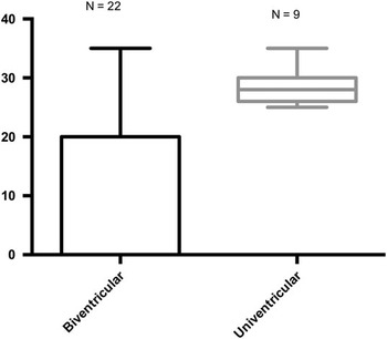
There was a significant difference in atrioventricular septal angles between the two groups, with a range of 7.4, and standard deviation of 11.1, in those undergoing biventricular repairs, as opposed to 28.6°, with standard deviation of 3.04, in those requiring functionally univentricular repair (p<0.0002). We were unable to achieve biventricular repair in any patient with an atrioventricular septal angle >25°, whereas the angle in the patient in whom biventricular repair was attempted but failed was 35° (Fig 3).
Graph showing the difference in atrioventricular septal angulation in the groups of patients undergoing biventricular as opposed to functionally univentricular repair.

Discussion
Multiplanar re-formatting has already shown its value as a tool for use in analysis of patients with complex congenitally malformed hearts, such as those with atrioventricular septal defect and common atrioventricular junction, as it gives new insight into the changes of intra-cardiac structures during the cardiac cycle.Reference Lang, Mor-Avi, Sugeng, Nieman and Sahn 9 – Reference Hlavacek, Crawford, Chessa and Shirali 11 Images can be manipulated and cut in different planes to allow best visualisation of the area of interest. The technique has also been shown to provide information additive to that gained by cross-sectional imaging alone in pre- and post-operative patients with atrioventricular septal defects, including those with ventricular imbalance. It has also been demonstrated to provide detailed information regarding the morphology of the left atrioventricular valve and accurately to show its function and mechanism of regurgitation when compared with surgical findings.
In this study, we now demonstrate that the values for angulation of the atrial septum relative to the ventricular septum at the crux of the heart predict accurately the presence of ventricular imbalance and hypoplasia. This single measurement, therefore, may guide the decision to opt for biventricular as opposed to functionally univentricular repair, and thereby lead to better surgical outcomes.
Extreme deviation of the atrial septum with respect to the ventricular septum in atrioventricular septal defect results in double-outlet atrium, with one atrium emptying into both ventricles.Reference Brancaccio, Amodeo, Rinelli, Filippelli, Sanders and Di Donato 12 In double-outlet right atrium, there is extreme leftward deviation of the lower portion of the atrial septum towards the left atrioventricular valve orifice, resulting in the systemic venous inflow bisecting the ventricular septum, with resultant biatrial filling of the left ventricle. The reverse is true in the case of double-outlet left atrium. Depending on the degree of angulation, the ventricle towards which the atrial septum is deviated is larger and the other ventricle is hypoplastic. Double-outlet atrium, of course, is always present when there is straddling of an atrioventricular valve, this being another situation known to be associated with hypoplasia of one of the ventricles.Reference Beroukhim and Geva 13 The atrioventricular septal angle, therefore, represents a continuum that, at its extreme, creates a double-outlet atrium.
In this light, we hypothesised that excessive flow to the dominant ventricle may have resulted in hypoplasia of the under-filled ventricle. Such a notion of under-filling of the hypolastic ventricle in unbalanced atrioventricular septal defect was first proposed by Phoon et al,Reference Phoon and Silverman 14 with the potential influence of postnatal depressed ventricular growth due to septal bowing being elaborated by Foker.Reference Foker, Berry and Steinberger 15 Surgical management of patients with such unbalanced atrioventricular septal defects poses enormous challenges, largely because of the current inability to recognise accurately those suitable for biventricular repair. Cohen et alReference Cohen, Jacobs, Weinberg and Rychik 16 have previously suggested calculation of an atrioventricular valvar index as one of the echocardiographic tools with which to differentiate between balanced and unbalanced forms of atrioventricular septal defect. The index is derived by calculating the ratio of the smaller area over the larger area, and then named right or left dominant. This ratio, however, by itself cannot totally discriminate between functionally univentricular versus biventricular repair. Another recent study used a modification of the atrioventricular valvar index, where the area of the left atrioventricular valvar component is always the numerator and the total atrioventricular valvar area is always the denominator. This method has a numerical spectrum and no longer specifies right or left dominance.Reference Jegatheeswaran, Pizarro and Caldarone 17 More recently, the angle of the right ventricular relative to the left ventricular inflows has been incorporated as an important factor defining echocardiographic measures of right ventricular dominant patients with unbalanced atrioventricular septal defects.Reference Cohen, Jegatheeswaran and Baffa 18
It has also been suggested, however, that right ventricular volume overload results in right-to-left septal bowing and contributes to the appearance of a hypoplastic left ventricle, although one that can potentially accommodate a greater volume.Reference Phoon and Silverman 14 , Reference Foker, Berry and Steinberger 15 Therefore, surgical decision-making depends not only on the atrioventricular valvar index but also on absolute and potential left ventricular volumes. Multiplanar re-formatting provides the ability to analyse the dynamic morphology of the common atrioventricular valve in multiple planes and in three-dimensions. Moreover, in patients with atrioventricular septal defect, the common valve is often at an angle relative to the crux of the heart, and thus it is not possible always accurately to assess the area of the valve committed to the corresponding ventricle on cross-sectional echocardiography. Evaluation of the atrioventricular septal angle can become a more effective tool in this setting. As the outcomes of surgery are dependent on the anatomical substrate before surgery, we suggest that our measurements have permitted us to identify an important factor in the pre-operative work-up. Selection of the proper surgical strategy for patients with unbalanced atrioventricular septal defect remains a significant challenge. There is a broad spectrum of ventricular imbalance, and patients with milder forms are the ones providing the greatest challenges in decision-making. There is no single measurement that can alone discriminate between balance and imbalance. Measurement of the atrioventricular septal angle, nonetheless, along with other echo indices and assessment of anatomic factors, can be remarkably helpful in accurate decision-making for this population.
Limitations
Ours was a retrospective analysis, and our cohort was small, especially concerning those with unbalanced atrioventricular septal defect. The atrioventricular valvar index and the area of the valvar orifices could not be measured in all patients, and therefore lack statistical significance for our group of patients.
Conclusions
Using multiplanar re-formatting of three-dimensional echocardiographic data sets, we have demonstrated variation in the relation of the angulation of the atrial septum relative to the ventricular septum that has an important impact on surgical decision-making. The wider this angle, the greater the likelihood of hypoplasia of one of the ventricles due to underfilling, and thus unfavourable outcomes after attempted biventricular repair.
Acknowledgement
None.
Financial Support
This research received no specific grant from any funding agency, commercial, or not-for-profit sectors.
Conflicts of Interest
None.
Ethical Standards
Ethical approval was obtained according to local requirements.