Introduction
Tonsillectomy produces an open wound that is healed by secondary intention. This favours the development of pain and secondary haemorrhage, which are the main forms of post-operative morbidity. Pain is caused by injury to the glossopharyngeal mucosa and/or fibres of the vagus nerve, leading to irritation followed by inflammation and pharyngeal muscle spasm, ischaemia and a prolonged pain cycle that is not completely resolved until the muscle becomes covered with mucosa at post-operative days 14–21. Haemorrhage is caused by the emergence of blood vessels in the tonsillar fossa.Reference Rakesh, Anand, Payal and Pranjal 1 , Reference Elwany, A Nour and Magdy 2
Since tonsil removal using digital blunt dissection was first described by Cornelius Celsus in Rome in the first century ad, many techniques have been developed and modified in an attempt to reduce post-operative morbidity. Traditionally, electrocautery and cold instruments (e.g. scissors, bows, guillotine) have been the most widely used tools. Electrocautery has the advantage of improved haemostasis and shorter surgery time, while the cold technique produces less thermal injury and is therefore believed to cause less post-operative pain and dehydration.Reference Magdy, Elwany, el-Daly, Abdel-Hadi and Morshedy 3 Other surgical techniques such as radiofrequency ablation, laser, microdebrider or harmonic scalpel have also been used to improve the procedure. Although various studies have evaluated their advantages and disadvantages, no consensus has been reached on the optimal technique to minimise tonsillectomy-associated morbidity.Reference Aksoy, Ozturan, Veyseller, Yildirim and Demirhan 4
Many surgical procedures, including tonsillectomy, are increasingly being performed in an out-patient setting to avoid the cost of a hospital stay. However, it is important to consider the degree of post-operative morbidity involved when changing surgical practice.Reference Hahn, Rungby, Overgaard, MØller, Schultz and Tos 5 Post-operative pain is one of the main problems after tonsillectomy. Strategies used to control pain include using the least aggressive surgical technique together with pharmacological analgesic therapy. Moreover, providing educational materials and information to patients may reduce both anxiety and post-operative pain, and increase patient satisfaction.Reference Echevarría 6
Histopathological analysis of the resected tonsil can quantify the collateral damage caused by each technique for correlation with clinical data.Reference Magdy, Elwany, el-Daly, Abdel-Hadi and Morshedy 3 This study aimed to compare the histological findings in tonsils removed by cold or electrocautery dissection and determine their relationship with post-operative complications (mainly pain and haemorrhage).
Materials and methods
Study design
This descriptive, observational, prospective study enrolled 79 adult out-patients who underwent tonsillectomy at the Otorhinolaryngology Department of a university hospital. Patients were divided into two groups based on the date and type of surgery: 39 patients who underwent cold dissection surgery from 3 February to 28 October 2011, and 40 patients who underwent electrocautery dissection from 4 October 2013 to 17 December 2014.
Both groups received information about the procedure, post-operative care and pain management. At telephone follow-up on post-operative days 4, 7 and 15, pain was assessed using a 0–10 scale in which 0 represents no pain, 5 represents moderate pain and 10 represents the worst possible pain. At post-operative follow-up, patients stated the number that best represented their pain intensity. The optimal score was three or below. All patients received post-operative combined drug treatment comprising 600 mg ibuprofen every 8 hours, 30 mg prednisone every 12 hours, and 20 mg omeprazole every 24 hours for 3 days, followed by a progressive reduction of prednisone while maintaining the dosage of all other medicines for 5 more days. Patients were given medications and dietary recommendations at the end of their hospital stay.
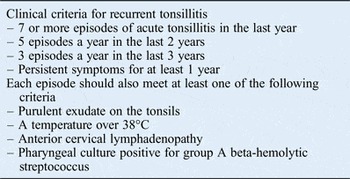
Both treatment groups were subject to the inclusion and exclusion criteria for tonsillectomy as recommended by the Spanish Society of Otorhinolaryngology and Head and Neck Surgery (Table I) and to the inclusion and exclusion criteria for ambulatory surgery as recommended by the Spanish Ministry of Health and Consumer Affairs.Reference Cervera Escario, Del Castillo Martín, Gómez Campderá, Gras Albert, Perez Piñero and Villafruela Sanz 7 , 8 In all patients, the indication for tonsillectomy was restricted to recurrent tonsillitis; no patients with peritonsillar abscess were included in the study.
Criteria for tonsillectomy due to recurrent tonsillitis

General anaesthesia comprised propofol and remifentanil for anaesthesia induction and remifentanil for maintenance. Ondansetron (4 mg) or dexamethasone (4 mg) was administered at the start of surgery, and analgesia comprising 80 mg corticosteroid (methylprednisolone), 1 g paracetamol and 50 mg dexketoprofen was administered before the end of surgery. Anaesthetic gases were oxygen, nitrous oxide and sevoflurane.
Data on patient age and sex and surgical technique were obtained from medical records. Data on pain, primary bleeding or haemorrhage, secondary bleeding or haemorrhage, fever, admission, re-admission, need for supplemental analgesia and number of emergency department visits were collected on post-operative days 4, 7 and 15 from both treatment groups using a telephone registration sheet.
The terms admission and re-admission were used as defined by the International Association for Ambulatory Surgery 9 : thus, admission is when a patient undergoes surgery, is not discharged and requires hospitalisation; and re-admission is when the patient undergoes surgery, is discharged and after a period at home, returns to hospital and requires hospitalisation.
The need to record all bleeding episodes and the difficulty in quantifying the volume of blood lost by patients at home led to the use of the following classifications. Primary bleeding was defined as an episode occurring within the first 24 hours following surgery that could be controlled by physical measures (e.g. applying an ice pack to the neck, gargling with frozen water). Primary haemorrhage was defined as an episode occurring within the first 24 hours following surgery that requires surgical attention. Secondary bleeding was defined as an episode occurring more than 24 hours after surgery that can be controlled by physical measures (as described above). Secondary haemorrhage was defined as an episode occurring more than 24 hours after surgery that requires surgical attention.
Surgical techniques
In both surgical techniques, the peritonsillar space was dissected between the tonsillar capsule and the muscle wall. Tonsillectomy by cold dissection technique was performed using Metzenbaum scissors, and ligation was performed using polyglactin 910 suture gauge 3-0. Haemostasis was supplemented with monopolar coagulation as needed. For electrocautery dissection, tonsillar dissection was performed with the monopolar electrosurgical pencil (Valleylab Force Fx 8cs; Covidien-Medtronic; Minneapolis, USA) at intensity 3 (0–9 scale) using a spatula-shaped terminal, the body of which was insulated to prevent damage to the surrounding tissues. All procedures were performed by otolaryngology trainees under the supervision of a senior staff member.
Histological analysis
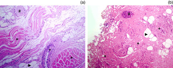
Histological analysis of the tonsils was performed by a pathologist who was blinded to the surgical technique. A total of 74 randomly selected tonsils (one for each patient, without side identification) were analysed after formaldehyde fixation and paraffin embedding. Sections (4 μm thick) were cut and stained with hematoxylin and eosin using standard methodology for analysis by optical microscopy. The following histological parameters were measured for each sample: (1) representativeness, scored as 0 (no or less than 1 mm of peritonsillar soft tissue) or 1 (at least 1 mm of peritonsillar soft tissue); (2) damage by cauterisation of the surgical bed, scored as 0 (less than 50 per cent of peritonsillar tissue), 1 (50–90 per cent) or 2 (more than 90 per cent); and (3) the presence of striated muscle, adipose tissue, nerves and blood vessels in the surgical bed, along with any damage to these structures caused by cauterisation (Figure 1).
Representative photomicrographs showing peritonsillar tissue (a) without electrocautery damage and (b) with electrocautery damage. (a) Image shows preserved muscle fibres (*), blood vessels (+), nerves (#) and adipose tissue (arrowhead). (b) Image shows disorganisation of the same structures, along with coagulated, intense eosinophilic tissue due to thermal damage. (H&E, ×200).

Statistical analysis
Statistical analysis was performed using IBM SPSS Statistics for Windows, version 20.0 (Armonk, New York, USA). Quantitative variables were compared using the student's t test and qualitative variables were compared using the χ2 test. A p value of less than 0.05 was considered statistically significant.
Results
Patient characteristics
Of the 40 patients who underwent cold dissection tonsillectomy, 1 was excluded for failure to comply with the prescribed treatment and 2 were excluded because their tonsils did not show adequate histological representation (as assessed by the pathologist). The final group comprised 37 patients (13 men and 24 women) aged between 15 and 44 years (mean 25 years). Of the 39 patients who underwent electrocautery dissection tonsillectomy, 2 were excluded because their tonsils did not show adequate histological representation. The final group comprised 37 patients (13 men and 24 women) aged between 14 and 41 years (mean 26 years). Thus, both groups were comparable with respect to age and sex.
Pain assessment
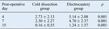
As shown in Table II, pain intensity was significantly lower in the cold dissection group than in the electrocautery dissection group on post-operative days 4, 7 and 15.
Relationship between pain score and surgical technique

Data are the mean ± standard deviation.
Relationship between surgical technique and complication frequency
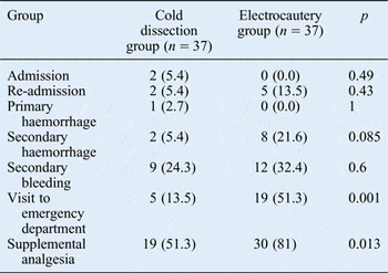
Overall, complications were more common in the electrocautery dissection group, including significantly more secondary haemorrhage episodes, emergency department visits and supplemental analgesia (Table III). Only one patient (in the cold dissection group) had a primary haemorrhage, and secondary haemorrhage was more common in the electrocautery dissection group (three out of four cases).
Relationship between pain score and complication frequency

Data are n (%).
Relationship between surgical technique and histological findings
There were no significant differences (p = 0.54) in the representativeness of peritonsillar soft tissue (present in 29 cases of cold technique and 32 of electrocautery dissection) or of muscle, vessel, nerve and adipose tissue, so these tissues could be evaluated in both groups. As shown in Table IV, cauterisation damage to the surgical bed was higher with electrocautery dissection than with cold dissection, and the difference almost reached statistical significance (p = 0.07). As expected, cauterisation of these structures was significantly more common when electrocautery was used than with cold dissection (p = 0.002).
Relationship between histological findings and surgical technique

Relationship among histological findings, pain and complications
The extent of tissue damage as determined by histopathological analysis was directly related to post-operative pain intensity, with a significant between-group difference on post-operative day 4 (Table V). There was also a significant association between pain scores on post-operative days 4 and 7 (p = 0.016 and p = 0.01, respectively) and cauterisation of anatomical structures, especially of the muscle.
Relationship between histological findings and pain scores

Pts = patients; post-op = post-operative
Patients with a greater degree of peritonsillar tissue injury had a higher incidence of secondary haemorrhage: the incidence was 28 per cent in patients with damage to more than 90 per cent of peritonsillar tissue, 4 per cent in those with damage to 50–90 per cent of peritonsillar tissue, and 10 per cent in those with damage to less than 50 per cent of peritonsillar tissue (p = 0.047). In addition, a higher degree of tissue damage was associated with a greater need for additional treatment for pain and a higher frequency of emergency department visits.
Discussion
Among the potential tonsillectomy complications, post-operative pain and haemorrhage are the most seriousReference Aydin, Taskin, Altas, Erdil, Senturk and Celebi 10 and are therefore the leading causes of post-tonsillectomy medical visits.Reference Bhattacharyya and Kepnes 11 As found in the present series, other studies reported the leading cause of emergency department visits to be uncontrolled pain, followed by bleeding or haemorrhage.Reference Aydin, Taskin, Altas, Erdil, Senturk and Celebi 10 , Reference Bhattacharyya and Kepnes 11 Different studies have linked haemorrhage to the surgical technique used. For example, Tomkinson et al. assessed 17 480 tonsillectomies, concluding that surgical technique, age and sex are the most important risk factors for severe post-operative haemorrhage.Reference Tomkinson, Harrison, Owens, Harris, McClure and Temple 12 A national UK audit found that tonsillectomy performed with so-called hot techniques (i.e. diathermy or coblation) had a post-operative haemorrhage rate three times higher than that of tonsillectomy performed with the cold technique.Reference Hahn, Rungby, Overgaard, MØller, Schultz and Tos 5 The present study similarly found that patients who underwent electrocautery dissection had a higher incidence of haemorrhage and higher pain scores compared with those who underwent cold dissection.
Sarny et al. used a questionnaire to evaluate all episodes of bleeding or haemorrhage and post-operative pain in 615 paediatric and adult patients who underwent tonsillectomy with cold dissection (mean hospital stay 3.9 days).Reference Sarny, Habermann, Ossimitz and Stammberger 13 Similar to the present study, these authors found that 25.7 per cent of patients had bleeding or haemorrhage episodes, 5.1 per cent of patients required surgical revision and adult patients reported moderate to severe pain. In the present study, adults underwent tonsillectomy in an out-patient setting. Bleeding or haemorrhage rates in patients who underwent cold dissection were similar to those in previous reports (27 per cent), but only 2.7 per cent of patients required surgical revision. In all, 70 per cent of patients scored pain levels as below 3 (on a 0–10 pain scale) in the first 4 post-operative days, which falls into the category of mild pain.
-
• There is no consensus on the optimal technique for decreasing tonsillectomy-associated morbidity
-
• Histological findings were compared in 37 patients who underwent cold dissection tonsillectomy and 37 who underwent electrocautery dissection tonsillectomy
-
• The relationship between histological findings and post-operative complications was assessed
-
• Cold dissection produced less tissue damage and was associated with fewer complications
Reports of histopathological analyses of the tonsils to determine the extent of thermal injury caused by different surgical techniques are scarce. Shah et al. compared plasma and monopolar electrocautery-mediated ablation in a series of 34 paediatric patients.Reference Shah, Galinkin, Chiavacci and Briggs 14 Histopathological analysis showed that electrocautery produced more thermal tissue injury. In a histopathological evaluation of 40 randomly selected tonsils extracted by cold dissection, monopolar electrocautery, laser or coblation, Magdy et al. showed that cold knife dissection produced less tissue damage.Reference Magdy, Elwany, el-Daly, Abdel-Hadi and Morshedy 3 In the present study, histopathological analysis of 74 tonsils extracted by cold dissection and electrocautery dissection similarly demonstrated that electrocautery dissection is associated with more tissue damage, thus explaining the higher incidence of post-operative pain and complications. Another parameter that should be considered when analysing peritonsillar tissue damage is the handedness of the surgeon because this can lead to different degrees of difficulty in dissecting the left or right tonsil (unless the surgeon is ambidextrous). However, this could not be investigated in the present study because only one randomly selected tonsil was studied for each patient.
Conclusion
Histological analysis of the tonsils showed that cold dissection tonsillectomy produced less tissue damage and less cauterisation of tissue structures, which was associated with less pain, a lower incidence of complications and therefore less need for supplemental analgesia and fewer emergency department visits compared with electrocautery dissection tonsillectomy. Therefore, our results suggest that cold dissection is the tonsillectomy technique of choice.