Introduction
Fascioliasis and amphistomiasis in ruminants in India are controlled exclusively by the use of chemical anthelmintics. Three crucial reasons for opting to seek alternative parasite control strategies are the increasing incidence of drug resistance, the desire to minimize residues in food and the need to reduce environmental degradation from chemical interventions (Sanyal, Reference Sanyal2001). Biological control methods using predacious fungi could be one such step in this direction (Sanyal, Reference Sanyal2005). Biological control is aimed at exploiting natural enemies to reduce the number of target pathogenic organisms to a level less than what would have occurred in the absence of the predators. Egg-parasitic fungi are characterized by their ability to colonize parasite cysts and eggs and are potential candidates for biological control. They generally attack by penetrating the eggs using specialized perforating organs or lateral branches from the mycelium (Stirling, Reference Stirling and Stirling1991). Two egg-parasitic fungi, Paecilomyces lilacinus and Verticillium chlamydosporium, which were isolated from an organic environment of animal origin from India (Sanyal et al., Reference Sanyal, De, Sarkar, Patel, Mandal and Pal2008), were screened in vitro for their suitability for possible field use (De et al., Reference De, Sanyal, Sarkar, Patel, Pal and Mandal2008). It has now largely been acknowledged that effective parasite control cannot be achieved persistently by a single method. The available classical and alternative technologies for animal parasite control need to be integrated properly. Although the use of anthelmintics may play a crucial role in such integrated control, their use should be minimized as far as practicable (Nari & Eddi, Reference Nari and Eddi2002).
Members of the benzimidazole group of anthelmintics are commonly and widely used to control fascioliasis and amphistomiasis of ruminants because of their broad spectrum of activity and wide availability at cheaper rates (Theodorides et al., Reference Theodorides, Gyurik, Kingsbury and Parish1976). It is known that benzimidazoles also have antimycotic properties that have been exploited by their use as fungicides on agricultural crops (Horsfall, Reference Horsfall1956). Benzimidazoles are reported to have a mycostatic effect on the nematode-trapping fungus Duddingtonia flagrans (Sanyal et al., Reference Sanyal, Chauhan and Mukhopadhyaya2004). As the egg-parasitic fungi, P. lilacinus and V. chlamydosporium, belong to the same class of Fungi Imperfecti as D. flagrans, it was considered essential to investigate the effect of benzimidazoles on these fungi so as to elucidate the specific implications for integrated parasite management. The present investigations, involving in vitro trials of the effects of albendazole and triclabendazole on the growth profiles of both the fungi, and in vivo trials in goats using intraruminal administration of these two benzimidazole anthelmintics and fed millet grain cultures of P. lilacinus and V. chlamydosporium, were conducted to meet this objective.
Materials and methods
Solid substrate fungal cultures
Wild strains of P. lilacinus and V. chlamydosporium isolated from Durg, Chhattisgarh, India, were established on 2.5% corn meal agar (CMA) Petri plates (90 mm disposable and radiation sterilized; Tarson India Ltd., Mumbai, India) containing 0.02% tetracycline (w/v), added after autoclaving of the media to prevent bacterial growth, and maintained at 26°C. The predatory activity of the fungi was checked by egg bait assay using a mixture of live eggs of Fasciola gigantica and Gigantocotyle explanatum (De et al., Reference De, Sanyal, Sarkar, Patel, Pal and Mandal2008). The fungi were then grown in solid substrate (millet grains) by putting a piece of corn meal agar medium containing fungal growth into a flask containing autoclaved millet grains (Sanyal, Reference Sanyal2000) and incubated at 26°C for 6 weeks, the flask being shaken twice weekly for 6 weeks to get uniform growth.
In vitro trial
Pure triclabendazole and albendazole, members of the benzimidazole anthelmintics, were obtained from Ciba-Geigy Ltd., Switzerland and Smith Kline Beecham India Ltd, Bangalore, India, respectively. They were added to the autoclaved molten media (2.5% solution of corn meal agar) separately, at a concentration of 0, 0.5, 1.5, 2.5, 3.5 or 4.5 ppm, before plating. For each concentration five replicates were prepared. An agar disc of 0.8 cm radius was scooped from the periphery of 2-week-old parent culture and placed at the centre of each medium plate (90 mm; Tarson Ltd) and incubated at 26°C. Two measurements of colony diameter of mycelial spread were recorded twice daily.
In vivo trial
Eight parasitologically naïve goats were kept indoors, divided into two groups of four animals each, on the basis of live weight and random allocation. They were maintained indoors and fed on grass by a cut-and-carry method and water ad libitum. After acclimatization to the specified diet for 7 days, each was fed on 20 g mixed fungal grain culture of P. lilacinus and V. chlamydosporium daily for 15 days. On the fifth day of fungus feeding, albendazole suspension (Albendos®, 2.5% w/v albendazole suspension; Dosch Pharmaceuticals, Mumbai, India) was administered intraruminally at 5.0 mg kg− 1 body weight to animals of one group and the other group was similarly treated with triclabendazole suspension (Fasinex®, 2.5% w/v triclabendazole suspension; Intervet India Ltd, Mumbai, India) at 10.0 mg kg− 1 body weight. Whole blood was collected in heparinized tubes (Vacutainer, Becton Dickinson Inc., California, USA) at 0, 4, 8, 12, 18, 24, 30, 36, 48, 72 h and at 0, 4, 8, 12, 18, 24, 30, 36, 48, 72, 96, 120, 144 and 168 h post-dosing with albendazole and triclabendazole, respectively. Plasma was separated and stored at − 20°C for further use. Simultaneously, faeces from each animal were collected per rectum and coproculture for fungal recovery from each sample was done from day 3 ( − 48 h) and 4 ( − 24 h) of fungus feeding, at the same times as blood sampling, and for 2 days after completion of blood sampling at 24-h intervals. To recover fungus from faeces, faecal samples were cultured in 2.5% corn meal agar plates baited with eggs of liver fluke and amphistome. Plates were examined under a stereoscope at weekly intervals for 4 weeks. Egg-parasitic fungi were detected from around parasitized eggs. Pure cultures were established by transferring mycelial growth to Petri plates containing 2% CMA. These isolates were maintained on this medium and identified by their morphology and morphometry (Samson, Reference Samson1974). Faecal recovery of fungus was scored as profuse recovery (++++), moderate recovery (+++), scanty recovery (++), negligible recovery (+) and no recovery (Nil).
Pure standards
Pure external standards of albendazole metabolites were supplied by Smith Kline Beecham India Ltd in the year 1997. Pure internal standards, ethyl oxfendazole and mebendazole, were supplied by Dr D.R. Hennessy, CSIRO McMaster Laboratory, Sydney, Australia in the year 1991. Pure external standards of triclabendazole metabolites were supplied by Ciba-Geigy, Switzerland in the year 1995. All the pure standards were stored as micronized powder in screw-capped glass vials at 4°C until further use.
Albendazole metabolites
For albendazole metabolites, solid-phase extraction of plasma, spiked with ethyl oxfendazole as internal standard, was performed using a C18 Sep-Pak cartridge (Waters Associates, Millford, Massachusetts, USA). Albendazole metabolites in extracted plasma were analysed through reverse-phase high-performance liquid chromatography using an isocratic mobile phase of 33% acetonitrile in 0.025 m sodium acetate with the detector module set at 292 nm (Sanyal, Reference Sanyal1997). Identification of the metabolites in plasma was done by comparison with the retention times of reference standards. The absolute recovery was 97% with the limit of detection being 0.02 μg ml− 1. The concentrations of each component in the plasma extracts were determined by comparing the detector response for each component in the sample with the corresponding peak in a standard mixture.
For triclabendazole metabolites, solid-phase extraction of plasma, spiked with mebendazole as internal standard, was performed following protein precipitation with acetone, using a C18 Sep-Pak cartridge (Waters Associates). Triclabendazole metabolites in extracted plasma were analysed through reverse-phase high-performance liquid chromatography using an isocratic mobile phase of 50% acetonitrile in 0.025 m sodium acetate with the detector module set at 292 nm (Sanyal, Reference Sanyal1995). The metabolites in plasma were identified by comparison with the retention times of reference standards. The absolute recovery was 99% with the limit of detection being 0.01 μg ml− 1. The concentrations of each component in the plasma extracts were determined by comparing the detector response for each component in the sample with the corresponding peak in a standard mixture.
Statistical analysis
Analysis of variance of the data was performed using GraphPad Prism 4 software (Version 4.02, 2004, GraphPad Software Inc., San Diego, California, USA).
Results
In vitro study
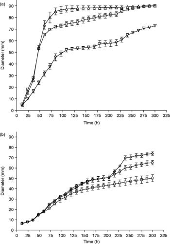
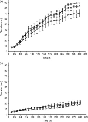
In vitro growth profiles of P. lilacinus and V. chlamydosporium under the influence of different concentrations of albendazole in corn meal agar plates are presented in figs 1 and 2, respectively. Verticillium chlamydosporium appeared to be more sensitive to albendazole than P. lilacinus. In fact, growth of P. lilacinus was more vigorous at 0.5 ppm concentration of albendazole at 60–168 h of incubation (P < 0.05; P < 0.001) compared to the controls (fig. 1). Significant growth inhibition (P < 0.001) of P. lilacinus occurred at a concentration of 1.5–4.5 ppm of albendazole at 36–300 h of incubation. In contrast, at 0.5 and 1.5 ppm concentrations of albendazole, growth curves of V. chlamydosporium behaved similarly, without any significant variation (P>0.05) except at 1.5 ppm concentration during 240–300 h of incubation (P < 0.01). However, significant growth inhibition (P < 0.001) of V. chlamydosporium occurred at a concentration of 2.5–4.5 ppm of albendazole at 72–300 h of incubation (fig. 2).
In vitro growth profiles (mean ± SEM) of Paecilomyces lilacinus in corn meal agar plates mixed with (a) 0 (□), 0.5 (△), 1.5 (▿) and (b) 2.5 (⋄), 3.5 (○) and 4.5 (□) ppm concentrations of albendazole and incubated at 26°C.

In vitro growth profiles (mean ± SEM) of Verticillium chlamydosporium in corn meal agar plates mixed with (a) 0 (□), 0.5 (△), 1.5 (▿) and (b) 2.5 (⋄), 3.5 (○) and 4.5 (□) ppm concentrations of albendazole and incubated at 26°C.

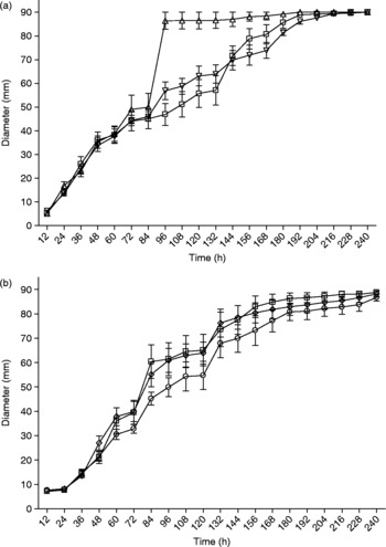
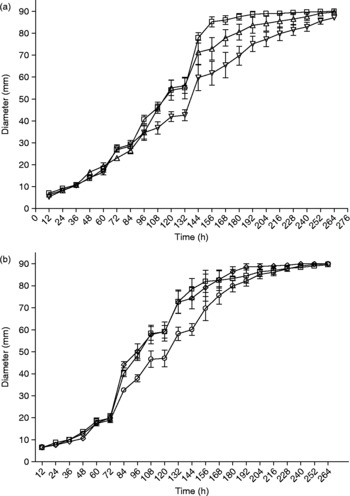
In vitro growth profiles of P. lilacinus and V. chlamydosporium under the influence of different concentrations of triclabendazole in corn meal agar plates are presented in figs 3 and 4, respectively. Triclabendazole appeared to have least inhibitory effect on in vitro growth of both P. lilacinus and V. chlamydosporium. Similar to albendazole, growth of P. lilacinus was more vigorous at 0.5 ppm concentration of triclabendazole from 96 to 132 h of incubation (P < 0.001). However, growth profiles of P. lilacinus at all the concentrations of triclabendazole became similar from 156 h of incubation onwards (fig. 3). On the contrary, growth curves of V. chlamydosporium behaved similarly at all concentrations, with significant inhibition here and there, while at 1.5 ppm triclabendazole significant growth inhibition (P < 0.001) was observed between 144 and180 h of incubation (fig. 4).
In vitro growth profiles (mean ± SEM) of Paecilomyces lilacinus in corn meal agar plates mixed with (a) 0 (□), 0.5 (△), 1.5 (▿) and (b) 2.5 (⋄), 3.5 (○) and 4.5 (□) ppm concentrations of triclabendazole and incubated at 26°C.

In vitro growth profiles (mean ± SEM) of Verticillium chlamydosporium in corn meal agar plates mixed with (a) 0 (□), 0.5 (△), 1.5 (▿) and (b) 2.5 (⋄), 3.5 (○) and 4.5 (□) ppm concentrations of triclabendazole and incubated at 26°C.

In vivo studies
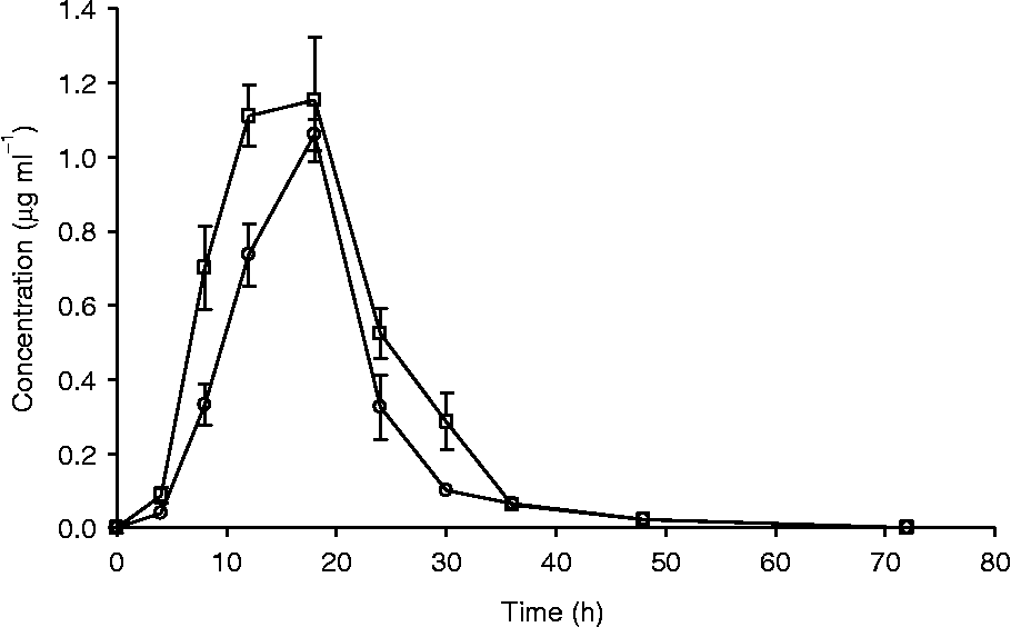
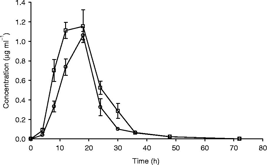
Plasma albendazole sulphoxide and albendazole sulphone profiles in goats following a single intraruminal administration of albendazole are presented in fig. 5. Albendazole sulphoxide, the anthelmintically active moiety, reached its maximum concentration of 1.15 μg ml− 1 at 18 h of drug administration, while the anthelmintically inactive albendazole sulphone moiety reached its maximum concentration of 1.06 μg ml− 1 at 18 h after drug administration (fig. 5). By 72 h after drug administration, no detectable levels of plasma albendazole metabolites could be observed.
Plasma concentrations (mean ± SEM) of albendazole sulphoxide (ABZ-SO; □) and albendazole sulphone (ABZ-SO2; ○) in goats following a single intraruminal administration of albendazole at 5.0 mg kg− 1 body weight.
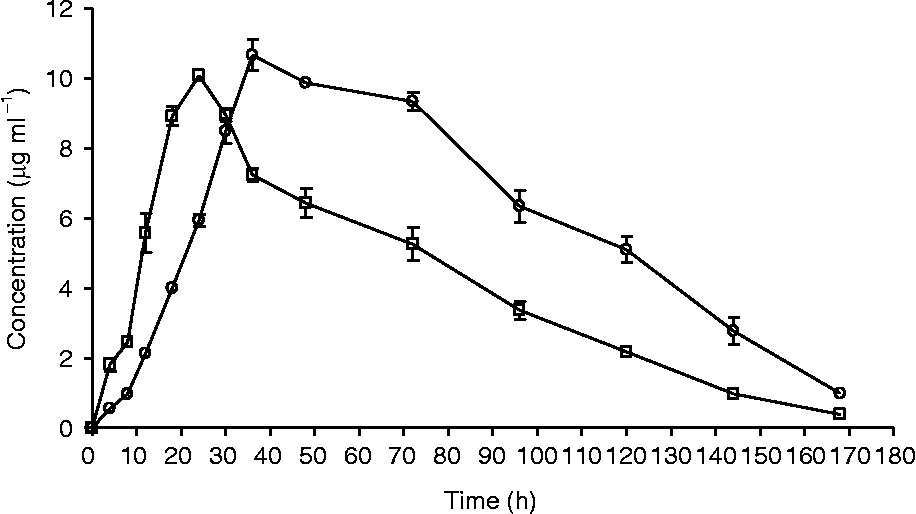
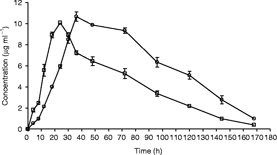
Plasma triclabendazole sulphoxide and triclabendazole sulphone profiles in goats following a single intraruminal administration of triclabendazole are presented in fig. 6. For triclabendazole, the anthelmintically active sulphoxide metabolite reached its maximum concentration of 10.09 μg ml− 1 at 24 h after drug administration, while the anthelmintically inactive sulphone moiety reached its maximum concentration of 10.67 μg ml− 1 at 36 h after drug administration (fig. 6). Detectable plasma levels of both the metabolites were observed up to 168 h after drug administration.
Plasma concentrations (mean ± SEM) of triclabendazole sulphoxide (TCBZ-SO; □) and triclabendazole sulphone (TCBZ-SO2; ○) in goats following a single intraruminal administration of triclabendazole at 10.0 mg kg− 1 body weight.
Fungal recovery
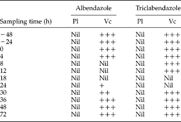
Results of re-isolation of egg-parasitic fungi from faeces of goats fed on fungal millet grain cultures of P. lilacinus and V. chlamydosporium before and following single intraruminal administration of albendazole and triclabendazole are presented in table 1. Paecilomyces lilacinus was not recovered from the faeces of goats at any sampling period. However, V. chlamydosporium was recovered from the faeces of goats at most of the sampling periods except during 8–18 h and 18–24 h after administration of albendazole and triclabendazole, respectively.
Re-isolation of egg-parasitic fungi from faeces of goats fed on fungal millet grain cultures of Paecilomyces lilacinus (Pl) and Verticillium chlamydosporium (Vc) before and following single intraruminal administration of albendazole and triclabendazole at 5.0 and 10.0 mg kg−1 body weight, respectively.
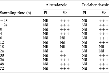
+++, Moderate recovery; ++, scanty recovery; + negligible recovery.
Fungus–drug interactions
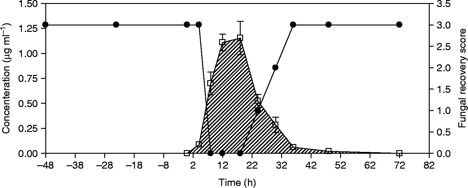
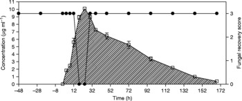
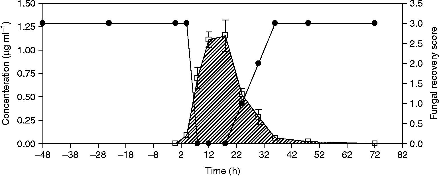
The plasma disposition profiles of albendazole sulphoxide and triclabendazole sulphoxide metabolites superimposed with scores for the recovery of fungus from faecal culture in goats are presented in figs 7 and 8, respectively. Moderate recovery of V. chlamydosporium could be obtained for the first 2 days before administration of albendazole and from 36 h after albendazole administration onwards. While no V. chlamydosporium could be recovered from the faeces of animals during 8–18 h of albendazole administration, scanty recovery of the fungus could be made on hours 24 and 30 after albendazole administration (fig. 7). Moderate recovery of V. chlamydosporium could be obtained for the first 2 days before administration of triclabendazole and at all hours post-administration of triclabendazole except during hours 18 and 24 (fig. 8).
Fungal recovery scores of Verticillium chamydosporium (●) superimposed with plasma profiles of albendazole sulphoxide (ABZ-SO; □) in goats fed on fungal millet grain cultures and administered a single intraruminal dose of albendazole at 5.0 mg kg− 1 body weight.
Fungal recovery scores of Verticillium chamydosporium (●) superimposed with plasma profiles of triclabendazole sulphoxide (TCBZ-SO; □) in goats fed on fungal millet grain cultures and administered a single intraruminal dose of triclabendazole at 10 mg kg− 1 body weight.

Discussion
The present in vitro experiments provided evidence that albendazole, a widely used benzimidazole anthelmintic against both gastrointestinal nematodes and flukes, possesses potent antimycotic properties, while the benzimidazole flukicide triclabendazole, does not. Albendazole previously displayed antimycotic effects when in vitro studies were conducted with the nematode-trapping fungus D. flagrans (Sanyal et al., Reference Sanyal, Chauhan and Mukhopadhyaya2004). De et al. (Reference De, Sanyal, Sarkar, Patel, Pal and Mandal2008) observed significant inhibition in colony diameters of P. lilacinus and V. chlamydosporium at 0.5–4.5 ppm concentrations of carbendazim, a benzimidazole fungicide used in agriculture. In the present study, the egg-parasitic fungus V. chlamydosporium appeared to be more sensitive to albendazole compared to P. lilacinus. Inherent susceptibility of V. chlamydosporium to benzimidazoles could only serve as a possible explanation. In contrast, triclabendazole, a widely used benzimidazole flukicide used exclusively against Fasciola gigantica and F. hepatica infections, did not appear to have any antimycotic properties under in vitro conditions.
The results provided ample evidence to suggest that V. chlamydosporium is able to survive gastrointestinal transit in goats, while P. lilacinus does not. The production of tough and resistant chlamydospores by V. chlamydosporium (Sanyal et al., Reference Sanyal, De, Sarkar, Patel, Mandal and Pal2008) suggests a possible explanation for this observed difference. Similarly, chlamydospores of the nematode-trapping fungus, D. flagrans, were reported to have the ability to tolerate the anaerobic and enzymatic environments of the gastrointestinal tract of both monogastric and polygastric animals (Sanyal, Reference Sanyal2005).
The present in vivo experiments provide evidence that albendazole exerts a potent antimycotic property at a plasma concentration of 0.70 μg ml− 1 of the active metabolite albendazole sulphoxide and the fungus could not be re-isolated from the faeces of the animals during 8–12 h of the sampling period (fig. 7). Similarly, V. chlamydosporium could not be re-isolated from the faeces of triclabendazole-administered goats during 18–24 h of the sampling period when the plasma concentration of triclabendazole sulphoxide was 8.92 μg ml− 1 of the active metabolite (fig. 8). These sulphide benzimidazole compounds are oxidized in the liver to form the anthelmintically active sulphoxide moieties and are recycled back to the intestine through enterohepatic circulation (Sanyal, Reference Sanyal1995). The resultant enteric levels of the anthelmintically active moieties possibly have antimycotic properties. Significant absorption of benzimidazoles occurs in the small intestine, most of the absorbed metabolites deriving from biliary secretions. As much as 60% of the dose of aromatic substituted benzimidazole drugs (fenbendazole, oxfendazole) are secreted in bile as free and conjugated metabolites (Hennessy et al., Reference Hennessy, Ali and Tremain1994). Approximately one-third of these metabolites are reabsorbed (Hennessy et al., Reference Hennessy, Steel and Prichard1993). The aliphatic substituted albendazole is less extensively secreted in bile (Hennessy et al., Reference Hennessy, Steel, Lacey, Eagleson and Prichard1989). Passage of biliary metabolites across the gastrointestinal tract wall provides significant contact with lumen-dwelling parasites.
For biological control using egg-parasitic fungi to work best, it appears important that the start of treatment should coincide with time(s) of the year just when an increase in long-lasting trematode egg numbers in the faeces is expected. With the right dosing and timing, it should be possible to significantly reduce the number of infective stages of the parasite to infect intermediate hosts, thereby providing the best environment for protecting livestock from the disease. In India, the parasitic burden in the host with resultant increase in faecal egg output increases with the advent of monsoon rain, which is also the strategic time of anthelmintic application (Sanyal, Reference Sanyal2001). As benzimidazoles are widely used anthelmintics in livestock to control parasitic trematodes, due precautions should be taken when integrating anthelmintic intervention and biological control to achieve sustainable control. While studying the nematode-trapping fungus D. flagrans, Sanyal et al. (Reference Sanyal, Chauhan and Mukhopadhyaya2004) suggested that a minimum wash-out period of 2 weeks would be required between the use of these two methods of parasite control. While a regime of parasite control involving the use of chemical anthelmintics cannot be implemented in an organic farming system, the combination of anthelmintics and biological control could be implemented in most conventional animal production systems.
Acknowledgements
This research was the result of the All India Network Project on Gastrointestinal Parasitism of Indian Council of Agricultural Research, New Delhi. Facilities provided by the Dean, College of Veterinary Science & AH, Anjora, Durg are thankfully acknowledged.