Introduction
The genus Oncicola Travassos, 1916 includes 24 parasite species, able to infect numerous vertebrates such as felids and canids (Amin, Reference Amin2013). In Brazil, the diversity of Oncicola spp. currently comprises O. sigmoides (Meyer, 1932), O. luehei (Travassos, 1917), O. macrurae (Meyer, 1931), O. magalhaesi Machado-Filho, 1962, O. micracantha Machado-Filho, 1949, O. paracampanulata Machado-Filho, 1963, O. oncicola (von Ihering, 1892), O. campanulata (Diesing, 1851), O. chibigouzouensis Machado-Filho, 1963 and Oncicola sp. (Vieira et al., Reference Vieira, Luque and Muniz-Pereira2008).
Morphological characters of this genus mainly include a small and elongated body; a proboscis with six longitudinal rows containing hooks with double roots, of which the basal ones are smaller; a smooth neck; very long lemnisci; large cement glands near the testes in the anterior portion of the body and large copulatory bursa (Travassos, Reference Travassos1917). Species of Oncicola have very similar morphology, so the use of light microscopic analysis alone can lead to doubtful classification. Genetic data of Oncicola species are also scarce: out of the nine references in GenBank, only one is identified to the species level.
In Serra da Capivara National Park (SCNP) in Brazil, dead specimens of the ocelot Leopardus pardalis (Linnaeus) were collected along roads or seized from hunters. The specimens were donated for helminthological analysis, and specimens of Oncicola venezuelensis Marteau, 1977, a species whose ultrastructural and genetic data were previously unknown, were found parasitizing the intestine of the dead animals.
The aim of this study was to describe the morphological and ultrastructural aspects of O. venezuelensis obtained by a combination of light and scanning electron microscopy, as well as to provide new genetic data for the species from a newly reported locality.
Materials and methods
Collection and examination of acanthocephalans
Serra da Capivara National Park is located in an isolated area of the north-east semi-arid region of Piauí state, Brazil (08°26′50.099″N to 08°54′23.365″S and 08°36′33.681″E to 08°46′28.382″W). Two adults of L. pardalis found dead in SCNP by staff of the Chico Mendes Institute for Biodiversity Conservation (ICMBio) were donated to the American Human Museum Foundation (FUMDHAM). For research purposes, FUMDHAM donated the digestive tracts of the animals to Oswaldo Cruz Institute/FIOCRUZ – Laboratory for Evaluation and Promotion of Environmental Health. The digestive tracts were tied with strings from anterior to posterior end, with separations between the oesophagus, stomach, and small and large intestines.
The parasites found in the gastrointestinal contents were washed in physiological saline (0.7%) and stored in 70% alcohol. In the laboratory, some acanthocephalans were stained with alcoholic chloride carmine, cleared in clove oil and mounted in Canada balsam. Measurements are given in micrometres, with ranges in parentheses. Drawings were made with a drawing tube and redrawn using Adobe Illustrator CS6.
For scanning electron microscopy (SEM), the acanthocephalans were washed in 0.1 m cacodylate buffer pH 7.2, post-fixed in 1% OsO4 and 0.8% potassium ferrocyanide for 1 h, dehydrated in a graded series of ethanol solutions (30–100%) for 1 h for each step, critical-point dried in CO2, sputter coated with gold and examined using a Jeol JSM 6390 microscope.
Molecular analysis
Genomic DNA was extracted using a QIAamp DNA Mini Kit (Qiagen, Hilden, Germany) according to the manufacturer's recommendations. The rRNA region spanning internal transcribed spacer (ITS)1, 5.8S and ITS2 was amplified by polymerase chain reaction (PCR) using the primers BD1 (5′-GTCGTAACAAGGTTTCCGTA-3′) and BD2 (5′-TATGCTTAARTTCAGCGGGT-3′) (Chen et al., Reference Chen, Zhang, Wen, Sun and Gao2010). The partial region spanning 28S rRNA was amplified by PCR using the set of primers C1 (5′-ACCCGCTGAATTTAAGCAT-3′) and D2 (5′-TGGTCCGTGTTTCAAGAC-3′) (Chisholm et al., Reference Chisholm, Morgan, Adlard and Whittington2001). PCR assays were carried out in a total volume of 15 μl containing 7.5 μl of 2 × GoTaq® Colorless Master Mix (Promega, Madison, Wisconsin, USA), 1.5 μl of each primer with final concentrations of 0.5 μm, 1.0−2.0 μl of cDNA sample and ultrapure water to complete. For ITS1, 5.8S and ITS2 regions, the reaction was performed under the following conditions: 94°C for 2 min; followed by 40 cycles at 95°C for 30 s, 56°C for 30 s and 72°C for 60 s; and 72°C for 7 min. For the partial 28S rRNA region, the reaction was done under the following conditions: 94°C for 2 min (initial denaturation); followed by 40 cycles at 95°C for 30 s, 55°C for 30 s and 72°C for 60 s; and 72°C for 5 min.
Amplicons were analysed, using 5 μl of PCR product, by electrophoresis in 2% agarose in Tris-borate EDTA gels, stained with SyberGreen (Invitrogen, Eugene, Oregon, USA) and photographed under UV transillumination. Amplified PCR products were purified with ExoSAP-IT (Affymetrix, Cleveland, Ohio, USA) and sequenced using the same primer set. DNA cycle sequencing reactions were performed using BigDye v.3.1 chemistry (Applied Biosystems, Foster City, California, USA). Sequences were edited in MEGA 6.0 (Tamura et al., Reference Tamura, Peterson, Peterson, Stecher, Nei and Kumar2011) and compared for similarities with sequences from GenBank using the BLASTN program from the National Center for Biotechnology Information (NCBI) (Altschul et al., Reference Altschul, Gish, Miller, Myers and Lipman1990). To examine the phylogenetic relationships, the nucleotide sequences were analysed using the CLUSTAL W algorithm (Tamura et al., Reference Tamura, Peterson, Peterson, Stecher, Nei and Kumar2011). The evolutionary model was selected using the program jModelTest (Posada, Reference Posada2008). The maximum-likelihood (ML) statistical method was used to construct trees, which were resampled by 5000 bootstrap replicates using the software MEGA 6.0 (Felsenstein, Reference Felsenstein1981; Tamura et al., Reference Tamura, Peterson, Peterson, Stecher, Nei and Kumar2011).
Results
The measurements and morphological characteristics of O. venezuelensis are in accordance with the data from Marteau (Reference Marteau1977) and Fuller & Nickol (Reference Fuller and Nickol2011).
Oncicola venezuelensis Marteau, 1977
Taxonomic summary
Description
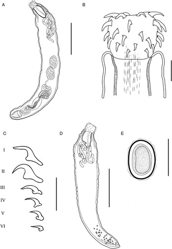
Male (based on three specimens). Total body length 9167 (5625–12,500) and width 1167 (750–1500) (fig. 1A). Proboscis 433 (400–500) long and 507 (370–600) wide (fig. 1B). Hooks: I, 120–125, distance between roots (dr) 100–110; II, 145–150, dr 100–115; III, 125, dr 105–110; IV, 105–110, dr 90–100; V, 95–105, dr 85–85; VI, 95–100, dr 80–85 (fig. 1C). Proboscis receptacle 960 (600–1180), single walled. Neck 220–270 (245) long by 200 wide. Anterior testis 930–1180 long and 500–600 wide. Posterior testis 1040–1250 long and 600–680 wide. Cement glands 600–700 long and 550–850 wide. Safftigen's pouch 1150–1200 long and 550–650 wide. Copulatory bursa 1500 long and 550 wide.
The morphology of Oncicola venezuelensis to show: (A) the male with globular proboscis, leminisci looped, testes in mid-trunk, cement glands, Safftigen's pouch and copulatory bursa reversed (scale bar: 2.5 mm); (B) globular proboscis and hooks (scale bar: 0.250 mm); (C) details of hooks I, II, III, IV, V and VI (scale bar: 0.250 mm); (D) female with proboscis, leminisci, uterine bell and genital pore subterminal (scale bar: 2.5 mm) and (E) egg of the female (scale bar: 0.050 mm).

Female (based on seven specimens). Total body length 9893 (6250–13,250) and width 1471 (1000–1950) (fig. 1D). Proboscis 540 (430–600) long and 670 (650–700) wide. Hooks: I, 127 (110–140), dr 122 (110–140); II, 103 (80–120), dr 89 (75–100); III, 88 (70–100), dr 73 (60–85); IV, 84 (70–90), dr 77 (65–85); V, 81 (70–100), dr 70 (60–85); VI, 62 (50–70), dr 52 (50–60). Proboscis receptacle 1060 (970–1200), single walled. Neck 300–340 (320) long by 400–550 (476) wide. Cerebral ganglion 110 long by 110 wide. Uterine bell 422 (350–500) long and 110 (100–130) wide. Uterus length 610 (400–720) and sphincter length 318 (200–400). Eggs 42 (30–55) × 26 (22–30) (fig. 1E).
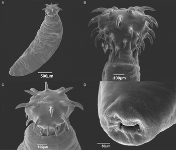
Scanning electron microscopic observation showed a trunk slightly flatted and rough, with a globular proboscis (fig. 2A) with a short and smooth neck (fig. 2B). The proboscis presented 36 hooks divided into six longitudinal rows of six hooks each (fig. 2C). The hooks differ in size and shape, progressively decreasing in size from apical to basal positions in the proboscis. Hooks I, II and III are ‘chisel-shaped’ at the tip. The female genital pore is subterminal (fig. 2D).
Scanning electron microscopy of Oncicola venezuelensis to show: (A) the entire body with proboscis; (B) the proboscis with hooks and neck; (C) the globular proboscis with 36 hooks, chisel-shaped at the tip; and (D) the genital pore in the female.

Molecular analysis
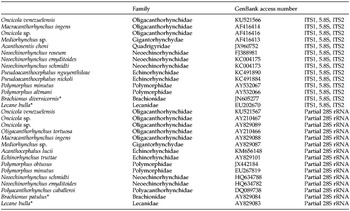
The genetic sequences were deposited in GenBank under accession numbers KU521566 (ITS1, 5.8S and ITS2 region; 681 bp) and KU521567 (partial 28S rRNA; 747 bp). The taxa used in phylogenetic analyses are listed in table 1.
List of the species of acanthocephalan parasites and outgroups used in the phylogenetic analyses of Oncicola venezuelensis using sequences of the ITS1, 5.8S and ITS2 and partial 28S rRNA genes.

*Rotifera outgroup.
The BLAST results for complete ITS1, 5.8S and ITS2 sequences indicated, first, 89% identity with 38% query cover and a maximum score of 324 for Macracanthorhynchus ingens (von Linstow, 1879) (AF416414), followed by 89% identity with 37% query cover and a maximum score of 309 for Moniliformis moniliformis (Bremser, 1811) (Moniliformidae) (AF416415) and, finally, 97% identity with 25% query cover and a maximum score of 291 for Oncicola sp. (AF416416). When the ITS1 region was analysed separately, the BLAST result indicated no significant similarity with sequences deposited in GenBank; the same was found for the ITS2 region. The 5.8S rRNA gene was indicated as having 99% identity with 99% query cover for M. moniliformis (AF416415), as well as 99% identity with 98% query cover for Oncicola sp. (AF416416) and a maximum score of 289 and 281, respectively.
The phylogenetic analysis of 5.8S rRNA gene sequences resulted in an inconclusive tree to separate Oligacanthorhynchidae from Gigantorhynchidae and Moniliformidae. Individual trees for each ITS1 and ITS2 rRNA region also resulted in inconclusive findings, with low statistical support for species of Oligacanthorhynchidae.
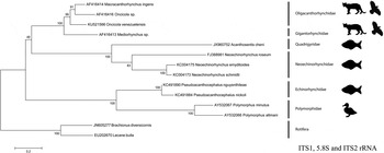
The tree using the complete ITS1, 5.8S and ITS2 rRNA better represented the phylogenetic relationship between the acanthocephalan families with higher statistical support when M. moniliformis was excluded (fig. 3). Our sequence of O. venezuelensis was aligned with 13 other sequences of different families of Acanthocephala. The best substitution model for the ITS1, 5.8S and ITS2 dataset was Tamura–Nei 3 parameters with gamma distribution (+G) and invariable sites (+I). ML tree inferred dataset yielded a tree with −ln of −3929.701. Based on the analysis, the Oligacanthorhynchidae clade was 99% supported and comprised our sequence of O. venezuelensis, Oncicola sp. and M. ingens and is closely related to Gigantorhynchidae (100% support). Other clades included species of Quadrigyridae Van Cleave, 1920 and Neoechinorhynchidae (Ward, 1917) clustered in a bootstrap resampling of 100%. Echinorhynchidae Cobbold, 1876 and Polymorphidae Meyer, 1931 appear, forming a clade with 100% support (fig. 3).
Maximum-likelihood tree of Oncicola venezuelensis based on the ITS1–5.8S–ITS2 region to show the relationship with other species of Acanthocephala. Numbers (%) on the branches indicate 5000 bootstrap replicates. The animals represent final host groups. GenBank accession numbers are shown and the scale bar indicates the nucleotide mutations per site.

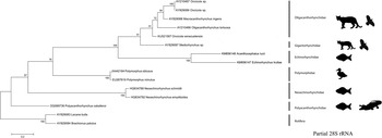
For partial 28S rRNA the BLAST results indicated 89% identity with 100% query cover and a maximum score of 917 for Oncicola sp. (AY210467). The sequence of the large subunit (LSU) was aligned with 14 other sequences. The best substitution model for the LSU dataset was Hasegawa–Kishino–Yano parameters, with gamma distribution (+G), and the ML tree inferred dataset yielded a tree with −ln of −4147.816. The ML tree showed the Oligacanthorhynchidae (91% supported), Gigantorhynchidae (91%), Echinorhynchidae (100%), Polymorphidae (100%), Neoechinorhynchidae (100%) and Polyacanthorhynchidae Golvan, 1956 as basal (fig. 4). The Oligacanthorhynchidae clade comprised two different sequences of Oncicola, M. ingens and Oligacanthorhynchus tortuosa (Leidy, 1850), and our sequence of O. venezuelensis is in a basal position with 91% bootstrap value. Like the ML tree based on the ITS1, 5.8S and ITS2 region, Gigantorhynchidae formed a cluster clade with Oligacanthorhynchidae (91% supported), well separated from the clade of Echinorhynchidae (100%).
Phylogenetic tree based on the maximum likelihood of the partial 28S gene, to show the relationship of Oncicola venezuelensis with other species of Acanthocephala. Numbers (%) on the branches indicate 5000 bootstrap replicates. The animals represent final host groups. GenBank accession numbers are shown and the scale bar indicates the nucleotide mutations per site.

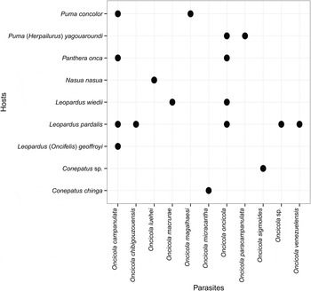
A scatter plot of Oncicola spp. reported from different carnivorous mammal hosts in Brazil shows that L. pardalis is the host parasitized by the largest diversity of species and O. venezuelensis is the fifth species of the genus now reported in this host in the country (fig. 5).
Host–parasite distribution of Oncicola species in Brazil.

Discussion
Oncicola venezuelensis was originally described from L. pardalis collected in Venezuela (Marteau, Reference Marteau1977), but recently it was also reported in the feral house cat, Felis catus Linnaeus in the US Virgin Islands (Fuller & Nickol, Reference Fuller and Nickol2011). Leopardus pardalis occurs from Texas to northern Argentina and north-western Uruguay (Oliveira, Reference Oliveira1994; Murray & Gardner, Reference Murray and Gardner1997). The host species is widely distributed in Brazil but the population has been decreasing due to deforestation (Oliveira et al., Reference Oliveira, Almeida and Campos2013). Therefore, Serra da Capivara National Park plays an important role in the preservation of the species.
Palaeoparasitological studies in Serra da Capivara National Park have reported the occurrence of the eggs of Oncicola sp. in feline coprolites up to 9000 years old, but their size was compatible with the O. campanulata species (60 × 43 μm) that have been found as parasites in Leopardus geoffroyi (d'Orbigny & Gervais), L. pardalis, Panthera onca (Linnaeus) and Puma concolor (Linnaeus) in Brazil (Sianto et al., Reference Sianto, Souza, Chame, Luz, Guidon, Pessis and Araujo2014). The new occurrence of O. venezuelensis in this area in north-eastern Brazil shows an interaction in the host–parasite geographical distribution.
In Brazil, the intermediate host of O. venezuelensis is unknown. Cystacanths have been recovered and described in Caribbean termites Nasutitermes acajutlae (Holmgren), in subcutaneous nodules of lizards Anolis cristatellus Duméril & Bibron and Anolis stratulus Cope, in the Indian mongoose Herpestes auropunctatus (Hodgson) and in pearly-eyed thrashers Margarops fuscatus (Vieillot) obtained in St. Thomas in the US Virgin Islands (Nickol et al., Reference Nickol, Fuller and Rock2006).
The species of Oncicola spp. parasitizing L. pardalis include O. oncicola, O. campanulata, O. chibigouzouensis, Oncicola sp. and now O. venezuelensis (Vieira et al., Reference Vieira, Luque and Muniz-Pereira2008; Fugassa, Reference Fugassa2015). Oncicola venezuelensis differs from these species by having lemnisci that can extend close to the testes and cement glands, and by the significantly smaller size of proboscis hooks and eggs. The long lemnisci sometimes remain looped in the area between the testes and the proboscis receptacle. This first scanning electron microscopic analysis of O. venezuelensis revealed the ultrastructural view of the shape and distribution of hooks in the proboscis, as well as the subterminal position of the female genital pore.
The analysis of the ITS1 rRNA sequence of O. venezuelensis showed no significant similarity with sequences deposited in GenBank, similar to the ITS2 region. These results for the internal transcribed spacers are probably due to the high level of variation for these regions associated with the small number of sequences for comparison.
For phylogenetic studies, the hypothesis that the 5.8S rRNA gene would best represent the phylogeny of acanthocephalan families was tested. Although the 5.8S rRNA gene generally has lower sequence variation compared to ITS1 and ITS2 regions, and is also known to be conserved like the 18S and 28S rRNA genes (Schlötterer et al., Reference Schlötterer, Hauser, von Haeseler and Tautz1994), our result was inconclusive in separating Oligacanthorhynchidae from Gigantorhynchidae and Moliniformidae, although we were able to separate other families.
Amin et al. (Reference Amin, Heckmann, Mohammed and Evans2016) analysed the 18S rDNA sequences of representatives of these families and found that Moniliformidae is sister to Oligacanthorhynchidae, stating that the order of the genera within the Oligacanthorhynchidae is not well resolved. In our analysis performed using the separate or complete ITS1, 5.8S and ITS2, the inclusion of M. moniliformis, which had greater similarity with our sequence, induced very low bootstrap support at internal nodes, with inconsistent data to separate species and families. When M. moniliformis was excluded, the bootstrap values increased, showing well-supported clades separating the families. Our sequence of O. venezuelensis nested with the representatives of the Oligacanthorhynchidae, and Macracanthorhynchus Travassos, 1917 as a sister genus. The Oligacanthorhynchidae appears grouped with Gigantorhynchidae. Both families are known to parasitize mammals and birds (Amin, Reference Amin1987; Bolette, Reference Bolette2007). In the phylogenetic reconstruction, Acanthosentis Verma & Datta, 1929 (Quadrigyridae) was considered sister to Neoechinorhynchus Stiles & Hassall, 1905 (Neoechinorhynchidae), which appears nested within the Quadrigyridae with high branch supporting values (100%). Neoechinorhynchidae typically use freshwater and marine fishes, as well as turtles, as definitive hosts, and Quadrigyridae generally use fishes (Amin, Reference Amin1987; Barger, Reference Barger2005). Representatives of both families are small, with a single-walled proboscis receptacle and hooks on the proboscis arranged in six distinct rows, with three hooks in each. Quadrigyridae can be differentiated for having a trunk entirely or anteriorly spined, while Neoechinorhynchidae have an unarmed trunk (Amin, Reference Amin1987). Echinorhynchidae was strongly supported as a separate clade from the Polymorphidae (100% supported). The first are typical parasites of freshwater and marine fishes, but also are able to parasitize amphibians and reptiles, and the latter are found in marine mammals, fish-eating birds and waterfowl (García-Varela et al., Reference García-Varela, de León, Aznar and Nadler2013).
In relation to the BLAST analysis of the partial 28S rRNA gene, O. venezuelensis and Oncicola sp. (AY210467), although presenting a high query cover (100%), showed low identity (89%) for this conserved gene, suggesting it is a different species. ML analyses for 28S rRNA showed results similar to those of the ITS1, 5.8S and ITS2 gene, in which Oligacanthorhynchidae forms a clade supported by a high value of bootstrap resampling (91%) and Gigantorhynchidae as sister group (100% supported). The general-view 28S rRNA tree showed that the Oligacanthorhynchidae, Gigantorhynchidae and Echinorhynchidae form a closely related clade with high support value (100%). Polymorphidae, Neoechinorhynchidae and Polyacanthorhynchidae form independent clades with low statistical support (<70%).
These new ultrastructural and genetic data of O. venezuelensis parasites of L. pardalis from a new geographic locality contribute to the improved understanding of the diversity and distribution of Oncicola species.
Acknowledgements
We are grateful to Instituto Chico Mendes de Conservação da Biodiversidade (ICMBio) and Fundação Museu do Homem Americano (FUMDHAM) for donation of the material. This study is part of the doctoral thesis of the first author, under the Programa de Pós-Graduação em Ciências Veterinárias, Universidade Federal Rural do Rio de Janeiro.
Financial support
This study was funded by the Coordenação de Aperfeiçoamento de Pessoal de Nível Superior (Parasitologia n.247/2012), PAEF/Instituto Oswaldo Cruz (n. IOC-008-FIO-15-4) and Conselho Nacional de Desenvolvimento Científico e Tecnológico (CNPq n. 449658/2014-7).
Conflict of interest
None.