As a result of spectacular improvements in diagnostics and treatment options, survival of patients with congenital heart disease (CHD) has increased dramatically. However, long-term morbidity and mortality are substantial as is the need for reinterventions.Reference Nieminen, Jokinen and Sairanen 1
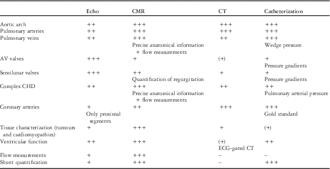
Imaging features prominently in the pre and postoperative management of patients with CHD. Diagnostic accuracy, burden to the patient, availability, and economics all have a role in the choice of the appropriate imaging modality in clinical practice. Echocardiography remains the mainstay of cardiovascular diagnostics, but other techniques are gaining in importance, particularly cardiovascular magnetic resonance (CMR) and computed tomography (CT; Table 1). Existing expert opinion-based guidelines, focused on adult populations, for the proper utilization of CMR are available.Reference Kilner, Geva, Kaemmerer, Trindade, Schwitter and Webb 2 Specific challenges apply to imaging children and adolescents with CHD. These include the even more pressing need to avoid the use of ionizing radiation, assessment of anatomy that is often complex and involves small structures, as well as imaging at fast heart rates. The current paper aims to express the consensus among experts in the field of paediatric CMR regarding its use in young patients with CHD. It is beyond the scope of this paper to discuss all technical details of CMR imaging in CHD, which have been addressed elsewhere recently,Reference Fratz, Chung and Greil 3 much more to describe the use of CMR in specific paediatric clinical situations.
Comparison of available imaging modalities for assessment of different conditions.

(+): limited application, major limitations; +: can be used in the assessment of this lesion, but has some significant limitations and better alternatives are available; ++: useful ommonly applied in the assessment of the lesion, and it may present some limitation; +++: technique of choice for the assessment of the lesion; – : cannot be adequately with this modality.
Settings and techniques
In contrast to scanning adults with ischaemic heart disease, the CMR approach to paediatric patients with CHD has to be individualized and nearly all sequences need to be adapted to the patient’s size, age, heart rate and, of course, the specific clinical question.Reference Fratz, Hess and Schuhbaeck 4 , Reference Tsai-Goodman, Geva, Odegard, Sena and Powell 5 Therefore, it is of critical importance that CMR studies of young patients with CHD are performed in centres with expertise in both the haemodynamic situation at hand and the CMR technique used.Reference Fratz, Hess and Schuhbaeck 4 , Reference Tsai-Goodman, Geva, Odegard, Sena and Powell 5 The presence of an expert CMR reader throughout the length of the study is typically required, as each heart is different and the sub-sequent imaging steps need to be individually tailored upon on the findings observed. High-quality results can be achieved only with a commitment to invest time and resources for sometimes lengthy scans and laborious post-processing of the data. Requirements regarding dedicated training for performing congenital CMR have been published.Reference Helbing, Mertens and Sieverding 6 Requirements for institutional accreditation, and particularly regarding a minimal case load, are available for adult general CMR in Europe, but are still lacking for paediatric heart disease and for CHD.Reference Plein, Schulz-Menger and Almeida 7
Basic sequences used in paediatric CMR
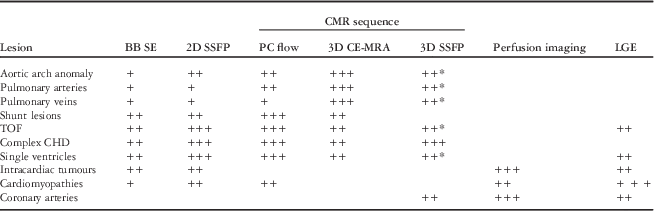
The sequences utilized in routine paediatric CMR can be divided into three main categories, following their purpose: to define morphology, to assess function, including ventricular volumes and blood flow, and to define myocardial tissue properties, including myocardial perfusion as well as fibrosis. Recommendations for sequences to be used for a given lesion are summarized in Table 2.
Summary of the sequences recommended for imaging different conditions.

+: may be used in the assessment of this lesion, but better alternatives available or does not provide additional information over other techniques; ++: useful technique, commonly applied in the assessment of the lesion; +++: needs to be part of any study in this lesion.
* If contrast needs to be avoided. BB SE, Black blood spin-echo.
Anatomical imaging
In spin-echo imaging pulse sequences (often referred to as ‘darkblood’ or ‘black-blood’ sequences), flowing blood appears dark whereas stationary tissues appear as varying shades of grey.Reference Finn, Nael, Deshpande, Ratib and Laub 8 The main advantage of spin-echo sequences is that they are less susceptible to artefacts caused by turbulent flow and metallic implants.Reference Garg, Powell, Sena, Marshall and Geva 9 Respiratory motion is usually addressed by breath-holding, multiple averages, or diaphragm tracking.
In steady-state free precession (SSFP) acquisitions, blood appears bright. Images can be displayed as a cine loop.Reference Finn, Nael, Deshpande, Ratib and Laub 8 Two-dimensional (2D) SSFP is the most frequently used sequence for anatomical imaging in CHD, as it allows views in any desired plane and adds qualitative information about function and valve motion. Image acquisition is usually performed during breath-holding. However, images can also be acquired during free-breathing in most paediatric patients. Cardiac motion is usually controlled by means of retrospective gating to ECG as this allows including the end-diastolic heart phase, as opposed to prospective ECG-triggering.
Contrast-enhanced magnetic resonance angiography (CE-MRA) uses intravenously administered gadolinium-based contrast agents to reduce T1 relaxation time of blood and improve the contrast between the blood pool and the surrounding tissue.Reference Holmqvist, Larsson, Stahlberg and Laurin 10 , Reference Meng and Grosse-Wortmann 11 The increased contrast (signal) allows adjusting the acquisition parameters for obtaining high-resolution images.Reference Masui, Katayama and Kobayashi 12 , Reference Valsangiacomo Buechel, DiBernardo, Bauersfeld and Berger 13 The dataset – a full volume composed of a stack of contiguous slices – can be reconstructed in any desired oblique plane (multiplanar reformats and maximum intensity projection images) or volume-rendered in a three-dimensional (3D) image. For optimal quality of the CE-MRA images, the sequence and particularly the timing of image acquisition need to be designed for the clinical question and the vessels that need to be assessed. Even in small children, high spatial resolution with submillimetre resolution can be achieved with CE-MRA in which the images are acquired during breath-holding.Reference Kellenberger, Yoo and Buechel 14 , Reference Macgowan, Al-Kwifi, Varodayan, Yoo, Wright and Kellenberger 15 Using this technique, even subtle variations in luminal diameters can be detected. Thus, CE-MRA has become widely accepted in the diagnosis of vascular disease in children, and cardiac catheterization can frequently be avoided.Reference Valsangiacomo Buechel, DiBernardo, Bauersfeld and Berger 13 , Reference Geva, Greil, Marshall, Landzberg and Powell 16 , Reference Greil, Powell, Gildein and Geva 17
CE-MRA is acquired without ECG-triggering and the reconstructed images represent an average appearance over the cardiac cycle. For this reason the edges of pulsatile vascular structures, such as the aortic root, as well as the coronaries appear blurry.
In time-resolved MR angiography techniques, the dynamic distribution of contrast medium into the pulmonary vasculature can be observed. Without dedicated timing of image acquisition, artefacts from motion are minimized and separation of arteries and veins is possible.Reference Goyen, Laub and Ladd 18
3D SSFP is an ECG-triggered pulse sequence with respiratory motion compensation by diaphragmatic navigatorsReference Piccini, Monney and Sierro 19 – Reference Razavi, Hill and Muthurangu 21 or navigation on the heart itself, called self-navigation.Reference Piccini, Monney and Sierro 19 These sequences can be combined with contrast media or not and produce a high-resolution 3D dataset of the whole heart and intrathoracic vasculature.Reference Sorensen, Korperich and Greil 20 – Reference Fenchel, Greil and Martirosian 22 Owing to the double gating (ECG and respiratory), cardiovascular structures close to the heart, such as the aortic bulb and the proximal coronary arteries, can be delineated very clearly.Reference Piccini, Monney and Sierro 19 , Reference Su, Chung and Muthupillai 23 , Reference Tangcharoen, Bell and Hegde 24 Image reconstruction is usually performed using multiplanar reformatting.
Functional imaging
Ventricular volumes and ejection fraction are assessed by acquisition of a stack of SSFP cine slices (as described above) covering the entire heart.Reference Finn, Nael, Deshpande, Ratib and Laub 8 The stack can be aligned in a ventricular short-axis plane parallel to the atrioventricular valves or perpendicular to the ventricular septum or in an axial plane transverse to the thorax.Reference Buechel, Kaiser, Jackson, Schmitz and Kellenberger 25 – Reference Fratz, Schuhbaeck and Buchner 28 Measurements should be performed consistently using the same orientation, as results from short axis and axial orientations are not interchangeable. On the stack of cine slices, end-diastolic and -systolic volumes and mass of both ventricles are derived by contouring the endocardial and epicardial borders. This method is based on Simpson’s summation of discs method with minimal geometrical assumption. This is of particular importance in the right ventricle or in functionally single ventricles. Normal values for both ventricles and both atria have been published in children,Reference Buechel, Kaiser, Jackson, Schmitz and Kellenberger 25 – Reference Robbers-Visser, Boersma and Helbing 27 , Reference Sarikouch, Koerperich and Boethig 29 and high reproducibility of the measurements has been demonstrated in children with CHD and with normal hearts.Reference Buechel, Kaiser, Jackson, Schmitz and Kellenberger 25 , Reference Mooij, de Wit, Graham, Powell and Geva 30 A consensus policy regarding ventricular segmentation, i.e. inclusion or exclusion of papillary muscles and trabeculations, leads to an even better reproducibility of the measurements.Reference Beerbaum, Barth and Kropf 31
Blood flow across a vessel can be measured accurately by using a velocity-encoded phase-contrast (PC) cine sequence. Typically, a slice is placed perpendicular to the vessel of interest. After contouring the vessel of interest, the volume of blood passing through the plane is calculated as the product of velocity and cross-sectional area.Reference Powell, Maier, Chung and Geva 32 In general, when assessing CHD, it is advisable to perform measurements in all large arteries, i.e. aorta, pulmonary trunk, and side branches, as well as in the caval veins and to compare the results for internal consistency.
First-pass myocardial perfusion imaging uses the dynamic inflow of gadolinium-based contrast medium into the myocardium to depict zones of decreased perfusion. Areas of reduced myocardial perfusion will remain dark, whereas normal perfusion shows a bright myocardial signal with the advent of gadolinium. The test can be performed at rest or under pharmacological stress for more clear depiction of ischaemia.Reference Finn, Nael, Deshpande, Ratib and Laub 8 , Reference Buechel, Balmer, Bauersfeld, Kellenberger and Schwitter 33 , Reference Taylor, Dymarkowski and Hamaekers 34 The option to perform perfusion imaging at rest and during stress in combination with superior spatial resolution compared with single photon emission computed tomography (SPECT) makes it an attractive technique.Reference Chiribiri, Bettencourt and Nagel 35
Stress imaging assesses ventricular function by 2D SSFP under pharmacological stress with dobutamin or exercise stress with CMR-compatible equipment. Stress imaging is performed in some centres in selected borderline clinical situations to test contractile reserve of a systemic chamber, such as in a right ventricle in systemic position or in univentricular hearts.Reference Robbers-Visser, Jan Ten Harkel and Kapusta 36
Late gadolinium enhancement (LGE), or myocardial delayed enhancement, is a technique demonstrating abnormal deposition of contrast agent within the myocardium late after contrast medium injection. The focal hyperenhanced areas represent regions of myocardial fibrosis.Reference Finn, Nael, Deshpande, Ratib and Laub 8 Fibrotic and necrotic areas in the myocardium appear bright on LGE images in contrast to the dark healthy myocardium. In children with CHD, LGE may result from scars after infarction, surgical scars in the myocardium, and/or from surgically placed patches.Reference Harris, Johnson, Weinberg and Fogel 37
In younger children, obtaining adequate temporal and spatial resolutions may represent a technical challenge, as the structures of interest are smaller and heart rates higher than in adults.Reference Kellenberger, Yoo and Buechel 14
Expert consensus key points
-
(i) Black-blood spin-echo, 2D SSFP, and 3D SSFP techniques as well as contrast-enhanced MR angiography are the most important sequences for anatomical imaging in CHD.
-
(ii) In children, higher spatial and temporal resolution is required to account for higher heart rates and smaller anatomical structures than in adults.
-
(iii) CMR is the current clinical gold standard for measurements of ventricular function and blood flow. Both are crucial components of a CMR examination in patients with CHD.
Sedation and anaesthesia
In younger children, typically before school-age, a CMR examination usually requires sedation or general anaesthesia. In young patients with complex CHD, in spite of the burden of general anaesthesia, CMR can be utilized for completing diagnosis before surgical repair or for planning other therapeutic interventions,Reference Tsai-Goodman, Geva, Odegard, Sena and Powell 5 , Reference Kellenberger, Yoo and Buechel 14 and purely diagnostic cardiac catheter examination with its related potential complications can usually be avoided.Reference Bergersen, Gauvreau, Lock and Jenkins 38 – Reference Bacher, Bogaert, Lapere, De Wolf and Thierens 40
The preferences and availability of sedation and/or general anaesthesia differ from centre to centre, and advantages and risks of each technique are discussed elsewhere.Reference Beekman, Hoorntje, Beek and Kuijten 41 , Reference Odegard, DiNardo, Tsai-Goodman, Powell, Geva and Laussen 42 When sedation or general anaesthesia is used, appropriate monitoring of the patient and presence of an expert team which is versed in the administration of drugs and equipped to deal with emergencies is mandatory.
Even though patients under anaesthesia, infants younger than 1 year, inpatients, and ICU patients are at higher risk for adverse events during CMR than the general outpatient population,Reference Dorfman, Odegard, Powell, Laussen and Geva 43 it has been shown that CMR can be safely performed even in critically ill infants, if carried out by a dedicated team.Reference Sarikouch, Schaeffler, Körperich, Dongas, Haas and Beerbaum 44 Careful analysis of the risks and benefits of a CMR examination under general anaesthesia needs to be performed before the procedure.
Certain patient groups, including those younger than 3 months of age, patients with a functional single-ventricle physiology, pulmonary hypertension, unrepaired cyanotic CHD, or airway pathology, may require hospitalization. 45
Expert consensus key points
-
(i) Dedicated staff and MR compatible equipment are necessary for performing general anaesthesia or deep sedation in children with CHD undergoing CMR.
-
(ii) In patients requiring anaesthesia or sedation, the risks and benefits of the procedure should be balanced carefully.
Safety and contraindications
Except for patients requiring general anaesthesia, the safety issues for CMR in children with CHD do not differ from those in general paediatric magnetic resonance imaging, and CMR is a safe imaging procedure. The main advantages of CMR are the lack of ionizing radiation and its non-invasiveness. Particularly, the use of radiation-free modalities is very important in children, as their risk for developing radiation-induced cancer is three to four times higher than in adults.Reference Picano, Vañó and Rehani 46 Correspondingly, CMR has a favourable benefit/risk profile compared with cardiac catheterization and CT.Reference Knuuti, Bengel and Bax 47
The most important step to reduce potential risks of CMR scanning is to establish a strict policy of screening before CMR for the patient and all accompanying persons, including parents and medical personnel.Reference Shellock and Spinazzi 48 This is particularly important to avoid the so-called missile effect caused by ferromagnetic objects drawn rapidly into the scanner by massive magnetic forces.Reference Chaljub, Kramer, Johnson, Johnson, Singh and Crow 49
Other potential safety issues related to CMR relate to hyperthermia, acoustic noise, implantable devices, and nephrogenic systemic fibrosis (NSF).
If used according to the manufacturer’s recommendations, CMR at 1.5 or 3 T does not pose any safety concerns with regard to energy deposition and hyperthermia. 50 Nevertheless, it is important to note that thermoregulatory mechanisms are immature in infants, abnormal in patients with cardiovascular disease, and can be influenced by some medications such as diuretics, calcium blockers, beta-blockers, amphetamines, and sedatives. Acoustic noise during CMR scanning can cause temporary or permanent hearing impairment. Adequate precautions to shield patients, including infants, should be taken at all times.
In general, most cardiovascular devices implanted nowadays are MR compatible and safe to be scanned. www.mrisafety.com provides specific safety information for specific devices and should be consulted before scanning.Reference Levine, Gomes and Arai 51 Cochlear implants and cerebrospinal fluid shunts are non-cardiovascular implants that may be present in CHD patients. CMR is contraindicated in patients who have a cochlear implant. Most cerebrospinal shunts are CMR compatible, but manufacturer-specific safety guidelines must be followed.
Cardiac pacemakers and implantable cardioverters/defibrillators (ICDs) are generally considered a relative contraindication to entering the MR environment.Reference Bhachu and Kanal 52 CMR-compatible cardiac pacemakers have been recently developed and are currently undergoing testing.Reference Rod Gimbel, Bello and Schmitt 53
Severe complications of gadolinium-derived contrast agents are rare in paediatric patients. Common side effects include extravasation of the contrast agent and allergic reactions from mild skin rash to cardiovascular decompensation. NSF is a rare but serious condition that consists of fibrosis of skin, joints, eyes, and internal organs.Reference Shellock and Spinazzi 54 The development of NSF has been linked to the biochemical structure of gadolinium-containing contrast agents in the presence of end-stage renal failure. Thus, all patients who are candidates for gadoliniumderived contrast medium administration must be screened for renal dysfunction, and in infants ionic macrocyclic contrast agents should be used.Reference Leiner and Kucharczyk 55 , Reference Thomsen 56
Expert consensus key points
-
(i) CMR in children and adolescents with heart disease has a high safety profile.
-
(ii) Lack of radiation is the most striking advantage over other advanced imaging modalities.
-
(iii) Main contraindications for CMR in childhood are cochlear implants, old-generation pacemakers, ICDs, and other noncompatible implants.
-
(iv) The use of gadolinium-derived contrast agents is contraindicated in children with end-stage renal failure, due to the risk for developing NSF.
Clinical applications
Aortic arch anomalies
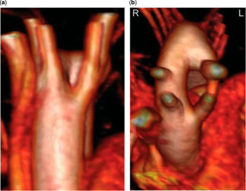
Common indications for performing CMR in anomalies of the aortic arch include vascular rings (Fig 1), interrupted aortic arch, truncus arteriosus communis, complex forms of aortic coarctation, and congenital connective tissue disorders, such as Marfan and Turner syndrome.Reference Eichhorn, Fink, Delorme and Ulmer 57 , Reference Eichhorn, Krissak and Rudiger 58 CMR can be performed at the time of diagnosis in order to refine an echocardiographic diagnosis and during follow-up after surgical correction or catheter intervention, when residual or recurrent stenoses and/or aneurysms need to be ruled out.Reference Eichhorn, Krissak and Rudiger 58 – Reference Greil, Kramer and Dammann 60 Stents in the aortic arch are not a contraindication for CMR; however, CMR is not suited to evaluate stent patency. The sequences used for imaging the aortic arch are summarized in Table 2.
Double aortic arch as shown in a 3D reconstruction from a contrast-enhanced MR angiography. View from the front ( a ) and from cranial ( b ).

The sensitivity of CMR techniques to detect vascular abnormalities is as good as that of conventional catheter angiocardiography (Table 1), so that cardiac catheterization can be reserved for selected cases and for catheter-guided interventions.Reference Kaiser, Kellenberger, Albisetti, Bergsträsser and Valsangiacomo Buechel 61
The ability to generate 3D anatomical images by CEMRA or 3D SSFP allows decision-making on the most appropriate treatment technique, i.e. cardiac surgery or catheter-guided intervention.Reference Kaiser, Kellenberger, Albisetti, Bergsträsser and Valsangiacomo Buechel 61 – Reference Prince, Narasimham and Jacoby 64 CT angiography is a sound alternative particularly when associated airway anomalies are suspected and/or when the clinical scenario is that of a young and sick infant or neonate.
In coarctation of the aorta, haemodynamic assessment is performed with a velocity-encoded PC cine sequence using high velocity encoding typically of at least 200 cm/s.Reference Oshinski, Parks and Markou 65 Flow measurements are usually performed in a perpendicular plane through the ascending aorta, proximal descending aorta (aortic isthmus, immediately distal to the stenosis), and at the level of the diaphragm. An increase in flow between the distal aortic arch and the descending aorta at the level of the diaphragm is indicative for significant collateral circulation. In addition, the velocity of the jet at the level of the stenotic isthmus, if PC measurement is performed accordingly, provides semi-quantitative information about the severity of vascular narrowing.
Expert consensus key points
-
(i) CMR is the first-line advanced imaging technique beyond echocardiography for the anatomical assessment of aortic arch anomalies.
-
(ii) Reconstructed 3D CMR images are helpful for planning interventions of the aortic arch.
-
(iii) CMR has an important role in follow-up after an intervention.
-
(iv) Blood flow measurements add functional information to the anatomical images.
The pulmonary arteries
The advantages of CMR imaging compared with other modalities are very good anatomical visualization of right ventricular outflow tract (RVOT), pulmonary bifurcation, and pulmonary side branches, combined with functional information. CMR is the only technique allowing quantitative flow measurements in the pulmonary arteries (Table 1).Reference Caputo, Kondo and Masui 66
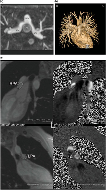
Anatomical imaging of the pulmonary arteries can be done by various angiographic techniques, as described above and in Table 2.Reference Eichhorn, Fink, Delorme and Ulmer 57 , Reference Knobel, Kellenberger, Kaiser, Albisetti, Bergstrasser and Buechel 67 CE-MRA has been validated against conventional angiographyReference Valsangiacomo Buechel, DiBernardo, Bauersfeld and Berger 13 and has also been demonstrated to reliably detect aorto-pulmonary collateral arteries in tetralogy of Fallot (TOF) with severe pulmonary stenosis or atresia.Reference Geva, Greil, Marshall, Landzberg and Powell 16 Furthermore, CMR provides clear visualization of the spatial alignment of the pulmonary bifurcation, as well as of the relationship between the pulmonary arteries and the surrounding structures, as the airways and other vascular structures such as the aorta (Fig 2).Reference Knobel, Kellenberger, Kaiser, Albisetti, Bergstrasser and Buechel 67
In a patient with transposition of the great arteries, who underwent the arterial switch operation with Lecomte manoeuvre, contrast-enhanced MR angiography demonstrates well the relationship between the pulmonary arteries and the surrounding structures, in this case the ascending aorta ( a ). Flow measurements in the right pulmonary artery ( b ) and in the left pulmonary artery ( c ) show a balanced lung perfusion with 50%/50% flow to the right and to the left. AO, aorta; MPA, main pulmonary artery; LPA, left pulmonary artery; RPA, right pulmonary artery.

Flow measurements by velocity-encoded PC cine add functional information to anatomical findings and are crucial for assessing the need for interventions (Fig 2). Flow redistribution in the pulmonary arteries has been observed not only in the presence of pulmonary artery stenosis, but also in pulmonary venous obstruction.Reference Roman, Kellenberger, Farooq, MacGowan, Gilday and Yoo 68 – Reference Kellenberger, Macgowan and Roman 70 Beside quantitative flow assessment, qualitative flow profiles may provide information about anomalous pulmonary resistance.
The presence of a stent in a pulmonary artery does not represent a contraindication for performing CMR imaging during follow-up assessment. Flow measurement can still be performed accurately, if PC cine images are acquired at a reasonable distance from the stent. Alternatively, pulmonary venous flow can be used as a surrogate for ipsilateral pulmonary arterial flow (Table 3).
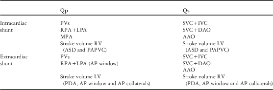
Flow measurements for the assessment of pulmonary (Qp) and systemic blood flows (Qs) in extraund intracardiac shunt lesions.

AAO, ascending aorta; AP, aorto-pulmonary window or collaterals; DAO, descending aorta; IVC, inferior vena cava; LPA, left pulmonary artery; LV, left ventricle; MPA, main pulmonary artery; PDA, patent ductus arteriosus; PVs, pulmonary veins; RPA, right pulmonary artery; RV, right ventricle; SVC, superior vena cava,
Expert consensus key points
-
(i) CMR combines a detailed visualization of the spatial alignment of the pulmonary arterial bifurcation and the side branches.
-
(ii) Through-plane flow measurements can accurately quantify differential lung perfusion in various conditions.
The pulmonary veins
CMR is considered the gold standard for assessing anomalous connection and stenosis of the pulmonary veins. CMR combines superb luminal anatomy, accurate quantification of blood flow patterns and volume and, importantly, information about the surrounding structures (Table 1).Reference Valsangiacomo, Hornberger, Barrea, Smallhorn and Yoo 71 , Reference Grosse-Wortmann, Al-Otay and Goo 72
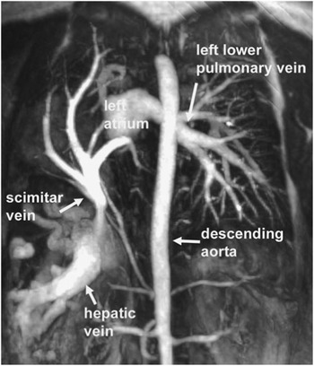
To obtain anatomical information about the vessel lumen in both pulmonary vein stenosis and anomalous connection, CE-MRA and/or 3D SSFP can be used (Fig 3). Alternatively to, or in conjunction with angiography, SSFP cine imaging along the vessel’s long axis can provide useful information, particularly if dynamic external compression is suspected.
Maximum intensity projection reconstruction of CE-MRA images in the coronal plane in a patient with Scimitar syndrome. All right-sided pulmonary veins drain via a common channel to a stenotic connection with the inferior vena cava (not opacified). The pulmonary venous blood from the right lung drains via collaterals to the dilated hepatic veins. The left-sided pulmonary veins drain normally to the left atrium (left upper pulmonary vein not shown).

Velocity-encoded PC cine is a powerful tool in the characterization of pulmonary venous pathology. It is used to measure right and left pulmonary arterial blood flow, to detect signs of pulmonary hypertension, and to unveil redistribution of blood flow away from affected areas of the lung.Reference Kellenberger, Macgowan and Roman 70
In anomalous pulmonary venous connection, it is important to detail the course and connection of each pulmonary vein, diagnose or rule out the presence of pulmonary venous obstruction, quantify the degree of left-to-right shunting (Qp/Qs), and describe associated lesions.Reference Roman, Kellenberger and Macgowan 69 , Reference Riesenkampff, Schmitt and Schnackenburg 73 A number of indicators during a CMR examination signal the presence of pulmonary vein stenosis.Reference Valsangiacomo, Hornberger, Barrea, Smallhorn and Yoo 71 , Reference Goo, Al-Otay, Grosse-Wortmann, Wu, Macgowan and Yoo 74 , Reference Valsangiacomo, Barrea, Macgowan, Smallhorn, Coles and Yoo 75 The most obvious sign of obstruction is morphologically narrowing of the pulmonary venous lumen. This is best imaged by targeted SSFP cine imaging and CE-MRA. In some instances, veno-venous collaterals to unobstructed pulmonary venous channels and/or to systemic veins are present. Furthermore, higher than normal flow velocity distal to a suspected narrowing and loss of the normal phasic velocity flow profile are suggestive of a haemodynamically significant obstruction.Reference Valsangiacomo, Barrea, Macgowan, Smallhorn, Coles and Yoo 75 Finally, as mentioned above, long-standing pulmonary venous narrowing leads to redistribution of pulmonary blood flow away from lung segments drained by the stenosed pulmonary vein, and results in an unbalanced lung perfusion, as easily detectable by velocity-encoded PC measurements in the branch pulmonary arteries.
Expert consensus key points
-
(i) CMR is considered the gold standard for assessing anomalous connection and stenosis of the pulmonary veins.
-
(ii) Combined blood flow measurements in the pulmonary veins and in the pulmonary arteries allow understanding the complex flow redistribution occurring in the presence of pulmonary venous obstruction and aorto-pulmonary collateral flow.
Shunt lesions
The exact quantification of intracardiac shunts is part of the comprehensive haemodynamic evaluation in many patients with CHD and often a determinant factor for surgery. CMR is an established modality for the non-invasive assessment of shunt location, flow direction, and magnitude.Reference Beerbaum, Korperich, Barth, Esdorn, Gieseke and Meyer 76 Anatomical detection of lesions resulting in shunt flows is typically performed with cine acquisitions. When obtained perpendicular or parallel to the intracardiac shunt direction, they provide a valuable assessment of the defect location and size of intracardiac shunt lesions throughout the cardiac cycle. Extracardiac shunts, such as major aorto-pulmonary collaterals and anomalous pulmonary venous connection, are readily delineated by CE-MRA.Reference Valsangiacomo, Hornberger, Barrea, Smallhorn and Yoo 71 , Reference Valente, Sena, Powell, Del Nido and Geva 77
Shunt calculation is one of the major strengths of CMR. As it provides accurate flow measurement in every desired vessel, CMR can overcome the well-known limitations of the traditional Fick’s method by oximetry as obtained with invasive techniques, or of Doppler echocardiography, which cannot quantify flow reliably.Reference Powell, Maier, Chung and Geva 32 , Reference Powell, Tsai-Goodman, Prakash, Greil and Geva 78
By using the velocity-encoded PC cine sequence, the ratio of pulmonary blood flow (Qp) and systemic blood flow (Qs) can be accurately quantified and reflects the size of shunt.
Independently of the location of the shunt, Qp is always the sum of pulmonary venous flow and Qs is always the sum of inferior and superior venae cavae flows, measured close to the heart.Reference Grosse-Wortmann, Windram and Yoo 79 Flow in the descending aorta may be used as a substitute for inferior vena cava flow and is technically easier to obtain. Depending on the anatomy, i.e. the location of the shunt, flows other than the pulmonary veins and the caval veins can be used as Qp and Qs, respectively (Table 3).
Another method to assess the magnitude of net shunts is to compare the selective cardiac output of the right with that of the left ventricle (Table 3). This method is commonly thought to be less accurate than velocity-encoded PC by many, owing to difficulties in accurately contouring the right ventricular (RV) endocardium including its trabeculations. Furthermore, this approach is inherently invalid in the presence of valvular regurgitation. For the purposes of ‘internal validation’, that is, to assess the accuracy of the flow measurements, Qp and Qs measured by different methods should be compared.
Expert consensus key points
-
(i) CMR can be used for the anatomical detection of intracardiac and extracardiac shunts.
-
(ii) Shunt calculation is one of the major strengths of CMR.
-
(iii) Comparison of shunt quantifications using various measurements is recommended for ‘internal validation’ of the data and to increase accuracy.
Tetralogy of Fallot
TOF has become one of the main indications for performing CMR during follow-up of CHD after surgical repair.Reference Kilner, Geva, Kaemmerer, Trindade, Schwitter and Webb 2 In young children, preoperatively, advanced imaging with CMR is only required in few selected cases with associated lesions such as situs anomalies, aortic arch anomalies, disconnected branch pulmonary arteries, and/or aorto-pulmonary collaterals. After initial surgical repair, typical residual findings during mid- and long-term follow-up include moderate to severe incompetence of the pulmonary valve, obstruction of the RV outflow tract, and/or branch pulmonary arteries. These findings cause chronic volume and/or pressure load of the right ventricle, with well-described and potentially lethal complications.Reference Gatzoulis, Balaji and Webber 80
Over the past two decades, CMR has been firmly established as the key imaging modality for serial follow-up in TOF patients.Reference Kilner, Geva, Kaemmerer, Trindade, Schwitter and Webb 2 CMR is the only technique that allows accurate quantification of pulmonary incompetence with measurement of regurgitation volumes and regurgitation fraction (both should be documented) by PC cine imaging (Table 1).Reference Rebergen, Chin, Ottenkamp, van der Wall and de Roos 81 , Reference Wald, Redington and Pereira 82
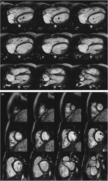
The volume and systolic function of both ventricles are determined by acquiring a stacked 2D-cine SSFP covering the ventricles in short-axis or axial orientationReference Buechel, Kaiser, Jackson, Schmitz and Kellenberger 25 , Reference Fratz, Schuhbaeck and Buchner 28 (Fig 4). This method has excellent reproducibility if performed in a standardized manner across institutions.Reference Beerbaum, Barth and Kropf 31 , Reference Sarikouch, Koerperich and Dubowy 83 Tricuspid valve regurgitation can be depicted using 2D SSFP, but is difficult to be correctly quantified due to the movements of the annulus plane during the cardiac cycle and to the usually co-existing pulmonary incompetence. Tailored 2D SSFP images through the RVOT, 3D SSFP, and/or CE-MRA give clear views of the anatomy of the RVOT and the pulmonary side branches. Additional functional information can be obtained by measuring the differential pulmonary perfusion and assessing the backflow separately in both pulmonary side branches.Reference Kang, Redington and Benson 84 , Reference Voser, Kellenberger and Buechel 85
2D SSFP of the ventricles in a patient with dilated RV after TOF repair as demonstrated in an axial stack ( a ) and in a short-axis stack ( b ).

All this information is generally used in the clinical management of these patients and considered of particular importance in timing decisions regarding pulmonary valve replacement.
In this context, RV size and function are the main factors to be considered in addition to clinical symptoms and findings. Although there is no universal agreement on cut-off volume, an indexed end-diastolic RV volume of 160ml/m2 and an end-systolic RV volume of 80ml/m2 have been shown to predict normalization of RV size after pulmonary valve replacement.Reference Oosterhof, van Straten and Vliegen 86 Proper selection of patients undergoing percutaneous pulmonary valve implantation requires accurate description of the geometry of the RVOT by CE-MRA or 3D SFFP and exclusion of anomalies of the coronary arteries by 3D SSFP.
The presence of myocardial scarring is detected by LGE of the myocardium, the extent of which has been found to be related to exercise intolerance, regional wall motion abnormalities, and propensity for arrhythmia.
Targeted 2D SSFP or CE-MRA may additionally detect dilatation of the ascending aorta, demonstrate arch sidedness (right aortic arch in 20% of patients with TOF), and exclude potential major aortopulmonary collateral arteries.Reference Kutty, Kuehne and Gribben 87
Expert consensus key points
-
(i) CMR is the key imaging modality for serial follow-up in TOF patients.
-
(ii) CMR enables the assessment of RV outflow tract, pulmonary bifurcation, and pulmonary arteries as well as quantification of RV volume and function and regurgitant blood flow in pulmonary regurgitation.
-
(iii) This information features prominently in surgical decision making around pulmonary valve replacement.
Complex CHD
Complex CHD frequently consists of a combination of situs anomalies, abnormal atrioventricular and/or ventriculo-arterial connections, and/or additional defects, including septal defects, ventricular looping anomalies, as well as malformations of the outflow tracts. In addition, malformations of the extracardiac thoracic vessels and tracheobronchial anomalies may be present.
Planning and performing CMR examinations in patients with complex CHD require thorough expertise in congenital malformations to avoid misinterpretation or incomplete results.
Dedicated CMR examinations provide a comprehensive picture of complex CHD, including anatomy and haemodynamics (Table 1). Three-dimensional reformatting algorithms may enhance surgical decision-making and planning of operative procedures.Reference Picano, Vañó and Rehani 46
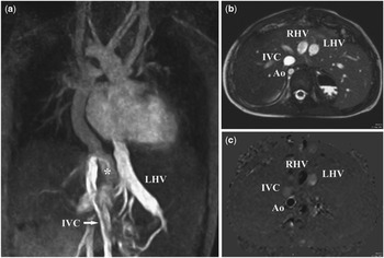
In complex CHD, views in axial, coronal, and sagittal orientations should always be at the beginning of every CMR exams to facilitate orientation within the thorax. Oblique imaging planes are tailored towards individual aspects of anatomy and for ventricular function and flow analyses. The abdomen should be covered in the localizer/scout images and by techniques, enabling the identification of vessel anatomy and size as well as anatomy of the upper abdominal organsReference Geva, Vick, Wendt and Rokey 88 (Fig 5).
Four-year-old boy with right atrial isomerism, bilateral superior caval veins, univentricular atrioventricular connection to a solitary ventricle, pulmonary atresia, and right aortic arch developed severe hypoxaemia after Fontan completion due to a non-included left-sided hepatic vein. ( a ) Multiple intrahepatic collateral channels underscoring a right-to-left shunt from the right hepatic vein (RHV; asterisk) to the left hepatic vein (LHV) and to the left-sided atrium. Contrast agent is applied via the left lower limb. ( b ) T2-weighted SSFP transverse plane "'3 cm below the diaphragm. Visceral heterotaxia. Both descending aorta (Ao) and inferior caval vein (IVC) are right-sided. RHV and LHV are arranged in nearly parallel fashion midline. ( c ) Flow measurement using velocity-encoded PC for quantification. Flow direction is encoded by optical density. Dark lumens indicate flow directed caudally. Reversely, bright lumens indicate flow directed cranially.

Turbo spin-echo sequences are particularly useful for the evaluation of tracheo-bronchial anomalies and their relationship to the pulmonary vascular tree which provides important clues to the thoracic situs and for the detection of anomalies of the great arteries.
The use of CMR in complex CHD including heterotaxy syndrome has been validated in numerous studiesReference Prakash, Torres, Printz, Prince and Nielsen 89 and has been demonstrated to provide superior delineation of the abdominal situs, of pulmonary and systemic venous malformations, and of the relationship of the heart to abdominal and mediastinal structures compared with echocardiography and cardiac catheterization.Reference Geva, Vick, Wendt and Rokey 88 , Reference Hong, Park and Ryu 90 – Reference Newman, Feinstein and Cohen 92 Thus, CMR has been recently recognized as the first-line imaging modality for imaging complex CHD.Reference Kilner, Geva, Kaemmerer, Trindade, Schwitter and Webb 2
Expert consensus key points
-
(i) CMR is an important adjunct to echocardiography for imaging complex CHD in children and in adults.
-
(ii) CMR provides complete information about situs, segmental cardiac connections, additional intracardiac and extracardiac malformations, and accurate haemodynamic information.
Single ventricles throughout staged palliation
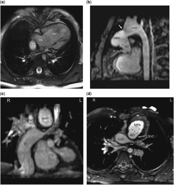
The palliative treatment strategy for functionally univentricular hearts may require between two and three or more procedures before achieving the stage of the Fontan-type circulation.Reference Gewillig 93 , Reference Fogel, Weinberg, Chin, Fellows and Hoffman 94 Strengths of CMR in the setting of single-ventricle circulations include the detailed assessment of potentially complex anatomy, particularly of the large vessels, functional imaging of the ventricles and large vessels throughout the stages, and assessment of flow, particularly of the pulmonary/Fontan circulation (Fig 6).
Two-year-old girl with hypoplastic left heart syndrome ( a ) and status post-bilateral cavopulmonary anastomosis and Fontan completion. 3D SSFP images reconstructed with an MIP technique illustrate a patent Damus–Kaye –Stancel (DKS) anastomosis (***) with the origin of the right coronary artery ( b ); the segment between the right and left pulmonary artery (**) was reconstructed at time of Fontan completion, as it was subatretic due to external compression of the dilated neo-aortic root (DKS) and presents now with recurrent narrowing ( c and d ). AO, aorta; C, extracardiac Fontan conduit; MPA, main pulmonary artery; LA, left atrium; LV, left ventricle; LSVC, left superior vena cava; RA, right atrium; RV, right ventricle.

Prior to the creation of a partial cavopulmonary connection, imaging of the ascending aorta and aortic arch, of the pulmonary arteries, and of the systemic and pulmonary veins are mandatory.Reference Fogel 95 Ventricular size and function, valve function, and the subaortic outflow tract should be studied. A combination of 2D and 3D SSFP, black-blood, phase-contrast sequences and/or CE-MRA can be used for these purposes (Table 2).Reference Prakash, Khan, Hardy, Torres, Chen and Gersony 96 A combined protocol using echocardiography and CMR may result in less complications when compared with cardiac catheterizations and similar long-term outcomes in properly selected patients.Reference Brown, Gauvreau and Powell 97 Imaging of an aorto- or ventriculo-pulmonary shunt can also be achieved with CMR by using 3D SSFP or CE-MRA.Reference Kellenberger, Yoo and Buechel 14
Before completion of a total cavopulmonary connection (TCPC), a similarly detailed work-up is required. In addition, the connection between the superior vena cava and the pulmonary arteries has to be visualized. The presence of significant aorto-pulmonary collateral vessels can be demonstrated by using CE-MRA. The difference of total pulmonary venous flow and the total branch pulmonary arterial flow allows quantifying the additional blood flow to the lungs due to aorto-pulmonary collaterals.Reference Grosse-Wortmann, Al-Otay and Yoo 98 The use of only CMR for planning the TCPC stage is being discussed;Reference Piccini, Monney and Sierro 19 however in most centres, haemodynamic studies before the completion of a Fontan-type circulation are still performed including cardiac catheterization (Table 1).
After completion of the Fontan circulation, CMR is recommended for serial follow-up of systolic and diastolic ventricular function, ventricular geometry, and serial quantification of valvular incompetence. Reference Fogel, Weinberg, Chin, Fellows and Hoffman 94 , Reference Fogel 95 , Reference Robbers-Visser, Kapusta and van Osch-Gevers 99 Stress imaging can be used for testing ventricular contractile reserve in selected cases.Reference Robbers-Visser, Jan Ten Harkel and Kapusta 36 Additional useful information provided by CMR during follow-up includes anatomy and function of the Fontan pathway, identifying obstructions, baffle leaks, thrombus formation, and collateral flow.
Expert consensus key points
-
(i) In patients with single ventricles, CMR is recommended after Fontan completion for serial follow-up of ventricular function and anatomical assessment of the Fontan pathway.
-
(ii) During staged palliation, CMR can be used to detect residual findings requiring additional interstage interventions.
Cardiac tumours
Paediatric cardiac tumours are rare and usually benign (75–90%). Rhabdomyomas and fibromas are the most frequent cardiac tumours in children. Among malignant tumours, metastatic involvement from non-Hodgkin lymphoma, leukaemia, neuroblastoma, nephroblastoma, or sarcoma is much more common than primary cardiac sarcoma or lymphoma.Reference Beghetti, Gow, Haney, Mawson, Williams and Freedom 100
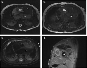
CMR fulfils all the diagnostic goals of tumour imaging in one single examination (Table 1).Reference Kiaffas, Powell and Geva 101 , Reference O’Donnell, Abbara and Chaithiraphan 102 Thus, a CMR examination should describe size and location of the tumour; evaluate any haemodynamic relevance, such as obstructions to inflow or outflow and impairment of myocardial and/or valvular function; describe the specific tissue properties of the mass, including signal/intensity ratio, infiltration of adjacent tissue, tissue appearance. Exact location in relation to the cardiac structures (endoluminal, floating, intramyocardial) should be included (Fig 7).
Embryonic rhabdomyosarcoma (**) in the apex of the right ventricle in a 3-month-old boy. SSFP ( a ), T1-weighted ( b ), and T2-weighted images ( c ) in an axial plane demonstrate different tissue characteristics in different sequences. Short-axis post-contrast images ( d ) show contrast medium enhancement particularly in the superficial tissue layers and less in the core of the tumour. LV, left ventricle; RV, right ventricle.

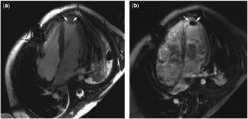
For a comprehensive evaluation of an intracardiac mass, the following imaging sequences and orientations are recommended: cine SSFP in an axial and oblique imaging plane across the tumour, T1-weighted TSE with and without fat suppression covering the tumour as well as the uninvolved ventricular myocardium, T2-weighted TSE, first-pass perfusion and LGE100 (Table 2). First-pass myocardial perfusion imaging may provide useful information about tumour vascularity. Moderate and strong enhancement is more indicative of malignant processes, whereas mild enhancement is found in up to 40–50% of benign tumours as well as in highly vascularized tumours.Reference Kiaffas, Powell and Geva 101 , Reference Beroukhim, Prakash and Buechel 103 LGE does not predict malignancy, but is very helpful in tissue characterization. When an intracardiac thrombus is suspected, LGE imaging with a long inversion time (600 ms) is recommended (Fig 8).Reference Weinsaft, Kim and Shah 104
Appearance of intracavity thrombus on 2D SSFP ( a ) and LGE images ( b ).

After having acquired all the different images, a correct diagnosis of the type of tumour can be derived by using the criteria proposed from a recent multicentre paediatric data collection.Reference Beroukhim, Prakash and Buechel 103 These criteria based on tissue characteristics shown by the different sequences used achieved a diagnostic accuracy of up to 97%, with a single correct diagnosis in 55% and a correct differential diagnosis in 42%, respectively.Reference Beroukhim, Prakash and Buechel 103 Diagnosis of malignancy can be at least suspected on the basis of tumour location, tissue inhomogeneity, and/or pericardial/pleural effusions with a sensitivity of 0.88 and specificity of 0.92.Reference Hoffmann, Globits and Schima 105
Expert consensus key points
-
(i) CMR is an important part of non-invasive tumour characterization by providing information on size, location, haemodynamic relevance, relationship to the adjacent structures, infiltration, as well as tissue properties.
Cardiomyopathies
The two most common forms of paediatric cardiomyopathy are dilated cardiomyopathy (DCM; annual incidence 0.57 cases per 100,000) and hypertrophic cardiomyopathy (HCM; 0.47 per 100,000 persons).Reference Wilkinson, Landy and Colan 106
In addition to echocardiography, recognized as first-line imaging modality in children with cardiomyopathy, CMR provides noninvasive myocardial tissue characterization. In particular, CMR can detect the presence and extend of myocardial oedema, scarring and replacement fibrosis with high spatial resolution. In adults, pattern of fibrosis on LGE images may point towards the aetiology of the disease, and may influence decision-making and patient management.Reference Gagliardi, Bevilacqua, Di Renzi, Picardo, Passariello and Marcelletti 107 , Reference Karamitsos, Francis, Myerson, Selvanayagam and Neubauer 108
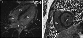
A wide range of abnormalities detectable with CMR have been related to the DCM phenotype, including coronary artery anomalies, idiopathic disease (majority of cases), or secondary causes, such as myocarditis or neuromuscular disorders. CMR is able to exclude anomalies of the origin and proximal course of the coronary arteries without the use of radiation and non-invasively.Reference Beerbaum, Sarikouch, Laser, Greil, Burchert and Korperich 109 In the acute phase of myocarditis, CMR can be used to assess global ventricular function, detect regional wall motion abnormalities (2D SSFP cine imaging), early myocardial inflammatory changes (T2-weighted imaging), as well as myocardial cell necrosis/fibrosis (LGE imaging) (Table 2).Reference Vashist and Singh 110 In chronic myocarditis, CMR can be used for monitoring biventricular functionReference Grosse-Wortmann, Roche, Yoo, Seed and Kantor 111 and demonstrate resolution of the inflammatory processes as well as late myocardial remodelling.Reference Gagliardi, Bevilacqua, Di Renzi, Picardo, Passariello and Marcelletti 107 The pattern of contrast distribution using LGE imaging is typically nodular, patchy, with a subepicardial and mid-wall myocardial distribution most commonly seen in the lateral and inferior walls of the left ventricle. In children, the extension of contrast is usually subepicardial, but may become transmural (Fig 9).Reference Vashist and Singh 110 The presence of pericardial effusion provides supportive evidence for myocarditis. CMR parameters significantly associated with poor outcomes in adult patients are: transmural myocardial late enhancement, global hypokinesia, LV dilation, and LV ejection fraction, 30%. In neuromuscular disorders, CMR provides information about the degree of myocardial fibrosis, inflammation and impairment of myocardial contractility and relaxation. In patients with Duchenne muscular dystrophy, LGE allows identification of subjects at risk for progressive heart failure.Reference Mavrogeni, Papavasiliou and Spargias 112
Focal myocarditis in a 4-year-old child who presented with chest pain and elevated cardiac enzymes. LGE images in a horizontal long-axis view ( a ) and in short axis ( b ). Enhancement is found in the mid-wall of the lateral wall (arrows). 2D SSFP cine showed mild hypocontractility of the lateral wall, correspondingly.

In HCM, CMR provides accurate quantification of ventricular mass and function of both ventricles. The distribution of hypertrophy can be best delineated by acquiring images in several planes, and particularly apical hypertrophy can be detected more accurately than with echocardiography. The presence of fibrosis on LGE imaging is a potential risk factor for ventricular arrhythmias in adultsReference Olivotto, Maron and Appelbaum 113 and is observed more rarely in children. If HCM is related to an inborn error of metabolism (8%), such as Fabry’s or Pompe’s diseases, CMR may be used to serially monitor the effects of enzyme replacement therapy.Reference Barker, Pasquali and Darty 114
In left ventricular non-compaction, definition of criteria is challenging and controversial. LGE may not be present in children.Reference Marin-Rodriguez, Ossaba-Velez, Maroto Alvaro and Sanchez-Alegre 115 Nevertheless, CMR has increased diagnostic accuracy compared with echocardiography, given that the anatomy of the cavity and trabeculae can be more clearly visualized.Reference Alhabshan, Smallhorn, Golding, Musewe, Freedom and Yoo 116
Assessment of myocardial iron load using
 ${\rm T}_{{\rm 2}}^{{\asterisk}} $
measurements is an established tool in the diagnosis of cardiac haemochromatosis in patients requiring repeated blood transfusions. The optimal timing for screening young patients by CMR is under debate, and depends on patient age and transfusion burden. Often, CMR can be postponed until 8 years of age so that anaesthesia is not required. A single measure of mid-septal
 ${\rm T}_{{\rm 2}}^{{\asterisk}} $
is sufficient and correlates well with the global
 ${\rm T}_{{\rm 2}}^{{\asterisk}} $
.Reference Pepe, Positano and Santarelli
117
CMR imaging of arrhythmogenic right ventricular cardiomyopathy (ARVC) in children is challenged by the evolving nature of the disease.Reference Fogel, Weinberg, Harris and Rhodes 118 , Reference Yoo, Grosse-Wortmann and Hamilton 119 Thus, repeated studies have been recommended. Morphological and regional wall motion abnormalities must be evaluated by careful analysis of SSFP cine images in different planes and orientations. CMR-based criteria for ARVC according to the revised Task Force Criteria include RV akinesia/dyskinesia, but only when combined with RV dilatation and/or reduced ejection fraction.Reference Marcus, McKenna and Sherrill 120 Imaging of fatty infiltration or LGE appears to be of low yield in children.Reference Yoo, Grosse-Wortmann and Hamilton 119
Expert consensus key points
-
(i) At time of diagnosis and during follow-up of patients with cardiomyopathies, the strengths of CMR are specific tissue characterization and accurate quantification of ventricular volume and function.
-
(ii) In dilatative cardiomyopathy, CMR can aid in determining the aetiology of the disease.
-
(iii) In HCM, CMR is used for exact quantification of myocardial mass, delineation of the distribution of hypertrophy, as well as for detection and quantification of myocardial fibrosis.
Coronary arteries/perfusion
In children, imaging of the coronary arteries (CA) and evaluation of myocardial perfusion with first-pass myocardial imaging as well as the presence of myocardial scars are indicated in suspected congenital anomalies of the CA, CA fistulas, after surgery for CHD involving CA transfer, before percutaneous pulmonary valve replacement with a valve veering stent, and in patients with vasculitis (e.g. Kawasaki, Takayasu arteritis, or after heart transplantation).Reference Buechel, Balmer, Bauersfeld, Kellenberger and Schwitter 33 , Reference Kostolny, Tsang and Nordmeyer 121 , Reference Greil, Seeger and Miller 122 Even though the capability to provide all this combined information makes CMR a unique modality, cardiac catheterization currently remains the gold standard for CA imaging in children (Table 1). ECG-gated multidetector row CT is an established non-invasive alternative for CA assessment because it is easy to use, generally available and exploits fast acquisition times compared with CMR in adults. However, important limitations in children remain radiation exposure and fast heart rate.Reference Brenner and Hall 123
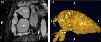
Magnetic resonance coronary artery (MRCA) imaging of the proximal and mid regions of the major epicardial CA by using 3D SSFP can be performed in infants and childrenReference Piccini, Monney and Sierro 19 , Reference Tangcharoen, Bell and Hegde 24 , Reference Greil, Desai and Fenchel 124 (Fig 10). As the patients growth and heart rate decreases, image quality increases. This technique can be used to detect an anomalous origin and proximal course of the CA.Reference Beerbaum, Sarikouch, Laser, Greil, Burchert and Korperich 109 Although the ability to asses CA in children with Kawasaki disease and to evaluate the vessel wall of the CA has been reported,Reference Greil, Seeger and Miller 122 the validity of MRCA for reliable detection of CA stenoses is unclear.
3D SSFP images of a 7-year-old boy with Kawasaki disease. A curved multiplanar reformat ( a ) and a volume-rendered 3D reconstruction ( b ) demonstrate five aneurysms (asterisks) of the right coronary artery. AO, aorta; IVC, inferior vena cava; RA, right atrium; RV, right ventricle; A, anterior; I, inferior; L, left; P, posterior; R, right; S, superior.

Assessment of myocardial perfusion with first-pass myocardial imaging has been validated in the adult population and has been demonstrated to have a diagnostic performance superior to SPECT.Reference Schwitter, Wacker and van Rossum 125 , Reference Schwitter, Wacker and Wilke 126 The clinical experience in children is limited, but good sensitivity and specificity have been demonstrated in comparison with X-ray coronary angiography.Reference Buechel, Balmer, Bauersfeld, Kellenberger and Schwitter 33 , Reference Taylor, Dymarkowski and Hamaekers 34 Performing first-pass imaging in children (see basic sequences) requires adapting the acquisition parameters to the higher heart rate and the small dimensions of the heart. Age and body size are still limiting factors, and infants below the age of 1 year are still considered suboptimal candidates. Nevertheless, myocardial perfusion imaging in older children may provide crucial additional dynamic information in addition to static conventional coronary angiography performed by catheterization. Moreover, myocardial perfusion imaging can be used for selecting patients, who really necessitate invasive coronary angiography.
LGE imaging provides information regarding the viability of myocardial tissue. Areas of late enhancement correlate well with areas of fibrosis (scars).Reference Kim, Fieno and Parrish 127 Frequent paediatric indications for LGE imaging include myocardial infarctions after surgery, CA thrombosis in Kawasaki disease, follow-up of different CHD, HCM, and myocarditis (Fig 9).Reference Taylor, Dymarkowski and Hamaekers 34 , Reference Rathod, Prakash, Powell and Geva 128 , Reference Babu-Narayan, Goktekin and Moon 129
Expert consensus key points
-
(i) In children, CMR can be used for imaging of the proximal segments of the coronary arteries and for assessment of myocardial perfusion.
-
(ii) High heart rate and small size remain limiting factors in young children.
Conclusions
CMR has become a widely accepted technique for a large number of different indications in children with heart disease. Specific cardiac, paediatric as well as imaging expertise is required and should be available in centres applying CMR in these patients, as the imaging approach requires careful tailoring to the specific question and individual patient.
Funding
S.S. was supported by the Competence Network for Congenital Heart Defects funded by the German Federal Ministry of Education and Research (BMBF) under grant numbers FKZ 01G10210 and 01Gi0601.
Conflict of interest
C.B.D. is consultant for Circle Cardiovascular Imaging Inc., Calgary, Canada.