Introduction
Sperm-mediated gene transfer has been routinely used to generate transgenic animals, including mice, rats and pigs. It involves pronuclear microinjection of foreign DNA attached to sperm. This method was first reported by Lavitrano et al. (Reference Lavitrano, Camaioni, Fazio, Dolci, Farace and Spadafora1989). Subsequent studies have worked out the mechanisms by which exogenous genes were taken up (Camaioni et al., Reference Camaioni, Russo, Odorisio, Gandolfi, Fazio and Siracusa1992; Lavitrano et al., 1992; Sin et al., 1993; Patil and Khoo, 1996; Tsai et al., 1997; Huguet & Esponda, Reference Huguet and Esponda1998) and have improved the efficiency (Huguet et al., Reference Huguet and Esponda1998; Farre et al., Reference Farre, Rigau, Mogas, Garci, Canai, Gomez-Foix and Guez-Gil1999). Uptake of foreign DNA by sperm can be achieved through simple incubation (Horan et al., Reference Horan, Powell, McQuaid, Gannon and Houghton1991), electroporation (Venugopal et al., Reference Venugopal, Anathy, Kirankumar and Pandian2004), liposome-mediated transfection (Wang et al., Reference Wang, Lin, Zhang and Chen2001; Reference Wang, Lin and Chen2003), restriction-enzyme-mediated integration (REMI, Shemesh et al., Reference Shemesh, Gurevich, Harel-Markowitz, Benvenisti, Shore and Stram2000), antibody-linked gene transfer (LB-SMGT, Chang et al., Reference Chang, Qian, Jiang, Liu, Wu, Chen, Lai, Lo, Hsiao, Drown, Bolen, Huang, Ho, Shih, Yao, Lin, Chen, Wu, Lin, Xu and Wang2002), testis-mediated gene transfer (TMGT; Ogawas et al., Reference Ogawa, Hayashi, Jada, Sato, Kurihara and Imaya1995; Sato et al., Reference Sato, Gotoh and Kimura1999; Huguet et al., Reference Huguet and Esponda2000; Yonezawa et al., Reference Yonezawa, Furuhata, Hirabayashi, Suzuki, Takahashi and Nishihara2001; Celebi et al., Reference Celebi, Auvray, Benvegnu, Plusquellec, Jegou and Guillaudeux2002; Sato et al., Reference Sato, Ishikawa and Kimura2002) and transgene-ICSI (Chan et al., Reference Chan, Luetjens, Dominko, Ramalho-Santos, Simerly, Hewitson and Schatten2000a,Reference Chan, Luetjens, Dominko, Ramalho-Santos, Simerly, Hewitson and Schattenb; Hirabayashi et al., Reference Hirabayashi, Ito, Sekimoto, Hochi and Ueda2001). Transgene-ICSI was invented by Perry et al. (Reference Perry, Wakayama, Kishikawa, Kasai, Okabe, Toyoda and Yanagimachi1999). It overcomes the low reproducibility that was usually seen with other sperm-mediated methods. Transgenic porcine embryos (Lai et al., Reference Lai, Sun, Wu, Murphy, Kuhholzer, Park, Bonk, Bill, Day and Prather2001; Nagashima et al., Reference Nagashima, Fujimura, Takahagi, Kurome, Wako, Ochiai, Esaki, Kano, Saito, Okabe and Murakami2003) were also produced in addition to mice and rat (Perry et al., Reference Perry, Rothman, de las Heras, Feinstein, Monbaerts, Cooke and Wakayama2001; Szczygiel et al., Reference Szczygiel, Moisyadi and Ward2003; Hirabayashi et al., Reference Hirabayashi, Kato, Ishikawa, Kaneko, Yagi and Hochi2005). Additional advantages of transgene-ICSI include the ability to transfer large size DNA to animal bodies (Osada et al., Reference Osada, Toyoda, Moisyadi, Akutu, Hattori, Sakaki and Yanagimachi2005), and the higher efficiency of this technique compared with pronuclear microinjection (Moreira et al., Reference Moreira, Giraldo, Cozar, Pozueta, Jimenez, Montoliu and Gutierrez-Adan2004).
To date, there has been no published report on transgene-ICSI research in rabbits. Transgenic rabbits have been used as human disease models and mammary bioreactors (Buhler et al., Reference Buhler, Bruyere, Went, Stranzinger and Burki1990; Murakami et al., Reference Murakami, Fujimura, Nomura and Imai2002). Due to their moderate body size, short breeding cycles, inducible ovulation, clear ooplasm and less fragile oocyte, rabbits hold great potential to become a model system for biotechnology and biomedical research. To generate a transgenic rabbit, pronuclear DNA microinjection is the most common method among several protocols. However, the currently used microinjection method in generating a transgenic rabbit has a low pregnancy rate (<40%), small litter size (0–2), low percentage of positive offspring and chimeric outcomes (Fan & Watanabe, Reference Fan and Watanabe2003). It is necessary to establish an efficient protocol for production of transgenic rabbits.
In the present study, we investigated the ability of frozen–thawed rabbit sperm in the uptake of exogenous DNA in a range of differently diluted solutions and at various pH. Furthermore, the experiment of transgenic embryo production by ICSI-transfected rabbit sperm was carried out. We observed embryo development and efficiency of ICSI-mediated gene transfer in rabbit. The effect of different DNA concentrations was also tested and reported in this paper. This is the first report on ICSI-mediated gene transfer technique in rabbits.
Materials and Methods
Reagents and media
All chemicals used in the experiments were purchased from Sigma Chemical Co. unless otherwise stated.
Experimental animals
New Zealand white female rabbits of 5–6 months old were used as oocytes donors. Male rabbits of the same strain and 10–12 months old were used as semen donors. Each animal was caged individually and housed in an air-conditioned room (23 ± 2 °C, 30–60% relative humidity), and was fed with a commercial diet with water ad libitum.
Labelling of the plasmid DNA probe
GFP plasmid was firstly linearized with restriction endonuclease NotI, labelled by tetramethyrodamine-6-dUTP (GIBCO) and purified according to the manufacturer's recommendation.
Sperm collection and binding sperm with fluorescent-labelled plasmid
Sperm was prepared as previously reported (Bredderman et al., Reference Bredderman, Foote and Yassen1964). Semen was centrifuged at 1000 g for 5 min and the supernatant was discarded. The sperm pellet was transferred to the bottom of 1.5 ml centrifuge tubes containing 0.5 ml prewarmed TCM199, DM (Wang et al., Reference Wang, Lin, Zhang and Chen2001), DPBS, CZB, or HCZB, respectively. After 30 min of incubation, the upper 100 μl of medium containing swim-up sperm was talken and the sperm were washed twice and diluted to a concentration of 106/ml with different solutions. The diluted sperm preparations were then submerged in liquid nitrogen for up to 2 min followed by thawing at room temperature. Fluorescently labelled plasmid was added to the thawed sperm preparation to a final concentration of 10 ng/ml. In each group, 300 sperm from six random areas were counted and the percentage of fluorescent cells was calculated under a confocal microscope. The experiments were repeated at least three times.
Preparation of transfected sperm for ICSI
Sperm was collected and treated as described above using TCM199. Just before use, sperm were thawed and mixed with plasmid GFP in either linear or circular form for 5 min. The finally concentration of DNA was 5 ng/ml, 10 ng/ml, and 20 ng/ml, respectively.
Preparation of oocytes
The donor rabbits were superovulated by intramuscular injection of 130 IU pregnant mare stimulating gonadotropin (PMSG), followed by equivalent dose of human chorionic gonadotropin (hCG, Ningbo Hormone Factory) 72 h later. Oocytes were collected by flushing each oviduct with 5 ml HEPES-buffered TCM199 containing 10% FCS at 14 h post-hCG administration. Collected oocytes masses were digested for 5 min in HEPES-buffered TCM199 containing 0.1% hyaluronidase, and cumulus cells were removed by repeated pipetting after incubation.
Intracytoplasmic sperm injection (ICSI)
A morphologically normal single sperm cell was picked tail first into the injection pipette from the sperm drop containing 8% polyvinylpyrrolidone (PVP). The pipette was moved into the injection medium where oocytes were placed. Sperm injection method was the same as previously described Li et al. (2008). Briefly, eggs were held at the 9 o'clock position with the first polar body positioned at 6 o'clock. The injection pipette was push forward at the 3 o'clock position until it entered cytoplasm. After sperm was directly injected into oocyte cytoplasm with as little operation medium as possible, the injection needle was withdrawn carefully. Finally the injected oocyte was released from the holding pipette. All of the micromanipulation work was performed at room temperature.
Activation of ICSI oocytes with binding GFP DNA sperm
Oocytes that survived after ICSI were treated for artificial activation using ionomycin, DMAP and CHX with working concentrations at 5 μM, 2 mM, and 10 μg/ml, respectively. ICSI oocytes were activated by two treatments of ionomycin for 5 min each at 1 h intervals. During the period between the two ionomycin treatments, oocytes were placed in TCM199 media containing DMAP and CHX for further assisted activation.
In vitro culture and evaluation of embryo development
Embryos were continually cultured in rabbit embryo culture medium (TCM199 supplemented with 10% fetal bovine serum, 1.25 mM sodium pyruvate, and 0.1 mM EDTA) at 38°C in a 5% CO2 incubator as previously reported (Wang et al., Reference Wang, Lin, Zhang and Chen2001). The survival embryos were examined for pronuclear formation 5–7 h after microinjection. Oocytes with two distinct pronuclei and a second polar body were considered fertilized. The embryos in culture were observed once every 24 h under an inverted microscope until 120 h of culture. Half of culture medium was replaced every 48 h. The positive transgenic embryos were identified by expression of GFP protein under a fluorescence microscope.
Statistical analysis
Chi-squared test was used to analyse all percentage data. The total cell number of blastocyst was analysed by using Student's t-test. Differences between groups were considered significant at p < 0.05.
Results
Efficiency of frozen–thawed rabbit sperm in uptaking exogenous DNA
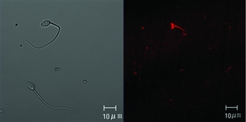
Fluorescence-labelled plasmid DNA was incubated with frozen–thawed sperm without cryoprotection. A microscopic examination revealed that the associated DNA was mainly located in the posterior area of the sperm head (Fig. 1).
Transfected sperm by DNA labelled with 6-dUTP.

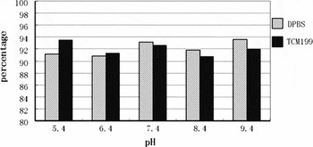
We next examined whether the pH of the freezing medium had any effect on transfection efficiency. Sperm were frozen–thawed in either DPBS or TCM199 with five pH gradients (5.4, 6.4, 7.4, 8.4, and 9.4). Transfection efficiency was tested after incubation with fluorescently labelled plasmid DNA. The result is shown in Fig. 2. No significant difference between any two treatment groups of different pH was observed (p > 0.05).
Effect of pH value on transfection efficiency of exogenous DNA (10 ng/μl).

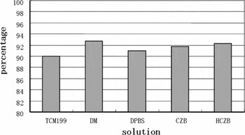
Five dilution media (e.g. TCM199, DM, DPBS, CZB and HCZB) were tested for their influence on the binding of DNA with frozen–thawed sperm. We can see the result in Fig. 3. There was no significant difference among the dilution media (p > 0.05). The rate of positive sperm binding DNA ranged from 90.0 to 92.7%.
Effect of dilute solution on transfection efficiency of exogenous DNA (10 ng/μl).

Development and GFP expression of embryos following ICSI with DNA binding sperm
Oocytes injected with DNA-bound sperms were competent in developing to blastocyst stage from fertilization in vitro. The extrusion of second polar body and pronucleus formation occurred 5–7 h after ICSI. Most of these zygotes can develop to 2–4-cell stage at 18 h. On the third day, embryos at the 8–16-cell stage and morula were present. Early blastocysts were observed after about 70 h of development. Expanded and hatched blastocysts were found on day 4 and day 5 (the injection day is d0).
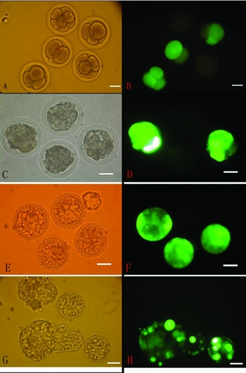
Some embryos were found expressing GFP as early as at 2-cell stage about 24 h after ICSI. The number of GFP-positive embryos increased after 50 h of culture. GFP-positive embryos at various developmental stages are shown in Fig. 4. We also found that positive embryos were not the same in fluorescence intensity.
Transgenic embryos at different developmental stages. (A, C, E, G) Bright-field pictures of developing ICSI produced GFP-positive embryos; (B, D, F, H) corresponding images collected under fluorescent light exposure (magnification × 200).

Rate of transgenic embryo with different DNA concentration
The effect of various DNA concentrations on embryonic development in vitro and EGFP expression after microinjection is shown in Table 1. The concentration of DNA (5, 10 and 20 ng/μl) had no significant effect on the formation rate of positive transgenic embryo at 54 h and 96 h after ICSI (p > 0.05). However, there were fewer GFP-expressing embryos at 30 h culture in the 5 ng/μl treatment group (p < 0.05), while no significant difference was observed between 10 ng/μl and 20 ng/μl DNA treatments.
The rate of GFP-expression embryos fertilized with sperm incubated with DNA of various concentrations.

a ,b Different letter superscripts in the same column indicate significant difference (p < 0.05).
Effect of linearity of DNA construct on production of transgenic embryos
We examined whether the form of plasmid (circular vs linearized) has any effect on transfection efficiency and zygote development (Table 2). There were no significant differences between circular plasmid and linearized plasmid in the rate of oocyte cleavage and the potential of in vitro development (p > 0.05). The cleavage rate of oocyte injected with non-transfected sperm was 91.9%, not higher than that injected with transfected sperm (p > 0.05). But the rate of blastocyst was significantly reduced when the sperm was transfected with the plasmid constructs (p < 0.05).
Effect of DNA construct on transgenic efficiency.

a ,b Different letter superscripts in the same column indicate significant difference (p < 0.05).
Discussion
It usually takes 30–60 min for intact animal sperm to complete transfection by foreign DNA (Chan et al., Reference Chan, Luetjens, Dominko, Ramalho-Santos, Simerly, Hewitson and Schatten2000a; Wang et al., Reference Wang, Lin and Chen2003). However, it takes only a few minutes to transfect sperm with damaged plasma membrane (Perry et al., Reference Perry, Wakayama, Kishikawa, Kasai, Okabe, Toyoda and Yanagimachi1999; Szczygiel et al., Reference Szczygiel, Moisyadi and Ward2003; Hirabayashi et al., Reference Hirabayashi, Kato, Ishikawa, Kaneko, Yagi and Hochi2005). The mechanisms by which foreign DNA associates with intact sperm and disrupted sperm are different. During transfection of fresh intact sperm, DNA molecules first bind to the plasma membrane and then become internalized possibly by the mediation of CD4. Internalization is an active process. In contrast, exogenous DNA molecules bind directly to sperm that have had the plasma membrane destroyed by TRITON-100 or freeze–thawed. The binding of negatively charged DNA may be facilitated by the positive charge of the sperm nucleus. There is no membrane barrier between them, so sperm can take up DNA quickly. Results of our experiment support this viewpoint. Observations under a light microscope indicate that rabbit sperm frozen in liquid nitrogen without cryoprotectants could have serious damage to the plasma membrane. Loss of membrane at the posterior area was present. It facilitates direct contact of exogenous DNA with the sperm nucleus and completes transfection. In the present study, we used dilution solutions that vary in composition (TCM199, DM, DPBS, CZB, HCZB) and pH (5.4, 6.4, 7.4, 8.4 and 9.4). The efficiency of transfection by exogenous DNA was not significantly different among these dilution solutions. The results suggest that plasma membrane of sperm can be efficiently damaged by freezing in liquid nitrogen without cryoprotectants.
The ICSI-mediated gene transfer method was developed by Perry and colleagues in 1999. They used three ways to destroy the plasma membrane of mouse sperm and mixed this with DNA for 1 min. The percentage of transgenic-positive embryo was 64–94% after fertilization by ICSI. Twenty per cent of the offspring were transgenic after embryo transfer. This technique is highly efficient (Moreira et al., Reference Moreira, Giraldo, Cozar, Pozueta, Jimenez, Montoliu and Gutierrez-Adan2004) and capable of transferring large DNA fragments (>200 kb). Transgenic mice and rats have been successful produced using this technique (Hirabayashi et al., Reference Hirabayashi, Kato, Aoto, Sekimoto, Ueda, Miyoshi, Kasai and Hochi2002, Reference Hirabayashi, Kato, Ishikawa, Kaneko, Yagi and Hochi2005; Kato et al., Reference Kato, Ishikawa, Kaneko, Yagi, Hochi and Hirabayashi2004). The positive embryo rates can be up to about 50% in pig (Nagashima et al., Reference Nagashima, Fujimura, Takahagi, Kurome, Wako, Ochiai, Esaki, Kano, Saito, Okabe and Murakami2003) and 34.6% in rhesus monkey (Chan et al., Reference Chan, Luetjens, Dominko, Ramalho-Santos, Simerly, Hewitson and Schatten2000b). In this study, our group describes the first ICSI-mediated transgenic research in rabbit. We observed GFP expression in the 2-cell-stage embryo (24 h after ICSI). Similar observations in embryos derived from ICSI with transfected sperm were reported by others (Chan et al., Reference Chan, Luetjens, Dominko, Ramalho-Santos, Simerly, Hewitson and Schatten2000a; Kaneko et al., Reference Kaneko, Moisyadi, Suganuma, Hohn, Yanagimachi and Pelczar2005). In our experiment, about 60% of the ICSI blastocysts expressed GFP. There is evidence that expression of GFP could certify integration of foreign gene (Kato et al., Reference Kato, Yamanouchi, Ikawa, Okabe, Naito and Tojo1999; Perry et al., 2002). We found that DNA concentration affects the time and the intensity of GFP expression in embryos after ICSI. When the higher DNA concentration was used, there was a higher percentage of GFP-expressing embryos at the 2–4-cell stage, and the fluorescent intensity was generally stronger in the high DNA group at the early developmental stage. This may be caused by variation in copy number of the incorporated DNA. It may be the case that more DNA molecules attach to the sperm nucleus when a high concentration was used. Thus more GFP proteins could be made at the early stage of embryonic development. It can also be seen from our experimental results that some embryos expressing green fluorescent protein could be examined at the early stage, and the number of transgenic embryo rose along with culture time. The positive embryos maintained GFP expression constantly up to the blastocyst stage. This observation is not completely consistent with a previous report (Kaneko et al., Reference Kaneko, Moisyadi, Suganuma, Hohn, Yanagimachi and Pelczar2005), in which positive embryos were also observed at the 2-cell stage, but without the progressive increase in the number of transgenic embryos from early stage to blastocyst stage.
Increase in fluorescence intensity of blastomeres has a negative impact on the development of embryos (Perry et al., Reference Perry, Wakayama, Kishikawa, Kasai, Okabe, Toyoda and Yanagimachi1999). Some of the EGFP-expressing embryos became arrested very early in their development and coinjection of DNA had a toxic effect on postimplantation embryos. Previous research has shown that abnormality of paternal chromosome influences development of embryos, and integration of exogenous DNA significantly can increase degeneration of paternal chromosome (Szczygiel et al., Reference Szczygiel, Moisyadi and Ward2003). Obviously, decreased developmental competence of embryos derived from ICSI with transfected sperm results from integration of a foreign gene. The present data support this view. It can be seen from Table 2 that the rate of blastocyst formation derived from oocytes fertilized with transfected sperm was significantly lower than of those fertilized with non-DNA bound sperm, and both linear and circular DNA had the same toxic effect on embryonic development. It is believed that chelating reagents can protect the sperm chromosome. However, our operation media contained neither of these, which might explain the decreased embryo development in vitro, a view consistent with that of Szczygiel et al. (Reference Szczygiel, Moisyadi and Ward2003).
In conclusion, freeze-thawing treatment of rabbit sperm without cryoprotectant efficiently increases the ability of exogenous DNA attachment. The influence of sperm dilution media pH within the range 5.4–9.4 was minimal. Oocytes fertilized with transfected sperm have a decreased development potential in vitro. The conformation of foreign DNA (linear or circular) did not affect transgenic efficiency in ICSI-mediated gene transfer. The present results demonstrated that genetically modified rabbit blastocysts could be produced by transgene-ICSI technique.
Acknowledgements
This work was supported by the research grant from High-Tech Research & Development Program of China (2002AA206311 and 2006AA02Z148).