Introduction
The cognitive changes associated with neurodegenerative diseases, such as Alzheimer's Disease (AD), lead to a progressive decline in the patient's ability to perform activities of daily living (ADL). Recent evidence has suggested that AD is a continuum, with the clinical symptoms of a major neurocognitive disorder (DSM-5: American Psychiatric Association, 2013) becoming apparent a decade or more after the biomarker-associated pathophysiological process begins in sporadic AD (Morris et al., Reference Morris2009; Rentz et al., Reference Rentz2010; Sperling et al., Reference Sperling2011; Knopman et al., Reference Knopman2012; Ellis et al., Reference Ellis2013; Villemagne et al., Reference Villemagne2013), and autosomal dominant AD (Bateman et al., Reference Bateman2012). Research concerning biomarkers in the early stages of neurodegeneration has suggested that functional impairment occurs before cognitive impairment (Jack et al., Reference Jack2010).
ADL can be stratified according to difficulty and complexity in three levels of functioning (Reuben et al., Reference Reuben, Laliberte, Hiris and Mor1990). Basic ADL (b-ADL) are defined as the activities meeting the basic physiological and self-maintenance needs. Instrumental ADL (i-ADL) are essential, together with b-ADL, to maintain independent living. Advanced ADL (a-ADL) are more sophisticated activities, beyond those necessary to live independently (De Vriendt et al., Reference De Vriendt, Gorus, Cornelis, Velghe, Petrovic and Mets2012, Reference De Vriendt, Mets, Petrovic and Gorus2015) such as, using (household) technology, driving, going on holidays, doing sports, practice hobbies, or arts (De Vriendt et al., Reference De Vriendt, Gorus, Cornelis, Velghe, Petrovic and Mets2012).
i-ADL may be impaired in the early stage of AD (Marshall et al., Reference Marshall2011a; Reference Marshall2014) and are more likely to be sensitive to the early effects of cognitive decline (Pérès et al., Reference Pérès2008). Traditionally, general cognitive functioning (other than the presence of memory complaints) needed to be preserved – as well as the capability to perform daily life activities independently – for a person to be classified as having a Mild Cognitive Impairment (MCI) (Petersen et al., Reference Petersen, Smith, Waring, Ivnik, Tangalos and Kokmen1999). In particular, when MCI was subsequently described as “a concept in evolution,” it was reported that very mild problems in i-ADL are generally consistent with MCI, while b-ADL should be preserved (Petersen et al., Reference Petersen, Caracciolo, Brayne, Gauthier, Vesna Jelic and Fratiglioni2014). Importantly, MCI associated with compromised i-ADL abilities has been found to predict progression to major neurocognitive disorders (Yoshita et al., Reference Yoshita2006; Ogama et al., Reference Ogama, Sakurai, Shimizu and Toba2014, Reference Ogama, Yoshida, Nakai, Niida, Toba and Sakurai2016; Jenkel et al., Reference Jekel2015). Moreover, there is increasing evidence for early i-ADL decrements in individuals with amnestic MCI (aMCI) (Farias et al., Reference Farias, Mungas and Jagust2005; Bangen et al., Reference Bangen2010; Luck et al., Reference Luck, Luppa, Angermeyer, Villringer, Konig and Riedel-Heller2011). This association is in line with the finding that aMCI represents an increased risk for major neurocognitive disorder, such as AD (Jungwirth et al., Reference Jungwirth, Zehetmayer, Hinterberger, Tragl and Fischer2012).
Impairment of daily life functions worsens with the clinical stage of AD, and increases caregivers’ burden (Kamiya et al., Reference Kamiya, Sakurai, Ogama, Maki and Toba2014). The following have been reported to be associated with i-ADL impairment: cognitive decline (Burton et al., Reference Burton, Strauss, Hultsch and Hunter2006; Cahn-Weiner et al., Reference Cahn-Weiner2007; Royall et al., Reference Royall2007; Tomaszewski et al., Reference Tomaszewski2009), depressive symptoms (Kondo et al., Reference Kondo, Kazama, Suzuki and Yamagata2008; Hybels et al., Reference Hybels, Pieper and Blazer2009; Nyunt et al., Reference Nyunt, Lim, Yap and Ng2012; Song et al., Reference Song, Meade, Akobundu and Sahyoun2014), female sex (Sahin et al., Reference Sahin2015), lower education (Sahin et al., Reference Sahin2015), older age (Sahin et al., Reference Sahin2015), physical dysfunction (Seidel et al., Reference Seidel, Brayne and Jagger2011; Gobbens et al., Reference Gobbens and van Assen2014; Albert et al., Reference Albert, Bear-Lehman and Anderson2015; Artaud et al., Reference Artaud, Singh-Manoux, Dugravot, Tzourio and Elbaz2015), and executive dysfunction (Burton et al., Reference Burton, Strauss, Hultsch and Hunter2006; Cahn-Weiner et al., Reference Cahn-Weiner2007). The evidence that executive dysfunction impacts i-ADL in subjects with AD was previously reported (Boyle et al., Reference Boyle, Malloy, Salloway, Cahn-Weiner, Cohen and Cummings2003; Pereira et al., Reference Pereira, Yassuda, Oliveira and Forlenza2008; Tomaszewski et al., Reference Tomaszewski2009). In this direction, a study by Marshall et al. (Reference Marshall2011b) further demonstrated a significant relationship between executive dysfunction and i-ADL impairment in normal ageing, MCI, and mild AD. Executive dysfunction and i-ADL impairment have been shown to predict progression from aMCI to clinical AD (Tabert et al., Reference Tabert2002). Moreover, they are thought to be associated with each other and prefrontal dysfunction (Tabert et al., Reference Tabert2002). Interestingly, a reduced awareness of i-ADL deficits brings patients with mild AD to overestimate their functional capacity. This aspect was previously associated with specific executive dysfunction – in terms of self-monitoring, set-shifting, response inhibition – and with the presence of mood changes (Amanzio et al., Reference Amanzio2011; Reference Amanzio, Vase, Leotta, Miceli, Palermo and Geminiani2013).
Since the association between functional impairment and executive dysfunction is important for diagnostic and prognostic purposes, we decided to further study this association taking into account specific aspects that, to the best of our knowledge, have not been analyzed in a single study: (1) A large group of participants had been carefully selected in order to represent subjects who have AD etiopathology as the cause of their impairments; (2) The cognitive deterioration had been studied, using an overall neuropsychological assessment, in order to analyze the contributions of different cognitive-behavioral sub-domains to functional dysfunctions; (3) Specific EFs have been analyzed in order to describe possible association with i-ADL disabilities. In particular, we conducted three multiple linear regression analyses in order to describe: (1) The role of global cognitive functioning and specific cognitive variables (selective attention, episodic memory, and language comprehension); (2) The role of specific executive functions (EFs), such as self-monitoring, set-shifting, and response inhibition; (3) The relationship with awareness of deficits, mood changes, and perspective taking in terms of Theory of Mind (ToM) of the first and second type (Premack and Woodruff, Reference Premack and Woodruff1978).
Methods
Participants
All the outpatients were enrolled at the Neurology Division of the “Città della Scienza e della Salute” Hospital and the Martini Hospital, both in Turin (Italy).
Participants were included in the study if they had minor or major neurocognitive disorders (DSM-5, American Psychiatric Association, 2013), such as MCI likely due to AD and AD. Participants were excluded from the study if they had; (1) major depression or dysthymia, based on DSM-5 criteria (American Psychiatric Association, 2013); (2) a Mini-Mental State Examination (MMSE: Folstein et al., Reference Folstein, Folstein and McHugh1975) score of <19 given that the neuropsychological measurement is not as reliable when problems of language comprehension occur; (3) were taking medications that could substantially impact cognitive functioning or antidepressants and/or anxiolytics. Cerebrospinal Fluid (CSF) diagnosis that did not provide in-vivo evidence of Alzheimer's pathology was considered a further exclusion criterion.
The patients underwent extensive clinical and neuroradiological investigations, including structural magnetic resonance and Positron Emission Tomography with 2-deoxy-2-[fluorine-18] fluoro-D-glucose. Lumbar puncture with CSF measurement (phospho-Tau, total-Tau and 1–42beta-amyloid) was performed on all patients (Innogenetics kits, Ghent, Belgium; see Table S1, available as supplementary material attached to the electronic version of this paper at www.journals.cambridge.org/jid_IPG). Diagnoses of MCI likely due to AD and of AD were based on the recommendations from the National Institute on Aging/Alzheimer's Association workgroups on diagnostic guidelines for AD (Albert et al., Reference Albert2011; McKhann et al., Reference McKhann2011), taking into consideration the core research criteria (Dubois et al., Reference Dubois2014).
Assessment of i-ADL
We verified the subjects’ level of autonomy in daily living in terms of instrumental activities (Lawton and Brody, Reference Lawton and Brody1969) in the presence of a reliable informant. The scale was administered by interview to a knowledgeable family member or caregiver who provided answers.
The Lawton i-ADL scale is an appropriate instrument to assess independent living skills (Lawton and Brody, Reference Lawton and Brody1969). From a clinical perspective, it is emphasized that the i-ADL scale may provide an early warning of functional decline, or signal the need for further assessment (Graf, Reference Graf2008). These skills are considered more complex than b-ADL. The instrument is most useful for identifying how a person is functioning at the present time and for identifying improvement or deterioration over time.
For all the above, the “Italian Society for Gerontology and Geriatrics” considers i-ADL scale as part of the Comprehensive Geriatric Assessment. Importantly, the Piedmont Welfare System considers i-ADL scale as having legal value during the health inspections at the Geriatric Assessment Units (D.G.R. n. 42–8390 10/3/2008).
The i-ADL scale (Lawton and Brody, Reference Lawton and Brody1969) evaluates functional autonomy in the performance of eight different functions; (1) using the telephone; (2) shopping; (3) preparing food; (4) housekeeping; (5) doing laundry; (6) using transportation; (7) handling medications; and (8) ability to handle finances. Each item was rated dichotomously (0 = less able, 1 = more able). Total scores range from 0 (low function, dependent) to 8 (high function, independent). The higher the score the lower the level of dependence. Each ability measured by i-ADL scale relies on either cognitive or physical function, though all require some degree of both.
Neuropsychological assessment
The neuropsychological evaluation involved a wide assessment of global cognitive deterioration using: the MMSE and the Alzheimer's disease assessment scale – cognitive sub-scale (ADAS-cog: Rosen et al., Reference Rosen, Mohs and Davis1984). The disease severity was assessed with the Clinical Dementia Rating Scale (CDR: Hughes et al., Reference Hughes, Berg, Danziger, Coben and Martin1982), selective attention with Attentional Matrices (AM: Spinnler and Tognoni, Reference Spinnler and Tognoni1987), episodic memory with the Recall of a Short Story test (Babcock: Spinnler and Tognoni, Reference Spinnler and Tognoni1987), and language comprehension with the Token Test (De Renzi and Vignolo, Reference De Renzi and Vignolo1962; TT: Spinnler and Tognoni, Reference Spinnler and Tognoni1987). EFs were analyzed by means of the six subscales making up the behavioural assessment of the dysexecutive syndrome (BADS) neuropsychological battery (Wilson et al., Reference Wilson, Alderman, Burgess, Emslie and Evans1996). These can be summarized as follows: (1) The rule shift cards (RSC) subtest evaluates the ability to respond correctly to a rule and to shift from the use of one simple rule to another more complex one; (2) the action program (AP) assesses skills in solving a closed-ended sequential problem; (3) the key search (KS) subtest evaluates the ability to explore planning in the visual spatial domain and to solve an open-ended problem; (4) the temporal judgment (TJ) subtest ranks cognitive estimation ability; (5) the zoo map (ZM) subtest evaluates planning, sequential behavior, and the use of external feedback in problem solving; (6) the modified six elements (MSE) subtest evaluates ability to divide attention, task scheduling, performance monitoring, and prospective memory.
Importantly, as Lezak et al. (Reference Lezak, Howieson and Loring2004) pointed out, the BADS is the only test battery that is able to offer an extensive overview of EF analyses. Indeed, the BADS has been considered helpful in detecting executive dysfunction in a variety of diseases and in AD (Wilson et al., Reference Wilson, Alderman, Burgess, Emslie and Evans1996; Amanzio, et al., Reference Amanzio, Geminiani, Leotta and Cappa2008; Espinosa et al., Reference Espinosa2009; da Costa et al., Reference da Costa Armentano, Porto, Nitrini and Dozzi Brucki2013). Moreover, performance on the BADS has already been found to be related to prefrontal activity (Rodrigues Gouveia et al., Reference Rodrigues Gouveia, Dozzi Brucki, Fleury Malheiros and Bueno2007).
ToM of the first and second type, which refers to the “ability to mentalize,” to understand the mental state of others and to predict behavior based on those states, was also assessed. As expressed by Premack and Woodruff (Reference Premack and Woodruff1978) “in saying that an individual has a ToM, we mean that the individual imputes mental states to himself and to others” (p. 515). In particular, ToM visual stories were used to assess perspective-taking abilities (TOM 1 and TOM 2: Amanzio et al., Reference Amanzio, Geminiani, Leotta and Cappa2008). The subject has to solve problems involving: first-order attributions of false belief (of the type “A thinks X”) and second-order attributions of false belief (of the type “A thinks B thinks X”).
Specific neuropsychiatric scales for rating mood changes were also used to describe the patients’ behavioral profile: hypomania with the Mania Scale (MAS: Bech et al., Reference Bech, Rafaelsen, Kramp and Bolwig1978); apathy and depression with the Hamilton Depression Rating Scale (HDR-S: Hamilton, Reference Hamilton1960).
Unawareness of deficits at the time of testing was analyzed using the Awareness of Deficit Questionnaire – Dementia scale (AQ-D: Migliorelli et al., Reference Migliorelli1995). The AQ-D is a scale of demonstrated reliability/validity for ranking the severity of unawareness of deficits in AD (Migliorelli et al., Reference Migliorelli1995). Thirty questions divided into two sections (the cognitive and the behavioral) make up the questionnaire. The cognitive part assesses cognitive function and performance in ADL and i-ADL. The behavioral part assesses changes in interests and mood. All the questions were asked to the patients and to their caregivers blinded to the patients’ responses. Scores range from 0 (never) to 3 (always), with the minimum and maximum total scores obtainable ranging from 0 to 90 (cognitive section range = 0–66; behavioral section range = 0–24). The total AQ-D score is given by the difference between the caregiver's and the patient's forms. Higher scores on the AQ-D scale indicate greater unawareness of the disease and a reduced awareness of deficits, meaning that caregivers rated the patients as more impaired than did the patients themselves (Migliorelli et al., Reference Migliorelli1995). Since this method is based on a subtractive index of perception (caregivers’ minus patients’ scores), the ruling out of any bias in the caregivers’ judgments is crucial (Amanzio et al., Reference Amanzio2011; Reference Amanzio, Vase, Leotta, Miceli, Palermo and Geminiani2013). For this reason, we have ensured that the caregivers (with a mean MMSE score of 27) had normal psychiatric and neurological evaluations and a negative history of neurological disorders.
Finally, b-ADL was assessed with the Katz et al. (Reference Katz, Ford, Moskowitz, Jackson and Jaffe1963) scale. The Katz Index of independence in ADLs is one of the most commonly used tools to asses basic ADLs (Milnac and Feng, Reference Milnac and Feng2016). It evaluates functional autonomy in the performance of six different functions: (1) bathing; (2) dressing; (3) toileting; (4) transferring; (5) continence; and (6) feeding. Each item was rated dichotomously (0 = dependent, 1 = independent). Total scores range from 0 (low function, dependent) to 6 (high function, independent). A score of 2 or less indicates severe functional impairment, 4 indicate moderate impairment, and 6 indicate full function. Clinicians rate individuals as either fully independent (no supervision, direction, or personal assistance needed) or dependent (needing supervision, direction, personal assistance, or total care) across the six skills (Milnac and Feng, Reference Milnac and Feng2016). This measure was originally created to assess the physical functioning among those who were in rehabilitation (Milnac and Feng, Reference Milnac and Feng2016).
Procedures
Patients were evaluated by performing a neuropsychological assessment during their hospital admission lasting one week. The participants were assessed in three experimental sessions held one day apart and each lasting one-hour, with a view to preventing fatigue and lack of adherence to the tasks.
Statistical analysis
Statistical analyses were performed using SAS/STAT® 9.3 (Freund et al., Reference Freund, Littell and Spector1986; Schlotzhauer and Littell, Reference Schlotzhauer and Littell1987). Normality assumption distribution of outcome variables was evaluated using the Kolmogorov–Smirnov test. Since the distribution of i-ADL scores was not normal, we have dichotomized the variable using the median value (=6). We then divided the sample into above the median (n = 62) and up to the median (n = 82). The binary variable derived was afterwards used in multiple logistic regression analyses.
We conducted three logistic regression analyses adjusted for gender in order to study whether the level on i-ADL scale could be associated with cognitive and behavioral measurements. Importantly, we applied the “one in ten rule” according to which, logistic regression analyses could be used with a minimum of ten events per predictor variable (Harrell et al., Reference Harrell, Lee, Califf, Pryor and Rosati1984; Reference Harrell, Lee and Mark1996).
Moreover, the selection of the three models was performed in line with the results obtained in the literature on i-ADL. In particular, i-ADL has been previously linked to general cognitive decline, specific cognitive functions, and neuropsychiatric domains (Marshall et al., Reference Marshall2011b).
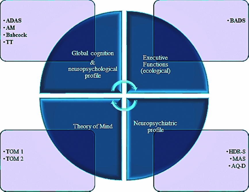
The final selected models considered i-ADL scale as the dependent variable and the following as independent variables (see Figure 1):
1. Model 1 – to address the role of global cognitive functioning and specific cognitive variables (global cognition, selective attention, episodic memory, and language comprehension): ADAS, AM, BABCOCK, TT.
2. Model 2 – to study the role of EFs with BADS sub-scales: BADS_RSC; BADS_AP; BADS_TJ; BADS_KS; BADS_ZOO; BADS_MSE.
3. Model 3 – to investigate the relationship with awareness of deficits, mood changes, and TOM: AQ-D, HDR-S, MAS, TOM 1 and 2.
The graph represents the independent variables and the neuropsychological domain they belong to.

Results
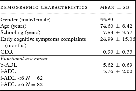
Over a 26-month period, 200 patients – complaining of different cognitive deficits and presenting for the first time at the out-dep of our clinics – were evaluated for their possible participation in the study. Based on the inclusion criteria, 144 hospital admitted patients (M/F = 55/89; mean age ± SD = 74.60 ± 6.42 years) took part in the study. In particular, 32 subjects with MCI likely due to AD, according to the CSF analysis, were included in the study (see Table S1). For those patients with major neurocognitive disorders, the CSF diagnosis provided in-vivo evidence of Alzheimer's pathology for 112 patients. The demographic and clinical data related to the patient population have been summarized in Table 1. In anamnesis, only age-related disorders and problems (i.e. slight sensory deficits, hypertension, hypercholesterolemia, gastritis, weight gain or loss, and deflection of mood). All of them are drug-naïve patients. Indeed, they had not taken antidepressants and/or anxiolytics and/or anti-cholinesterase drugs before the neuropsychological evaluation. The neuropsychological assessment reflected the diagnoses made by the CSF, biomarkers and neurological exams. A total of 139 of the 144 patients obtained a CDR score of between 0 and 1, indicating a low-level of cognitive impairment (see Table 2).
Demographic and clinical characteristics

CDR = Clinical Dementia Rating Scale; SD = Standard Deviation.
b-ADL and i-ADL: higher scores indicate better performance.
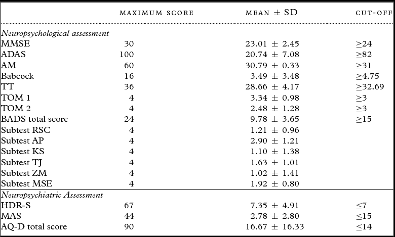
Neuropsychological and neuropsychiatric assessment synopsis. Wherever there is a normative value, the cut-off scores are given in the statistical normal direction. Cells in grey indicate the absence of a normative cut-off

MMSE = Mini-Mental State Examination; ADAS = Alzheimer's disease assessment scale; AM = Attentional Matrices; TT = Token Test; TOM = Theory of Mind; BADS = Behavioral Assessment of Dysexecutive Sindrome; RSC = Rule Shift Cards; AP = Action Program; KS = Key Search; TJ = Temporal Judgment; ZM= Zoo Map; MSE = Modified Six Elements; HDR-S = Hamilton Depression Rating Scale; MAS = Mania Scale; AQ-D = Awareness of Deficit Questionnaire – Dementia scale.
MMSE: lower scores indicate more severe cognitive impairment. ADAS: higher scores indicate more severe cognitive impairment. AM, Babcock, TT, TOM tasks and BADS: higher scores indicate better performance. AQ-D: higher scores indicate more severe unawareness. HDR-S and MAS scales: higher scores indicate more severe symptoms.
Considering the functional assessment, participants obtained a mean b-ADL score of 5.62 and a mean i-ADL score of 5.76, showing a low level of impairment on basic and instrumental ADL.
Association between i-ADL scale and neuropsychological variables
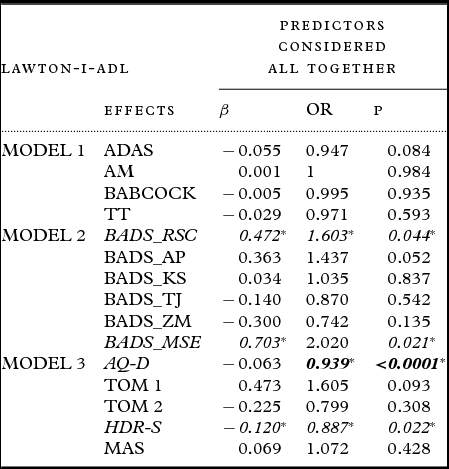
The results are presented as Odds Ratio (OR) and 95% confidence intervals (see Table 3). After adjusting the analysis for gender, i-ADL scores were influenced by BADS_RSC and BADS_MSE in model 2 and by AQ-D and HDR-S in model 3. Specifically, the worsening of performance at the BADS_RSC and BADS_MSE increases the probability that a participant has a dysfunction in i-ADL. Likewise, the chances of a subject being dysfunctional increases with the worsening of mood deflection and poor awareness. On the contrary, the level on the i-ADL scale was not influenced by global cognition, attention, memory, or language comprehension (in model 1).
Results for the logistic regression analysis applied in order to estimate the effect of neuropsychological and neuropsychiatric aspects on i-ADL. Outcomes were adjusted for the gender and are presented as Odds Ratio (OR) and 95% CI

*p < 0.05.
ADAS = Alzheimer's disease assessment scale; AM = Attentional Matrices; TT = Token Test; BADS = Behavioral Assessment of Dysexecutive Sindrome; BADS_RSC = subtest Rule Shift Cards; BADS_AP = subtest Action Program; BADS_KS = subtest Key Search; BADS_TJ = Temporal Judgment; BADS_ZM = Zoo Map; BADS_MSE = Modified Six Elements; AQ-D = Awareness of Deficit Questionnaire – Dementia scale; TOM = Theory of Mind; HDR-S = Hamilton Depression Rating Scale; MAS = Mania Scale.
Discussion
Our study is a first novel attempt to investigate possible association among i-ADL functioning, EFs and specific cognitive and behavioral variables, using an overall neuropsychological battery, in a selected patient population on the basis of CSF examination. Thus, newly diagnosed drug-naive MCI likely due to AD and AD patients provide an ideal population in which to study abnormalities in everyday functioning. Although we considered patients with different degrees of cognitive impairment, our sample was homogeneous in terms of etiopathogenesis, severity of symptoms (CDR = 0.90 ± 0.33, attesting a mild level of disease) and mood changes. Most importantly, our attempt to consider these kinds of patients in the same sample was justified by the regression analysis approach we used and by the international guidelines on aging that consider patients with cognitive impairment to lie on a continuum between MCI and mild AD (Petersen and Negash, Reference Petersen and Negash2008; Albert et al., Reference Albert2011; Dubois et al., Reference Dubois2014).
Based on the results, we obtained, there appear to be no straightforward associations between i-ADL scores and specific aspects of neuropsychological functioning, such as global cognition, long-term verbal memory, language comprehension, and selective attention (in model 1). On the contrary, we observed a significant association between i-ADL functioning and two BADS sub-scales (in model 2), and between i-ADL and AQ-D and HDR-S (in model 3), respectively.
Our findings showed that i-ADL was associated with executive dysfunction. In particular, the ability to inhibit a response, self-monitoring, and set-shifting in terms of cognitive flexibility (measured through the MSE and the RSC) seem to be key skills for i-ADL, as demonstrated by the logistic regression analysis. As we previously reported (Amanzio et al., Reference Amanzio, Vase, Leotta, Miceli, Palermo and Geminiani2013), being a modified version of Shallice and Burgess’ Six Elements Test (Shallice and Burgess, Reference Shallice and Burgess1991), the MSE was designed to assess the supervisory attentional system hypothesis. Specifically, MSE relies on the ability to inhibit a dominant response (i.e. perform the tasks in the given order), favoring the correct answer (i.e. alternating the execution of the tasks between all the proposed types). The MSE test also measures the ability to self-monitor performance and switch from task to task. In the same direction, the RSC subtest is a further measure of cognitive flexibility, involving the ability to move between different sets of responses. The RSC also measure abilities to shift and inhibit response and monitoring behavior (Cools et al., Reference Cools, Brouwer, De Jong and Slooff2000).
The three specific above-mentioned cognitive abilities – monitoring (updating), inhibition, and set-shifting – are defined in terms of basic EFs (Miyake et al., Reference Miyake, Friedman, Emerson, Witzki, Howerter and Wagner2000). These sub-components of executive control are considered mutually interacting (Miyake et al., Reference Miyake, Friedman, Emerson, Witzki, Howerter and Wagner2000). Not surprisingly, all of them, if compromised, seem to be involved in functional disabilities. Interestingly, we previously demonstrated that executive dysfunction in terms of inhibition, self-monitoring, and set-shifting resulted associated with a reduction in the awareness of functional disabilities of mild AD patients (Amanzio et al., Reference Amanzio, Vase, Leotta, Miceli, Palermo and Geminiani2013).
According to our results, planning or problem solving abilities – as higher-level subcomponent of EFs – did not seem to have any relationship with i-ADL. In particular, we found no positive results with BADS subtests, such as ZM, KS, and AP. Indeed, the ZM has been demonstrated to be useful in detecting planning impairment in AD. AD patients seem to have more problems developing logical strategies and executing complex predetermined plans (Piquard et al., Reference Piquard, Derouesné, Lacomblez and Siéroff2004; Allain et al., Reference Allain2007). KS is a more abstract task than the ZM, examining a person's ability to prepare an efficient plan of action in the context of a routine event. It is important to point out that these two tasks are considered to evaluate similar EFs (Wood and Liossi, Reference Wood and Liossi2007), related to the dorsolateral frontal lobe region (Millar et al., Reference Millar, Griffiths, Zermansky and Burn2006). In the same direction, AP assess the ability to develop an action-plan in order to solve a novel problem (Murakami et al., Reference Murakami2015). Finally, we found no relationship between i-ADL and TJ measuring cognitive estimation ability (Murakami et al., Reference Murakami2015).
Considering the neuropsychiatric profile and taking the IADL scores into account, we observed an involvement of mood changes, in terms of depression. This finding is consistent with the literature on minor and major neurocognitive disorders (Boyle et al., Reference Boyle, Malloy, Salloway, Cahn-Weiner, Cohen and Cummings2003; Marshall et al., Reference Marshall2011b). Moreover, patients with a reduced awareness of illness seem to have more difficulties in i-ADL. As we have previously demonstrated in AD patients, if the executive system does not function correctly, the comparator mechanism of self-monitoring does not detect mismatches between the current and previous performance states stored in the personal database and produces a reduced awareness for the instrumental domain (Amanzio et al., Reference Amanzio, Vase, Leotta, Miceli, Palermo and Geminiani2013).
Finally, although AD patients may display TOM impairment primarily mediated by hippocampal degeneration (Synn et al., Reference Synn2018), we did not observed an association between functional impairment and mentalizing performance in our patients. Our results support the hypothesis that performances on social cognition tests are not a good indicator to differentiate patients with adequate i-ADL functioning from mild dysfunctioning patients.
Future prospective studies will be helpful in order to further characterize the role of neuropsychological processes in the progression of i-ADL dysfunction.
Limitations section
The study here presented has been carefully designed and reached its aims; however, some critical aspects have to be outlined. The first aspect regards the tool used to assess the level of independent living skills, which could represent a possible confounding factor.
The evaluation of i-ADL may appear quite straight-forward. Despite this, procedures and tools can vary considerably. To date, a variety of examination tools measures the older adult's ADL and IADL performance, but there is no established gold standard for such assessment because few scales have been comprehensively evaluated (Capezuti et al., Reference Capezuti, Malone, Khan and Baumann2017). Indeed, measures differ in their capability to establish level of dependence and the kind of assistance needed for each evaluated activity. Although Lawton i-ADL scale has low psychometrical properties (eventually affecting our results), it was developed to assess the more complex ADLs necessary for living in the community. This scale is part of the comprehensive geriatric assessment and is considered appropriate for use with older adults admitted to a hospital by the Italian legislation.
A second aspect concerns the results that have not to be considered generalizable for patients with different etiopathogenesis other than AD. However, our study was necessary to better define the associations between functional deficits and specific neuropsychological variables in a highly selected sample of patients.
Finally, we have focused our study in few predictors of IADL to assure a good power calculation. The selected predictors are in line with the international literature. Further studies would be necessary in order to analyze other factors.
Conclusion
Our results suggest the importance of considering EF dysfunctions in reduced i-ADL functionality in patients who have AD etiopathology as the cause of their impairments. The findings support the hypothesis that patients with different level of cognitive impairment, such as MCI likely due to AD and AD, exhibit i-ADL dysfunction in the context of overlapping EFs, reduced awareness of deficits and mood changes. A complete neuropsychological evaluation – based on specific assessment of the ability to inhibit a response, self-monitoring, and set-shifting – might be able to identify those MCI patients, with reduced i-ADL functionality, at greater risk of developing a major neurocognitive disorder, such as AD. Finally, those patients with functional limitations in their daily living and reduced awareness may represent an important target population for tailoring specific interventions with important clinical implications, in terms of adherence to treatments and prognosis.
Conflict of interest
None.
Description of authors’ roles
M. Amanzio designed the study, supervised the data collection, and wrote the paper. She took part in the review and critique processes as PI. She also organized the study and participated in the statistical analyses (execution and organization, review, and critique). S. Palermo supervised the data collection and the neuropsychological assessment, participated in the statistical analyses, participated in writing the paper, and created the infographics. R. Rosato was responsible for the statistical design of the study and for carrying out the statistical analysis and participated in writing the paper. E. Rubino performed the neurological assessment (execution) and took part in the organization of the study and in the diagnostic phase (organization and diagnosis). M. Zucca and M. Bartoli performed the neuropsychological assessment (execution). D. Leotta and I. Rainero supervised the neurological assessment, took part in the organization of the study, and participated in writing the paper (organization, review, and critique).
Acknowledgments
The study was approved by the Ethics Committee “A.O.U. Città della Salute e della Scienza di Torino - A.O. Ordine Mauriziano - A.S.L. Città di Torino” as part of the core research criteria followed by the Neurological Units. All subjects gave their informed written consent to participate in the study.
This study has therefore been performed in accordance with the ethical standards laid down in the 1964 Declaration of Helsinki and its later amendments.
Supplementary material
To view supplementary material for this article, please visit https://doi.org/10.1017/S1041610218000455