Stenting is increasingly used for the treatment of aortic coarctation.Reference Forbes, Garekar and Amin 1 , Reference Holzer, Qureshi and Ghai 2 Results of coarctation stenting are encouraging but important complications may occur both acutely and during follow-up.
Stent fracture is a rare complication occurring during follow-up after stenting for aortic coarctation.Reference Holzer, Qureshi and Ghai 2 Similarly, aortic pseudoaneurysm is a rare, potentially serious complication after stenting for aortic coarctation, occurring both at the time of the procedure or during follow-up.Reference Forbes, Garekar and Amin 1 , Reference Holzer, Qureshi and Ghai 2
We report the case of a patient in whom an aortic pseudoaneurysm and fracture of a bare Cheatham-Platinum stent (NuMED, Inc., Hopkinton, New York, United States of America) developed after stenting for aortic coarctation. These complications were effectively managed by percutaneous implantation of a stent graft.
Case report
Aortic coarctation was diagnosed in an 11-year-old boy with headaches and systemic arterial hypertension. MRI delineated the morphology of the thoracic aorta. Minimum lumen diameter of the aortic coarctation was 3.5 mm, the aortic arch diameter measured 12 mm, and the descending thoracic aorta at the level of the diaphragm was 14 mm. Numerous arterial collaterals were present.
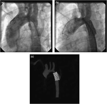
Aortography confirmed the MRI findings of typical aortic coarctation (Fig 1a). The pull-back pressure gradient was 65 mmHg. Aortic stenting was performed with a 34-mm-long bare Cheatham-Platinum stent hand-crimped on a 16-mm balloon-in-balloon (NuMED, Inc.). Final aortography revealed over-distension of the aortic isthmus with complete relief of the aortic coarctation (Fig 1b). Aortic wall injury was seen along the lower half of the stent. No residual gradient was detected during pull-back pressure measurement. After the procedure, systemic arterial hypertension was managed by intravenous infusion of sodium nitroprusside.
( a ) Aortography (lateral projection) delineating the tight aortic coarctation with numerous arterial collaterals. ( b ) Aortography (lateral projection) after stent implantation revealing over-dilatation of the aortic isthmus and aortic wall injury. ( c ) CT showing aortic wall injury along the posterolateral aspect of the stent. In comparison with ( b ), the distance between the left subclavian artery and stent is increased.

A month after the procedure, routine CT scan was performed, which delineated the widely patent stent. The aortic wall was thickened up to 5 mm along the posterolateral aspect of the stent, reflecting aortic wall injury (Fig 1c). In addition, the distance between the left subclavian artery and the stent increased since implantation.
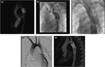
During follow-up, the patient was completely asymptomatic. Systemic arterial hypertension was effectively managed by enalapril. At regular follow-up visits, physical findings were unremarkable. Echocardiographic examinations revealed mildly accelerated blood flow velocity through the stent without recoarctation. Chest radiograph was performed for the first time 6 years after stent implantation and revealed dilatation of the aorta at the level of the stent. Both ends of the stent were fractured and two small parts of the stent were displaced. A CT scan was performed and revealed a widely patent, fragmented stent (Fig 2a). Posterolaterally along the stent, a large aortic pseudoaneurysm was seen communicating with the aortic lumen both above and below the stent.
( a ) CT demonstrating fracture and dislocation of both ends of the stent and large aortic pseudoaneurysm. ( b ) Aortography (lateral projection) revealing the fractured stent with dislocations and a large pseudoaneurysm communicating with the aortic lumen both above and below the stent. ( c ) Fluoroscopy (lateral projection) during positioning of the stent graft across the fractured stent and pseudoaneurysm. ( d ) Final aortography (lateral projection) following deployment of the stent graft demonstrating adequate position of the stent graft anchored proximally within the origin of the left subclavian artery and aligned along the aortic lumen well below the pseudoaneurysm. ( e ) CT demonstrating both proper placement of the stent graft and fragments of fractured stent aligned closely along the stent graft confirming resolution of the pseudoaneurysm.

Diagnostic cardiac catheterisation was performed via the right femoral artery. Aortography confirmed the CT findings and pull-back pressure measurement excluded recoarctation (Fig 2b). We were able to pass a pigtail catheter easily both through the stent and the pseudoaneurysm. We placed the pigtail catheter through the stent into the aortic arch for control aortographies. After preparation of percutaneous sutures, an 18 Fr sheath was introduced into the left femoral artery. A Gore Tag Thoracic Endoprosthesis (W. L. Gore & Associates, Inc., Flagstaff, Arizona, United States of America) with labelled diameter of 21 mm and length of 10 cm was positioned both across the stent and the pseudoaneurysm. Several aortographies were performed to allow precise positioning of the stent graft (Fig 2c). We aimed to position the stent graft to anchor its proximal bare end within the origin of the left subclavian artery. The stent graft was uneventfully deployed, and the final aortography confirmed adequate stent graft position (Fig 2d). The arterial entry point was sutured percutaneously.
A CT scan was repeated 1 month after stent graft implantation and confirmed a widely open aortic lumen, tight alignment of the stent graft to the aortic wall, and complete exclusion of the aortic pseudoaneurysm. Further follow-up CT scan was performed 1.5 years after stent graft implantation, demonstrating fragments of the fractured stent closely aligned along the stent graft, thus confirming elimination of the pseudoaneurysm (Fig 2e).
Discussion
Stent fracture is a rare complication occurring during follow-up after stenting for aortic coarctation.Reference Forbes, Garekar and Amin 1 , Reference Holzer, Qureshi and Ghai 2 Several mechanisms were suggested to predispose to fracture of stents implanted for aortic coarctation. The aortic isthmus is positioned between fixed – ascending aorta and aortic arch – and movable – descending thoracic aorta – aortic segments.Reference Carrozza, Santoro, Russo, Caianiello and Calabro 3 Mechanical stress on the stent is particularly high following implantation in a tight aortic coarctation. Increased pulsatility due to significant aortic regurgitation was also suggested as a possible contributing factor.Reference Ledesma, Jauregui and Ceron 4
Aortic pseudoaneurysm is also a rare complication of coarctation stenting occurring both acutely and during follow-up.Reference Forbes, Garekar and Amin 1 , Reference Holzer, Qureshi and Ghai 2 The performance of aggressive pre-stent angioplasty and a high balloon-to-coarctation ratio are suggested to predispose to aortic wall injury. Structural changes of the aortic wall contribute to aortic wall injury.
In our patient, aggressive dilatation of the stent was performed during the implantation procedure. The ratio between selected diameter of balloon-in-balloon (16 mm) and minimal coarctation diameter (3.5 mm) was high (4.5 mm). In addition, the stent was expanded past the maximal diameter of the aorta at the level of the diaphragm (14 mm). Final aortography, therefore, revealed overly dilated aortic isthmus and aortic wall injury along the lower half of the stent. A CT angiography performed 1 month after the procedure demonstrated thickened aortic wall along the posterolateral aspect of the stent. Thus, aggressive dilatation of the stent caused acute aortic wall injury in our patient and during follow-up, initial aortic wall injury progressed to a large aortic pseudoaneurysm. Retrospectively, we believe that staged approach to stent dilatation might prevent the occurrence of aortic wall injury. In contrast, the mechanism of stent fractures in our patient is not clear. In particular, it is difficult to explain how fractures occurred at the margins of the stent. In addition, the distance between the left subclavian artery and the stent increased during follow-up in our patient. We speculate that aortic wall injury allowed distal migration of the stent detected by CT angiography 1 month after the implantation procedure.
Careful follow-up is of paramount importance following stenting for aortic coarctation, particularly in patients with aortic wall injury. In our patient, aortic wall injury was detected by aortography immediately following stent implantation, and therefore CT angiography was indicated after the procedure to precisely delineate aortic wall injury. Thereafter, follow-up CT angiographies were indicated to assess the progression of aortic wall injury. In addition, even yearly chest radiographs would allow timely recognition of stent fracture and aortic pseudoaneurysms with a reasonable burden of ionising radiation. In contrast, regular echocardiographic examinations were performed in our patient and were inadequate to delineate either aortic pseudoaneurysm or stent fracture.
Implantation of balloon-expandable covered stent is mostly used for the treatment of a fractured stent.Reference Kenny, Margey, Turner, Tometzky, Walsh and Martin 5 Stent-in-stent implantation using covered stents provides stabilisation of the fractured stent and offers protection in case of progression of aortic wall injury. Balloon-expandable covered stents are particularly suitable for the management of stent fractures associated with restenosis. In contrast, self-expanding covered stents – that is, endovascular stent grafts – are widely used for treatment of aortic pseudoaneurysms and dissections. Their use was also described for the management of aortic aneurysms associated with aortic coarctation.Reference Kenny, Margey, Turner, Tometzky, Walsh and Martin 5 , Reference Khavandi, Bentham and Marlais 6 Thus far, self-expanding covered stents were used for the management of aortic aneurysms associated with post-operative coarctations; however, their use has not been reported for the treatment of aortic pseudoaneurysms and fractured stent as described in this case. A large aortic pseudoaneurysm without significant aortic obstruction was the predominant lesion in our patient, and therefore we decided for implantation of a self-expandable stent graft. Our patient was grown-up at the time of stent graft implantation, and therefore development of significant recoarctation is less likely in the future; however, in this case, we would perform balloon dilatation.
In conclusion, we report a case of aortic pseudoaneurysm and a fractured bare Cheatham-Platinum stent following stenting for aortic coarctation. These complications were effectively treated by percutaneous implantation of a self-expandable covered stent – that is, endovascular stent graft. Staged approach to stent dilatation might prevent the development of aortic wall injury, whereas careful follow-up evaluations would allow early detection of these complications. Further follow-up of our patient is mandatory due to the lack of long-term data.
Acknowledgement
The authors thank Samo Vesel, MD, PhD, for his assistance during the management of the patient and preparation of the manuscript.
Financial Support
T.P. was supported in part by the Slovenian Agency for Research grant No. P3-0343.
Conflicts of Interest
None.
Ethical Standards
This case report does not involve human and/or animal experimentation.