I. INTRODUCTION
Sintering describes the diffusion-driven process for the densification of metals and ceramics to generate dense polycrystalline microstructures.Reference German1, Reference Kang2 The consolidation process is commonly described through three primary stages following the initial particle contact formation. The first stage involves neck formation and growth, whereas the second and third stages relate to densification before and after the isolation of the pores, respectively. Activation of diffusion and, hence, sintering can be achieved by the addition of minor phases,Reference Chiang, Birnie and Kingery3, Reference Munir and German4 elevated temperatures,Reference Chiang, Birnie and Kingery3 applied isostatic or uniaxial pressure, or the application of external electromagnetic fields.Reference Olevsky, Kandukuri and Froyen5–Reference Munir, Anselmi-Tamburini and Ohyanagi7 Often, a combination of the above parameters is applied. Common consolidation routines are hot isostatic pressure (HIP) sintering, microwave sintering, and electric field-assisted sintering (EFAS), which includes spark plasma sintering (SPS), field-assisted sintering technology (FAST) and pulsed electric current sintering. Currently several hundred publications and patents per year report that EFAS techniques have the potential to increase the rate of densification, reduce sintering temperatures, shorten holding times,Reference Olevsky, Kandukuri and Froyen5, Reference Orrù, Licheri, Locci, Cincotti and Cao6, Reference Chen, Anselmi-Tamburini, Garay, Groza and Munir8 and suppress grain growth while achieving full density.Reference Chen, Anselmi-Tamburini, Garay, Groza and Munir8–Reference Kodash, Groza, Cho, Klotz and Dowding11 The resulting microstructures often provide unique macroscopic properties such as improved mechanical properties,Reference Nygren and Shen12, Reference Basu, Lee and Kim13 oxidation or corrosion resistance,Reference Basu, Lee and Kim13, Reference Yue, Zhang, Liu and Wang14 optical transmission,Reference Su, Wang, Chen, Shen, Nygren, Cheng and Yan15 and electrical properties.Reference Zhou, Zhao, Zimmermann, Aldinger and Nygren16, Reference Chen, Khor, Chan and Yu17 Despite the abundance of sintering studies reported in the literature and the wide application of EFAS, the atomic scale consolidation mechanisms that occur in the presence of electrical fields and relatively high heating rates during EFAS remain mostly unclear.Reference Chen, Anselmi-Tamburini, Garay, Groza and Munir8
The presence of surface films covering individual powder particles can have a significant influence on the kinetics of neck formation during the initial stage of sintering. For instance, several authors have reported studies on insulating surface oxide layers covering metallic nanoparticles during EFAS processing.Reference Kodash, Groza, Cho, Klotz and Dowding11, Reference Aldica, Khodash, Badica and Groza18–Reference Tokita25 For SPS, it was initially suggested that the electrical field generates plasma at the point of contact or interface between the nanoparticles that “cleans” nanoparticle surfaces of contaminants and oxide layers prior to neck formation.Reference Groza and Zavaliangos20, Reference Tokita25 However, Hulbert et al. were unable to find evidence of plasma formation under typical SPS conditions.Reference Hulbert, Jiang, Anselmi-Tamburini, Unuvar and Mukherjee26 Groza et al.Reference Kodash, Groza, Cho, Klotz and Dowding11, Reference Aldica, Khodash, Badica and Groza18–Reference Groza and Zavaliangos20 have shown that densification of metal powder agglomerates is enhanced with EFAS processing at medium heating rates and have suggested that discharges between oxide-covered metal particles lead to dissociation of current-limiting surface oxides. The reported observations of “surface cleaning” through field-mediated dielectric breakdown of the surface oxides are therefore considered efficient mechanisms to enable current-assisted materials transport for densification during EFAS. However, oxide removal from the particles’ surfaces can also be triggered in the absence of electrical fields and currents through oxide dissolution in the metal particles or conventional reduction-oxidation (redox) reactions at elevated temperatures. MunirReference Munir24 has demonstrated that the presence of stable surface oxides at higher temperature and limited oxide solubility in the metal particles may create an incubation time, i.e., retardation of the onset of sintering. For the cases of nickel and several other metals, Munir states that the presence of surface oxide layers does not affect the kinetics of sintering for relatively large particles with thin oxide layers.Reference Munir24
The nearly ubiquitous use of graphite dies and plungers during EFAS operations requires identification of the effect of carbon on particle surfaces and, hence, the sintering process. It was previously demonstrated that carbon materials such as graphite reduce nickel (II) oxide (NiO) at temperatures above 900 °C,Reference Sharma, Vastola and Walker27, Reference Baukloh and Springorum28 whereas other carbon-based materials can create reducing environments at temperatures as low as 450 °C.Reference Gandia and Montes29 It is therefore expected that the presence of carbon during the first stage of sintering may result in reduction reactions of NiO to form metallic contacts between individual particles at relatively low temperatures. During EFAS, such mechanisms may therefore complement dielectric breakdown of the oxide due to the applied electrical potential and, hence, accelerate densification. The present study investigates such redox reactions between individual nickel nanoparticles in the absence of any electrical fields.
Transmission electron microscopy (TEM) experiments are ideal for determining the morphology, structure, and chemical composition of nanometric powder particles and their agglomerates with high spatial resolution. In situ TEM techniques can be used to study consolidation mechanisms through heating of the TEM sample while simultaneously imaging morphological changes of powder agglomerates.Reference Asoro, Kovar, Shao-Horn, Allard and Ferreira30–Reference Ristau, Tiruvalam, Clasen, Gorskowski, Harmer, Kiely, Hussain and Brust34 Other in situ TEM techniques allow the investigation of consolidation mechanisms by exposing particle agglomerates to electrical currents.Reference Holland, Thron, Bonifacio, Mukherjee and van Benthem35, Reference Hummelgard, Zhang, Nilsson and Olin36 The early stage sintering of platinum (Pt) due to thermal annealing during in situ scanning transmission electron microscopy (STEM) has been reported in the literature. Asoro et al. have measured dihedral angles between individual particles supported on amorphous carbon as a function of temperatureReference Asoro, Kovar, Shao-Horn, Allard and Ferreira30 whereas Simonsen et al.Reference Simonsen, Chorkendorff, Dahl, Skoglundh, Sehested and Helveg31 observed thermally activated Ostwald ripening of Pt catalyst particles on SiO2 substrates. Ida et al. used in situ heating experiments in the TEM to demonstrate that a variation of the oxygen partial pressure has a significant influence on the sintering behavior of copper nanocrystals embedded in a polymer matrix.Reference Ida, Sugiyama, Chujyo, Tomonari, Tokunaga, Sasaki and Kuroda33 However, mechanisms for “surface cleaning,” such as the removal of metal oxide films through redox reactions prior to neck formation were not considered.
In this article, we report a systematic study of the reduction reaction of nickel oxide films that cover metallic nanoparticles of nickel. In situ thermal annealing of Ni nanoparticles inside the TEM in the presence and absence of amorphous carbon was used to directly demonstrate that the reduction of the surface oxide occurs at lower temperature when carbon is present. It was also found that common concentrations of carbon impurities in the regime of a few tens of a weight percent remain insufficient to produce similar effects. This study therefore provides the first direct experimental evidence that the utilization of graphite sample holders during EFAS processing has the potential to lower the processing temperatures for initial neck formation during the first stage of sintering.
II. METHODS AND TECHNIQUES
Nickel nanoparticles with diameters of approximately 20 nm were utilized for the presented study. The powders were obtained from Umicore (Brussels, Belgium) with impurity levels of 0.3 wt% C, 0.011 wt% S, 1.2 wt% O, and 0.003 wt% Fe as reported by the company. In situ TEM was carried out using a JEOL JEM 2500SE (JEOL Inc., Peabody, MA). TEM instrument operated at 200 keV. An aberration-corrected JEOL JEM 2100F/Cs scanning transmission electron microscope (STEM) instrument was used for the acquisition of electron energy-loss spectroscopy (EELS) data. For in situ heating, a single tilt Protochips Aduro sample holder was utilized. For this experimental setup, Ni nanoparticles were dispersed in ethanol and drop-casted onto a microelectromechanical system (MEMS) device that serves as sample support with a built-in heating element [see Fig. 1(a)]. The particles were supported by an electron-transparent holey amorphous carbon film that itself was placed on holey silicon carbide (SiC) membranes as sketched in Fig. 1(b). The utilized MEMS devices were purchased from Protochips Inc. (Raleigh, NC). For the investigation of particle “sintering” in the absence of amorphous carbon [see Fig. 1(c)], the MEMS devices were cleaned prior to drop casting for 90 min in plasma consisting of 80% argon and 20% oxygen using a Fischione model 1020 Plasma Cleaner (E.A. Fischione Instruments, Inc., Export, PA). To fully remove any remaining amorphous carbon, the MEMS devices were subsequently heated to 1000 °C for 8 h in air. Plasma cleaning and subsequent annealing leads to growth of thick SiO2 layers on both surfaces of the SiC membrane. Once attached to the TEM specimen holder, the heating element embedded on the MEMS device is electrically contacted and temperature is controlled by the applied electrical current based on a pyrometer calibration carried out prior to the in situ experiments.
Experimental setup for in situ TEM. (a) is a picture of the MEMS device that serves as sample holder for in situ heating experiments and is electrically contacted on the sample holder. (b) and (c) are sketches of the experimental geometry along the dashed line in (a) with oxide-covered Ni particles placed on MEMS devices either on amorphous holey carbon films or directly on insulating SiO2/silicon carbide membranes, respectively.

While the nanoparticles were observed by TEM, the temperature was gradually increased from room temperature to 700 °C in 25 °C increments. The temperature was kept constant at each incremental step until no further morphological changes were observed. TEM micrographs were recorded through video acquisition.
STEM-EELS experiments were carried out with a Gatan Tridiem parallel electron energy-loss spectrometer (Gatan, Inc., Pleasanton, CA) attached to the JEOL JEM 2100F/Cs. EELS line profiles of the C K, O K, and Ni L2,3 absorption edges across the Ni particles were recorded for spatially resolved chemical characterization.
III. RESULTS
A. Ni on amorphous carbon
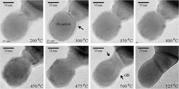
Figure 2 shows a series of bright field TEM micrographs recorded from adjacent Ni particles suspended on an amorphous carbon film. For temperatures up to 350 °C individual particles can be distinguished that are in contact with each other. Each particle, however, is surrounded by some diffuse image contrast with average intensities brighter than those from the volume of the metal particles, and darker than those observed from the amorphous carbon support. These contrast features represent continuous films covering the surfaces of the individual particles. Faceting of the nickel particles is observed with increasing temperature whereas the diffuse image contrast begins to disappear at temperatures above 350 °C. Once a temperature of 475 °C is reached, the surface layer surrounding the Ni particles can no longer be resolved, as indicated by the dashed line marking the perimeter of one of the particles in the corresponding micrograph.
Series of HRTEM images recorded at different temperatures during in situ heating experiments. At low temperatures individual Ni particles can be discerned that are covered with a surface layer (see, e.g., arrow at 300 °C). The diffuse image contrast disappears above 450 °C, as indicated by the dashed line marking the particle perimeter in the image recorded at 475 °C. Starting at 500 °C the formation of grain boundaries (GBs) can be observed.

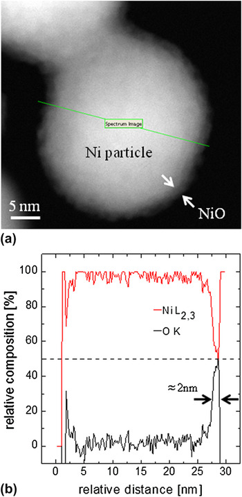
To identify the nature of the observed surface layer, EELS intensity line profiles for the O K and Ni L2,3 edges were recorded across one of the particles prior to in situ annealing. Figure 3(a) shows a high-angle annular dark-field STEM survey image of the particle in which the length and direction of the recorded profile is marked. The relative chemical composition was calculated from integrated relative EELS intensities of the O K and Ni L2,3 edges, and is plotted as a function of relative distance across the particle. The EELS intensities comprise nearly 100% nickel throughout the volume of the particle, whereas oxygen contributes insignificantly to the EELS spectrum. Within 2 nm of the particle surface the Ni:O intensity ratio is close to 55:45, indicating the presence of nearly stoichiometric nickel oxide covering the particle surface.
HAADF-STEM image of a nickel nanoparticle that is covered with a continuous surface layer (a). EELS spectra were recorded across the particle along the green line labeled “Spectrum Image.” The resulting relative composition profile obtained from integrated EELS intensities for the O K- and Ni L2,3-edges is plotted in (b). The surface layer is approximately 2 nm thick and comprises almost stoichiometric NiO.

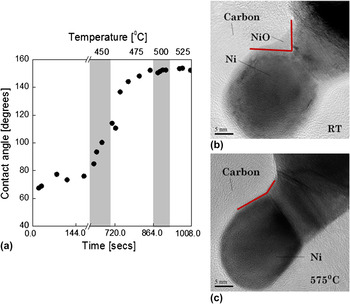
The TEM image series in Fig. 2 reveals neck formation at the contact area between the adjacent particles. Above 450 °C, a straight line with dark image contrast is observed at the contact area between two particles (marked by arrows in Fig. 2), indicating the formation of relatively flat grain boundaries (GBs). At 525 °C, several additional parallel lines with dark contrast occur in the bright field TEM image. To illustrate neck growth, the contact angle between adjacent particle surfaces was measured as a function of temperature and is plotted in Fig. 4(a). For temperatures below 350–450 °C, the angle between Ni/oxide interfaces of the neighboring particles was used to determine contact angles. Figure 4(a) shows that the contact angle remains nearly constant around 75° below 450 °C, and rapidly increases between 450° and 475 °C. Once a temperature of 500 °C is reached, the contact angle assumes a value of around 150° and remains unchanged for the duration of the experiment. Figures 4(b) and 4(c) provide a direct comparison of TEM images recorded from the particles before and after the in situ annealing experiment, respectively. The different contact angles are marked in both micrographs.
(a) Contact angle between two adjacent Ni particles as a function of annealing temperature. The angles were obtained from TEM micrographs recorded during in situ annealing. Each data point corresponds to at least local metastable equilibrium that was reached for each individual temperature setting. TEM micrographs in (b) and (c) show the investigated particle contact before and after in situ annealing, respectively.

B. Ni in the absence of amorphous carbon
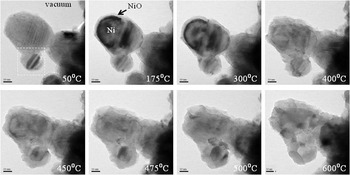
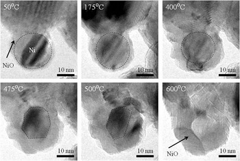
To test the influence of the presence of amorphous carbon on the reduction of the nickel oxide covering the nanometric particles, in situ heating experiments were repeated with nickel particles supported only by oxide-covered SiC films [see Fig. 1(c)]. For these experiments, the amorphous carbon support films were removed from the Aduro MEMS devices using the plasma cleaning routine described above. For particles extending over the holes in the supporting membranes, the absence of residual carbon after plasma cleaning and lack of carbon contamination during in situ TEM imaging was confirmed by EELS measurements of the C K edge intensities before and after the annealing procedure. Figure 5 shows a series TEM bright field images recorded at different temperatures. Similar to Fig. 2, the Ni particles were found to be covered by roughly 2-nm thick nickel oxide films. During in situ annealing, the oxide surface layers remain present for temperatures as high as 700 °C. During thermal annealing, the dark image intensities in Fig. 5 that arise from diffraction contrast disappear with increasing temperature. Above 300 °C the formation of interparticle contacts is observed, which indicates neck formation and growth. It can furthermore be observed that continuous NiO surface layers rearrange to polycrystalline films or partial agglomerates on the particle surfaces.
Series of HRTEM images recorded at different temperatures during in situ heating experiments in the absence of any carbon. At low temperatures individual Ni particles are covered with almost stoichiometric NiO. Above 300 °C NiO agglomerates and the metallic Ni cores of the particles can no longer be observed. The area marked by dashed lines is reproduced in Fig. 6. Scale bars for each image represent 10 nm.

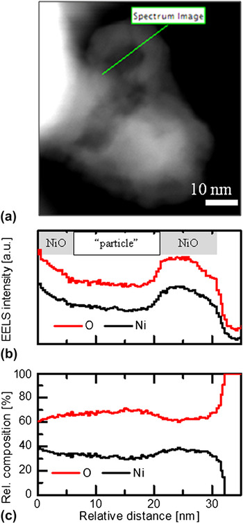
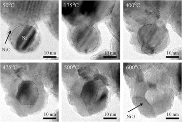
The area marked by the dashed lines in Fig. 5 is reproduced with larger magnification in Fig. 6. The oxide-covered smaller particle is in contact with significantly larger crystals that are also covered by surface oxides. With increasing temperature, the diameter of the metal core of the small particle decreases whereas the particle changes its overall shape. The TEM diffraction contrast of the metal core changes with temperature indicating possible rotation of the metal core with respect to the optical axis. At 600 °C, no pure Ni core was observed at the position where the small particle was originally located. Instead, agglomerated NiO was left behind. Figure 7(a) shows a high-angle annular dark field (HAADF) STEM image of the same particle. The incoherent HAADF image intensities suggest the presence of a shell of agglomerated NiO. To confirm this observation spatially resolved EELS intensity line profiles of the O K and Ni L2,3 edges were recorded [Fig. 7(b)]. The integrated EELS intensities plotted as a function of relative distance reveal increased nickel and oxygen signals from the remaining shell when compared with the former center of the particle. The relative composition profiles plotted in Fig. 7(c) exhibit significantly less than 50% of nickel across the entire particle with a further decreased nickel composition in the former center particle.
Series of TEM micrographs recorded during in situ annealing. At 50 °C a relatively small Ni particle is covered by a continuous surface oxide. With increasing temperature the metal core changes its diffraction contrast and becomes smaller. At 600 °C an empty NiO shell is left behind.

HAADF-STEM image (a) of the remainder of the small particle shown in the previous figure. Intensities from the incoherently scattered electrons reveal a remaining shell of NiO across which a “Spectrum Image” was recorded. The corresponding EELS intensity profiles for the O K- and Ni L2,3 ionization edges and the relative compositions are plotted as a function of relative distance in (b) and (c), respectively.

IV. DISCUSSION
A. Reduction–oxidation reactions
The in situ annealing experiments of Ni nanoparticles with a surface oxide layer of nearly stoichiometric NiO revealed the reduction of NiO at temperatures between 350° and 450 °C in the presence of carbon, whereas the oxide remained stable at temperatures as high as 700 °C in the absence of carbon. An interpretation of the experimental observations requires a detailed thermodynamic evaluation of the possible reaction equations under consideration of the oxygen partial pressures.
The dissociation reaction of NiO and the respective standard Gibbs free energy change is provided by Eq. (1).

The partial pressure for oxygen inside the TEM column is approximately p O2 = 5 × 10⁻⁹ Torr. Following Eq. (2), a calculation of the reaction equilibrium temperature for NiO reduction under such experimental conditions reveals that dissociation of NiO into solid Ni and gaseous oxygen will occur for temperatures above 920 °C.Reference Gaskell37
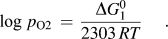
In Eq. (2)R is the universal gas constant, T is temperature in Kelvin and p O2 is the partial pressure in units of atm.
As indicated above, the presence of carbon could be playing a significant role in the observed reduction-oxidation reactions. The equilibrium temperatures and the Gibbs Free energies ΔG 0 for the following possible reactions between carbon and oxygen were considered for determining the mechanisms for NiO reduction in the presence of carbon.


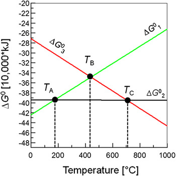
The Gibbs free energies for the reactions in Eqs. (1), (3), and (4) are plotted as a function of temperature in the form of an Ellingham diagram in Fig. 8. By plotting the variation in the molar Gibbs free energy of oxidation as a function of temperature of the various reactions, the relative stabilities are easily observed before and after the intersections of a given pair of reactions. Equations (1), (3), and (4) were subsequently used to calculate the temperatures for which the ΔG 0 curves intersect, i.e., ΔG 03 = ΔG 01, ΔG 04 = ΔG 01, and ΔG 04 = ΔG 03. Hence, the following three reactions are at equilibrium for the given temperatures:
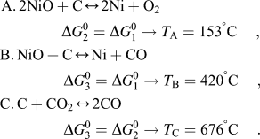

Under the given experimental conditions, i.e., vacuum environment inside the TEM with p O2 ≈ 5 × 10⁻⁹ Torr, the reduction of NiO and formation of gaseous CO2 (case A) becomes energetically favorable above 153 °C compared with unaltered NiO layers. This reaction is known as direct conversion. Once a temperature of 420 °C is reached, NiO will reduce to Ni with the formation of CO (case B), which in turn immediately transforms to CO2 below 676 °C (case C) in a process known as indirect conversion.
For the case of Ni nanoparticles supported by amorphous carbon, the TEM micrographs do not reveal any significant morphological changes below 350 °C. Instead, the complete removal of the oxide layer is observed at approximately 450 °C. As a result faceting of the nanoparticles is observed at this temperature (see Fig. 2) indicating that the associated free surface energies for nickel are smaller compared with that of the Ni/NiO interface. The disappearance of the faceting at 475 °C indicates that the roughening temperature for nickel was reached, i.e., consistent with previous reports in the literature.Reference Conrad, Aten, Kaufman, Allen, Engel, Dennijs and Riedel38, Reference Maiya and Blakely39
Above 500 °C, a stable contact angle between adjacent particles of around 150° is observed [see Fig. 4(a)]. Considering free surface energies for Ni of 1.9 J/m2,Reference Maiya and Blakely39 the corresponding grain boundary energy for the observed particle geometry is estimated as 0.98 J/m2. This value is slightly larger than general grain boundary energies estimated by Li et al.Reference Li, Dillon and Rohrer40 due to the rather unusual two-particle geometry present in the reported experiment.
NiO reduces to metallic Ni in the presence of carbon through a multistep reduction-oxidation process. NiO starts reducing at low temperature according to case A while the reduction reaction is likely kinetically limited. With increasing temperature, more oxygen becomes available through continuous reduction of NiO. Gaseous CO is formed following case B. According to the Gibbs free energies plotted in Fig. 8 carbon monoxide immediately oxidizes to CO2 and escapes into the vacuum of the TEM instrument. The experimentally observed temperature ranges for reduction of the metal oxide are in excellent agreement with the thermodynamic calculations. This result was somewhat unexpected considering that the Gibbs free energy calculations are strictly valid only for bulk materials and do not consider the kinetics of the redox reactions for Ni-NiO core-shell structures with nanometer dimensions. However, the relatively large surface-to-volume ratio for nanometric powder particles significantly increases the amount of work required to completely desorb the surface oxide layer. This purely thermodynamic argument may be complementary to the kinetic limitation discussed above.
B. Role of carbon during consolidation
TEM imaging during thermal annealing of Ni particles dispersed in amorphous carbon revealed that the surface oxide layer starts to disappear around 350 °C, whereas the complete removal of nickel oxide was observed for temperatures between 450° and 475 °C. Above these temperatures, rapid neck formation occurred as shown by the observed changes of the contact angle (see Fig. 4).
For the oxygen partial pressures inside the TEM instrument, a reduction of NiO to metallic Ni is expected only above 920 °C. In addition, annealing experiments carried out in the absence of additional carbon reveal that the oxide surface layers agglomerate into polycrystalline NiO, which remains stable for temperatures as high as 600 °C. The experimental observations hence provide conclusive and direct experimental evidence that the presence of amorphous carbon is highly effective in reducing surface oxides from nickel particles at relatively low temperatures. Oxides are transferred into the gas phase and can escape the particle agglomerate when placed in a vacuum. The TEM data indicate that, in the absence of amorphous carbon, oxygen is not transferred into the gas phase but rather remains as agglomerated NiO (see Figs. 5–7). This result further demonstrates that the underlying SiO2/SiC membrane does not contribute to the reduction of NiO.
In the case of neighboring particles with significantly different sizes [see Fig. 7(a)], sufficient driving force exists to cause Ni self-diffusion from the smaller particle through NiO to be absorbed by the larger particle, i.e., Ostwald ripening.Reference Hassen41 For similarly sized particles, however, some neck formation is observed although not as extensive as in the presence of carbon (see Fig. 5). Such behavior is consistent with an earlier study by Munir,Reference Munir24 who reported that NiO does not limit the transport of nickel between particles during conventional sintering. It is therefore concluded that the presence of carbon promotes metal neck formation and growth with subsequent densification after reduction of NiO. In the absence of carbon, however, NiO is retained and inhibits the formation and/or growth of pure metal necks. The experimental observations hence suggest that the rate limiting process for neck formation and growth between the oxide-covered nickel particles is the oxygen- rather than Ni mobility in NiO.
The observed coalescence of nickel particles via in situ TEM does not represent sintering in the traditional sense since the particle arrangement during the experiment is not three-dimensional and only small particle agglomerates are considered. It does however suggest that the role of the oxide in nanometric metal sintering has implications for the natural competition between coarsening and densification. The TEM results and subsequent thermodynamic analysis of possible redox reactions demonstrate that the presence of amorphous carbon shifts the equilibrium temperature for the reduction of NiO to significantly lower temperatures. The oxygen partial pressure at the surface of the nanoparticles is mostly controlled by the presence or absence of carbon and not the dynamic vacuum conditions in either the TEM or a macroscopic SPS experiment. Based on the in situ TEM results it is therefore reasonable to predict that the presence of carbon in traditional sintering experiments will enable lower consolidation temperatures during the early stages of sintering, and, hence, accelerate densification. In addition, the presence of the metal oxide, often considered to limit grain boundary mobility during consolidation, promotes the coarsening of the nanometric powder at the expense of densification. The level of carbon impurities of 0.3 wt% for the metal powder used in this study further indicates that the addition of amorphous carbon to the green powder may be necessary to activate the redox processes observed in this study.
V. CONCLUSIONS
This study reports in situ TEM experiments, in conjunction with thermodynamic calculations, on the reduction of surface oxides on nickel nanoparticles. In the presence of amorphous carbon, the equilibrium temperature for the reduction of the surface oxide on nickel nanoparticles is significantly reduced by several hundred centigrade as compared with the case when no carbon is present. Experimentally observed reduction temperatures are in excellent agreement with predictions from thermodynamic calculations considering redox reactions between NiO and carbon. The chemical reduction of the surface oxide was found to precede neck formation between individual particles during in situ thermal annealing. In the absence of carbon, particle coalescence occurs at significantly higher temperatures through Ostwald ripening concurrently with agglomeration of remaining NiO. It is concluded that the presence of “free carbon” during consolidation processing, i.e., sintering, of polycrystalline microstructures can significantly alter the required temperatures necessary during the initial stages of densification.
ACKNOWLEDGMENTS
This study was partially funded through UC Davis start-up funds and a young faculty early career award by the U.S. National Science Foundation (DMR-#0955638). M.M. acknowledges financial support from the Japan Student Services Organization (JASSO). T.B.H. and A.K.M. were supported by the Office of Naval Research (Program Manager Dr. L. Kabacoff) under Grant No. N00014-10-1-0632