




No CrossRef data available.
Article contents
- Abstract
- Nomenclature
- Definition
- Morphology and histology of isolated left ventricular noncompaction
- Comparative vertebrate morphology
- Left ventricular noncompaction: an acquired or congenital cardiac malformation
- Incidence
- Genetics of left ventricular myocardial noncompaction
- Association with congenital cardiac disease
- Diagnostic techniques to image isolated left ventricular noncompaction
- Clinical findings in patients with isolated left ventricular noncompaction
- Ventricular noncompaction and neuromuscular disorders
- Determinants of severity and timing of presentation of patients with left ventricular noncompaction
- Differential diagnosis
- Treatment and outcome
- Conclusions
- Acknowledgements
- References
The morphological spectrum of ventricular noncompaction
Published online by Cambridge University Press: 13 July 2005
Article contents
- Abstract
- Nomenclature
- Definition
- Morphology and histology of isolated left ventricular noncompaction
- Comparative vertebrate morphology
- Left ventricular noncompaction: an acquired or congenital cardiac malformation
- Incidence
- Genetics of left ventricular myocardial noncompaction
- Association with congenital cardiac disease
- Diagnostic techniques to image isolated left ventricular noncompaction
- Clinical findings in patients with isolated left ventricular noncompaction
- Ventricular noncompaction and neuromuscular disorders
- Determinants of severity and timing of presentation of patients with left ventricular noncompaction
- Differential diagnosis
- Treatment and outcome
- Conclusions
- Acknowledgements
- References
Abstract
An abstract is not available for this content. As you have access to this content, full HTML content is provided on this page. A PDF of this content is also available in through the ‘Save PDF’ action button.
Information
- Type
- Continuing Medical Education
- Information
- Copyright
- © 2005 Cambridge University Press
The recently recognized myocardial malformation known as ventricular noncompaction is a fascinating disorder, characterized by the presence of an extensive trabeculated myocardial layer reinforcing the luminal aspect of the compact portion of the ventricular wall. Usually described thus far in the left ventricle, it is typically associated with abnormal systolic function, albeit also recorded with restrictive physiology and segmental hypokinesis.1–5 Seen most frequently in the structurally normal heart, increasing recognition shows that the lesion can also accompany diverse forms of congenital cardiac disease (Figs 1 and 2).1–10 When seen in the left ventricle, it is currently considered an unclassified form of cardiomyopathy, being neither dilated, hypertrophic, nor restrictive.2, 3, 11, 12 Although once considered rare,13–15 it is now being seen with increasing frequency, from the fetus to the adult.16–29 Diagnosis has now moved from the autopsy table to recognition during life, albeit the debate continues with regard to the features displayed by angiography, echocardiography, computed tomography, and magnetic resonance imaging that permit unequivocal recognition. In this review, we will consider myocardial noncompaction from the stance of comparative vertebrate biology, as well as discussing the genetic, developmental, clinical, and imaging characteristics of patients with noncompaction, seen both in isolation and in the setting of congenital cardiac disease. In the latter setting, we must ask whether noncompaction is a homogeneous process that, in its many clinical and pathological expressions, can be explained on the basis of a developmental abnormality. In this respect, it is of interest to note that, in the evolution of the vertebrates, myocardial noncompaction is advantageous, and indeed necessary, for the circulatory function of some fish, and yet its presence is decidedly disadvantageous for man.30–32
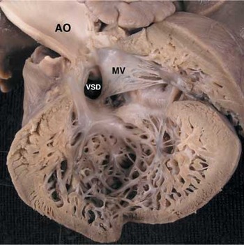
Figure 1. Left ventricular noncompaction in an infant with complex congenital heart disease and isomerism of the left atrial appendages. The left ventricular wall shows extensive intertrabecular spaces and many deep recesses. Note that there is also a ventricular septal defect (VSD). AO: aorta; MV: mitral valve.
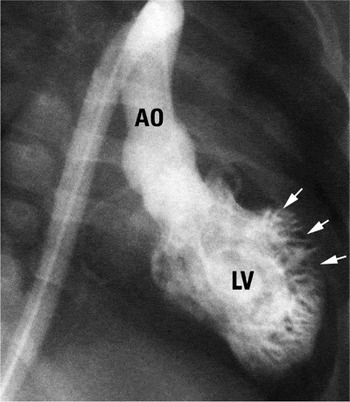
Figure 2. The angiographic appearance of left ventricular noncompaction in a neonate with critical aortic stenosis. The left ventriculogram (LV) in right long axis oblique views shows an extensive noncompacted layer (arrows) containing numerous trabeculations. AO: aorta.
Nomenclature
It was Dusek et al.33 who provided one of the earliest substantial descriptions of the entity that probably represents what we now call ventricular noncompaction. They called it postnatal persistence of spongy myocardium with an embryonic blood supply, albeit that other individual examples of the same lesion had already been published. The five hearts described by Dusek et al.,33 however, are far from the isolated cases that provoke discussion nowadays. All came from infants, and none was “isolated”. One case involved a cardiac fibroma, another came from a neonate with critical aortic stenosis, a third had pulmonary atresia with intact ventricular septum and so-called obliterative fibroelastosis of the coronary arteries, while the final two cases had abnormal origin of the left coronary artery from the pulmonary trunk. In their discussion of these then peculiar findings, the authors observed that the persistent trabecular network forming the luminal aspect of the ventricular wall resembled the characteristic arrangement of the myocardium of cold-blooded vertebrates. Subsequent to this initial description, many others, noting the presence of these changes, have again invoked the concept of evolutionary persistence of the spongy myocardium typically seen in the ventricular walls of some fish, amphibians, and reptiles.21, 30–32 Since the original description of the spongy arrangement, many other terms have been used to describe the abnormal morphology, including spongiform cardiomyopathy, myocardial sinusoids, hypertrabeculations, embryonic myocardium, anomalous ventricular myocardial patterns, dysplastic cardiac development, isolated left ventricular abnormal trabeculation, myocardial dysgenesis, ventricular dysplasia, honey-combed ventricle, as well as still other miscellaneous offerings.1, 5–10, 32–35 Until the last decade, it was the designation of “spongy myocardium” that held sway, although less so more recently. It is over the past 10 years or so that the lesion has come to be described in terms of myocardial noncompaction, or hypertrabeculation,1–5 rather than spongy myocardium. Indeed, Pignatelli et al.29 now suggest that the very concept of the “spongy myocardium” has fallen into disfavour, and has largely been abandoned, because it suggests that the basic morphogenetic abnormality may represent an arrest of the normal compaction of the loosely interwoven mesh of trabeculated myocardial fibres seen in the embryo. If this is the case, then as we will discuss, perhaps the concept has been discarded prematurely. Looking back at this change of emphasis, nonetheless, Chin et al.1 should be credited with being amongst the first to introduce the concept of “isolated noncompaction of left ventricular myocardium”. Description of the abnormal endomyocardial layers seen in this setting, however, transcends the issues of nomenclature or semantics. The description must be morphologically correct. Thus, as we have discussed above, some have used the designation of “myocardial sinusoids” to account for the appearance. This is unequivocally incorrect. As Gittenberger-de Groot36 has shown, myocardial sinusoids are thick-walled and distended intertrabecular myocardial spaces that connect with the subepicardial coronary arteries through a more-or-less distended capillary bed. Such lesions are typically seen in the setting of pulmonary atresia with intact ventricular septum. Indeed, deep recesses extending from the right ventricular cavity are a feature of this lesion when there is overgrowth of the ventricular cavity by mural hypertrophy. This can produce a “spongy” appearance, and one such case was included in the original series of Dusek et al.33 These changes, however, are markedly different from the entity now described as noncompaction (Fig. 1). Histological examination also reveals a lesser form of the acquired changes typical of pulmonary atresia with intact septum in the setting of hypoplasia of the left heart.37, 38 As with pulmonary atresia with intact septum, these lesions are again morphologically distinct from left ventricular noncompaction. Tsang and Chiu39 have made an extensive historical review of so-called myocardial sinusoids in the context of developing strategies for myocardial revascularization, but these are also distinct from the lesion to be discussed in our review. Due to the ongoing uncertainty regarding its relationship to “spongy myocardium”, we will describe the entity only as ventricular noncompaction.
Definition
The ventricular walls of both ventricles in the normal heart are made up predominantly of a compacted layer of myocardial fibres set in a matrix of supporting connective tissue.40 The luminal surfaces of the ventricles, nonetheless, show evidence of marked trabeculation, particularly at the ventricular apexes, with the trabeculations being appreciably coarser in the morphologically right than in the left ventricle (Figs 3 and 4). Careful examination of the normal ventricles also reveals marked variability in the distribution of the trabeculations, with the area around the bases of the papillary muscles of the mitral valve in the left ventricle, for example, showing a greater proportion of the wall to be trabecular rather than compact. The proportion of the normal ventricular wall made up of interlacing trabeculations never exceeds in thickness the part that is normally compacted, albeit that our recent findings have shown a spectrum of noncompaction in otherwise normal hearts.41 It is also the case that, early in development of the human heart, the area of the wall that is compacted makes up the minority of the full thickness, the larger part of the wall showing a trabecular arrangement remarkably similar to the abnormal pattern we now describe as ventricular noncompaction (Fig. 5). In postnatal life, the abnormal pattern can be recognized when the thickness of the trabecular layer exceeds that of the compact layer. Thus far, however, attention has been focused on the excessive number of abnormally conspicuous ventricular myocardial trabeculations, with deep intertrabecular recesses, confined primarily to the apical and mid-ventricular segments of the left ventricle.1–5, 12, 18, 23, 29 As we will discuss, there is both genetic and morphological heterogeneity in patients exhibiting myocardial noncompaction.42 Undue focus on these trabeculations has resulted in the appearance of definitions based on the number of prominent coarse trabeculations seen within a given area of myocardium, such as that proposed by Boyd et al.43 subsequent to their study of the left ventricular architecture of the normal heart. Such excessive trabeculations have been noted extending apically beyond the papillary muscles, albeit that they have the same echogenicity as the myocardium. The abnormal trabecular layer has been noted to move synchronously with the compact myocardium, and those observing the lesion have pointed out that it should not be attached to the papillary muscles,1–5, 12, 18, 23, 29 but as we have already emphasized, the persisting trabecular layer in the normal heart is always thickest adjacent to the papillary muscles. Some have incorrectly described these spaces within the trabecular layer as persisting sinusoids, and considered them to be enlargements of the coronary vessels. In fact, the recesses neither communicate nor connect with the coronary circulation. The recesses are covered by endocardium, and are directly continuous with the overall endothelial lining of the ventricular cavity (Fig. 6). They can serve as harbingers for focal formation of thrombus, especially when left ventricular function is compromised.1–5, 12, 18 When seen in sectional format, the end result is to produce a bilayered arrangement (Fig. 6), with the trabecular layer being at least as thick, or even thicker, than the compact layer of the ventricular wall.1–5, 12, 18 It is, of course, also necessary to distinguish this abnormal bilaminar appearance from the normal presence of false tendons and criss-crossing apical trabeculations, these being normal variants of left ventricular anatomy.44–55 These normal trabeculations never produce a discrete trabecular layer rivalling in thickness the compact layer.

Figure 3. The normal right ventricle has been opened to show its inlet, apical trabecular, and other components. The coarse apical trabeculations form the luminal surface of the compact myocardium.

Figure 4. The left ventricle is shown from the same heart that contained the right ventricle seen in Figure 3. The apical trabeculations are characteristically fine and criss-crossing when compared to the coarse trabeculations of the right ventricle (Fig. 3). The trabeculations, nonetheless, are no more than the luminal surface markings of the layer of compact myocardium.

Figure 5. This series of sections, from human embryos (A) at Carnegie stage 16, (B) at stage 18, and (C) immediately after the close of the embryonic interventricular foramen, show that, during development, there is an extensive trabecular layer which forms the greater part of the thickness of the ventricular wall when compared to the extent of the compact layer. It is also the trabecular layer which becomes compacted to form the papillary muscles of the atrioventricular valves (asterisks).
At this stage, the World Health Organization has still to recognize isolated left ventricular noncompaction, defined in the fashion suggested above as presence of an unexpectedly prominent trabeculated myocardial layer within the ventricular wall, as a distinct and separate form of cardiomyopathy. It is still considered, therefore, to represent an unclassified cardiomyopathy.11 It is possible that this may eventually change.2, 3, 12, 13 And while some consider left ventricular noncompaction in the human to be a developmental abnormality,1–5, 30–33 this view is not universally supported. Others consider the condition to be acquired postnatally, at least in some patients.5 Our own opinion was initially to support the concept of a postnatally acquired disease, but as we will show, there is now much evidence to support the concept that noncompaction does, indeed, represent failure to resorb the extensive trabeculated part of the ventricular wall seen during normal embryological development, and illustrated in Figure 5.
Morphology and histology of isolated left ventricular noncompaction
Hearts exhibiting the features of unequivocal left ventricular noncompaction possess a luminal meshwork of interlacing thin and thick endocardial bands, tendons, filaments, and trabeculations that intermingle with each other to form a discrete trabeculated layer of the ventricular wall that extends from the midportion of the left ventricle to its apex. Within this trabecular layer, it is frequently possible to find thick and prominent coarse trabeculations separated by deep intertrabecular recesses.1–5, 18, 23, 30, 33, 35, 56–58 Histological examination shows that the deep recesses are lined by endothelium (Fig. 6), which extends close to the surface of the persisting compacted ventricular layer. The histological examination also shows that the trabeculations are covered by excessive fibrous and elastic tissue, perhaps suggestive of some degree of subendocardial ischaemia (Fig. 6).1, 59–62 This arrangement is readily distinguished from the normal but prominent left ventricular trabeculations observed especially in the hearts from patients with left ventricular hypertrophy in and out of the setting of hypertrophic cardiomyopathy.43–55
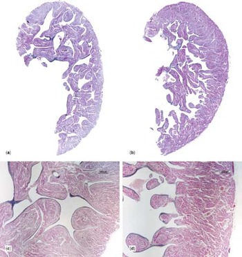
Figure 6. The sections shown in panels (a) and (b) are whole mounts of right and left ventricle, respectively, from the heart shown in Figure 1. There are many intertrabecular spaces with many deep recesses. Higher power histological sections, as seen in panels (c) and (d) show deep and numerous intertrabecular recesses that extend almost to the epicardial surfaces. Note the fibroelastic layer that enclosed the trabeculations (Movat pentachrome stain).
Comparative vertebrate morphology
The heart is the first organ to form and function in the vertebrate embryo.63–72 In this respect, Kirby69 has written: “Heart development in all vertebrates from fish to humans follows the same general pattern: fusion of the myocardium and endocardium in the ventral midline to form a simple tubular heart, onset of function, looping to the right side, chamber specification and formation, and finally development of specialized conduction tissue, coronary circulation, and mature valves”. It is important to remember that the fish ventricle is not septated.66–68 The ventricle is also undivided in all Amphibia.66, 67 The ventricular cavity in Amphibia, like in the fish, is compartmentalized by a number of parallel trabeculations of about equal height.68 The situation is more complex for the three major groups of reptiles than that observed in either fish or amphibians.66–68 In both Squamata (snakes and lizards) and Chelonia (turtles), the horizontally positioned ventricles are partially but not completely septated. The atriums connect with the left ventricle in both of these groups and blood crosses the incompletely partitioned ventricular septum from left-to-right. In the crocodilian heart, in contrast, the ventricular septum is vertically positioned and complete.66–68 The right atrium of the crocodile opens into the right ventricle, and the left atrium into the left ventricle. The fully formed four-chambered heart of the bird and human appear similar although there are developmental differences.66, 67 Hearts of man, mouse, and rats are very similar in structural design,63 although the right ventricle of the rat is less trabeculated than the human right ventricle. In the consideration of myocardial noncompaction in the different groups of vertebrates, it may be important to remember in which vertebrate species the ventricles are septated, and in those species that the ventricles are not septated, or incompletely so. The coordination of patterning and morphogenesis is the key to eventual successful formation of the definitive organ. Trabeculation is just one such process. In its earliest incarnation, the heart is a simple tube, composed of two concentric layers: an outer muscular and an inner endothelial layer. During the morphogenetic process of myocardial trabeculation, most of the cardiac jelly is replaced by sponge-like muscle.63–71 The complex process of trabeculation is not confined just to ventricular myocardium,73–79 although it is the ventricles which are the focus of our review. Atrial myocardium also passes through a process of trabeculation, albeit occurring somewhat later than ventricular trabeculation.63, 80 The highly organized developmental process of ventricular trabeculation changes at every stage of cardiac morphogenesis.81 Some consider the trabeculated ventricular myocardium as “finger-like projections”,82 but Sedmera and Thomas take issue with this view. Their investigations76 show that the embryonic trabeculated ventricular myocardium resembles sheets of a sponge-like structure, with no “finger-tips”, features clearly revealed either by scanning electron microscopy or other techniques involving three-dimensional reconstruction. The arrangement is also to be seen in sections taken from the developing human embryo, where prior to the completion of septation, the ventricular walls possess a relatively thin compact myocardial portion, and an appreciably thicker trabecular layer (Fig. 5). When this arrangement persists in the postnatal heart, then as has been stated in numerous publications,1–5, 12, 17–19, 30–33, 63–71, 73–81 the situation strongly resembles the spongy myocardium seen in nonmammalian vertebrates, including fish, amphibians, and reptiles. In adult homiotherms, nonetheless, the entire ventricular wall is made up of compact musculature, fed with blood through the coronary arterial supply.83–86 In adult poikilotherm animals, in contrast, the cardiac musculature consists primarily of the spongy type, possessing an outer compact layer which is typically thin.87–96 Almost 30 years ago, Basile et al.97 wrote citing Poupa: “This type of morphological organization of the myocardium seems to represent a complex transitory step during the evolution of vertebrates, the spongy myocardium of the most primitive poikilotherms being replaced in the higher species by a compact coronary-supplied muscle”. The spongy-like myocardium is supplied predominately by diffusion of blood from the ventricular lumen that flows into the intertrabecular spaces.83–97 Thus, in the opening paragraph of Grant's paper, “The comparative anatomy of the cardiac coronary vessels”, referring to the development of the coronary vessels in the rabbit,98 he states: “It is there shown that the heart is at first nourished by the blood contained in the intertrabecular spaces of the myocardium and that this intertrabecular system is modified during the course of development and incorporated with the coronary vessels”. In some fish, the compact and the spongy layers of the myocardium are not completely compartmentalized. Tota et al.92 have demonstrated using India-ink injections and neoprene casts that, in some fish, there is an extensive network of arterio-luminal vessels connecting the coronary bed with the lacunary circuit of the spongy myocardium. They suggest that this is the first evolutionary step in the development of the thebesian system. Thus, in certain species of fish, the rich spongy trabecular myocardium is necessary, indeed, advantageous for diffusion of gases.
It is fishes that show the highest overall morphological diversity among all the vertebrates. In fish, the cardiac anatomy, and ventricular shape, varies considerably according to the workload and the circulatory demands. Morphological studies reveal a direct relationship between lifestyle, myoarchitecture, and the model of ventricular blood supply in teleost fish, these being the typical bony fish with almost completely ossified skeletons.93–95 In the developmental hierarchy of the fish, not only is there evolution in the shape of the heart, but also in the architecture of the myocardium.93–95 In this respect, Ostadal,84 more than three decades ago, wrote how “changes in the inner structure of the myocardium during phylogenetic development are accompanied by changes in its blood supply”. The ventricle of the teleost fish may be saccular, tubular, or pyramidal.87–97 Sedentary species tend to have saccular or tubular ventricles, this shape being suited to a large stroke volume with low systemic impedance, while the active fish have a pyramidal ventricle, with a better developed compact layer. Like all vertebrates, nonetheless, the piscine myocardium requires a circulatory supply to provide oxygen. There are two mechanisms to provide the supply of oxygen in fish hearts, and they are developed to different degrees amongst different species. One method is for the oxygen to be derived by diffusion from the relatively oxygen-poor venous blood that bathes the ventricular endocardium via the trabeculations and lacunar spaces. The second method is to supply oxygen-rich blood through a coronary arterial circulation to the compact myocardium.84, 87–97 In those fish with the primarily lacunary form of blood supply, it is in large part both the gradient in tensions of oxygen between the luminal blood and the myocardium, as well as the distance over which diffusion must take place, that determines the degree of oxygenation of the cardiac ventricular mitochondrions.91, 99, 100 Cyclostomes, and most inactive fishes, have none or few coronary arterial vessels, and thus use exclusively diffusion of the venous supply of oxygen to feed the myocardium. As discussed, their ventricles tend to be tubular or saccular. Other fish, including also the elasmobranches, which have cartilaginous skeletons, use both diffusion though the trabeculations and supply through coronary arteries to supply oxygen in their pyramidal hearts.87–97 Hence, abundant trabeculations are the essence of the tubular or saccular hearts, with the ventricular myocardium having a very thin compact layer in the absence or near absence of any coronary vasculature. The hearts of fish such as the tuna, in contrast, have thick and well-vascularized compact myocardial layers, with a spiral arrangement of the cardiac myocytes.90, 97 Santer and Greer Walker101 attribute these differences to be conditioned by lifestyle, with the active species having the thicker compact layers.
The spongy, or predominantly trabeculated, myocardium as found in most teleosts seems to be particularly suitable for performing volume work. A good example is the heart of the icefish, an Antarctic species of the family Channichthyidae. These fish, once named by Norwegian whalers as the “bloodless fish”, are found primarily in the Southern Oceans surrounding Antarctica. They lack the ability to synthesize the respiratory pigments.102 The blood of these Antarctic fishes, therefore, is completely devoid of not only haemoglobin but also, for the most part, lacks red blood cells.102–107 These fish were also initially thought additionally to lack myoglobin, in both their cardiac and skeletal muscle. More recent studies, nonetheless, have shown that there is variable expression of myoglobin, and its messenger ribonucleic acid, within the ventricular myocardium in some of the species.106–111 The skeletal muscle is devoid of myoglobin. In those species lacking both haemoglobin and myoglobin, the larger part of the volume of the cardiac myocytes is occupied by mitochondrions. The nearly colourless blood of these teleosts carries oxygen only in physical solution, and thus the arterial capacity is reduced to 0.67 volumes per cent.102–106 There are also marked differences between the species in the amount of compact muscle as opposed to trabecular myocardium.99, 100, 103, 105, 106 The evolution of the icefish to the haemoglobinless phenotype is likely to have occurred due to direct deletion of genes, or through a multi-step process involving transcriptional inactivation of the adult and the embryonal globin complexes, followed by elimination of the nonfunctional genes themselves.110, 111 In those icefish that maintain an expression of myoglobin, it is unclear whether this respiratory pigment has a role in facilitating the transport or storage of oxygen at cold body temperatures.107 In this regard, however, there is some experimental evidence derived from study of isolated and perfused hearts that those species expressing myoglobin retain a functional advantage.107 It is likely that the high mitochondrial densities within hearts from those species lacking oxygen-binding proteins may help maintain the flux of oxygen by decreasing the distance required for diffusion between the ventricular lumen and mitochondrial membrane.99, 100 Still, the ventricle of the icefish is characterized primarily by trabeculated muscle, albeit with some myocardial pseudo-hypertrophy. It functions as a specialized volume pump moving large stroke volumes at a low heart rate, but is not able to produce high pressures, and thus cannot tolerate high systemic impedance. The icefish heart, in its adaptation to the stable low temperature and high content of oxygen of the Antarctic waters, demonstrates a relatively increased heart weight, high ventricular compliance, and low systemic impedance.102–106 Interestingly, the volume of blood in the icefish ranges from 6 to 7 times that of its body weight, this being from 2 to 4 times higher than seen in other teleosts.102–106 This dramatic increase in volume of blood ensures that an adequate amount of oxygen is carried in solution in the plasma.104, 106 Loss of haemoglobin in the icefish, therefore, correlates with increased trabeculation of the ventricle, this conferring a distinct evolutionary advantage.103–112
The most active teleost fish, such as the tuna, have cardiac ventricles with greater thicknesses of compact myocardium.87, 90, 92–95, 97, 101 The increased ratio of compact myocardium gives the ventricles of these fish the potential to act as pressure pumps. They can move small volumes of blood at a relatively high rate and pressure. The thickness of the trabecular layer, therefore, represents a compromise between minimizing the distance over which diffusion must take place, and maximizing the cross-sectional area for development of tension.87 Interestingly, in some fish with intermediate ratios of compact to spongy myocardium, diffusion of blood from the spongy myocardium permits survival following ligation of the entire coronary arterial system, thus demonstrating the functional adaptation of this form of cardiac muscle.87
In higher vertebrates, of course, the heart has developed from a simple tube into a complex organ with four chambers, specialized for efficient pumping at pressure. During this evolutionary change, there has been a concomitant change in the level of myocardial organization. The emergence of trabeculations in the luminal layers of the ventricles during their development permits the myocardium to increase its mass prior to the appearance of any discrete coronary circulation. After looping of the heart tube, these trabeculations are first seen within the inner myocardial layers (Fig. 7) as the ventricles balloon from the greater curvature.73–80 With ongoing development, the epicardial layer of the apical pouches becomes solidified to form the compact component of the ventricular myocardium (Fig. 5). The luminal layer at this stage, nonetheless, retains its trabeculations, producing patterns that are specific both for the right and the left ventricles, and for different species. In the chick, these changes begin between stages 16 and 17, in the mouse at 10.5 days of gestation (Fig. 7), and in humans at the end of the fourth week of gestation, thus corresponding to Carnegie stage 12 (Fig. 5). According to Icardo and Fernandez-Teran,78 trabeculation is the expression of three closely related events, namely the formation of endocardial outgrowths that eventually invade the myocardium, followed by the development of large intercellular spaces between the myocytes, and completed by a decrease in the thickness of the cardiac jelly. These authors, along with Sedmera et al.,75–77, 80 have discussed in detail the developmental aspects of trabeculation in the embryonic chick heart. Sedmera et al.113, 114 have also studied the ventricular myoarchitecture in the chick embryo under different conditions of loading. They found that a decrease in loading was associated with formation of a thinner compact layer, and persistence of the trabecular layer. Hu et al. have also studied the formation of trabeculations in the heart of the zebra fish, this species lending itself neatly for study by the developmental biologist both because of its optical clarity and the large number of mutants in its make-up.115 There is extensive early ventricular trabeculation in the zebrafish that persists into adulthood. Passive myocardial stiffness can change subsequent to remodelling of the myocardium, which includes an increase in the thickness of the trabecular layer during development.115 The genetic regulation of cardiac trabeculation, nonetheless, remains far from completely understood. King et al.116 have studied the expression of Peg 1 in the developing mouse heart, and have shown that this gene is involved in cardiac trabeculation. Peg 1, or Mest, is an imprinted gene widely expressed in the mouse embryo, and is found primarily in cells of mesodermal lineage. Analysis of myocardial morphology in viable mice lacking the Peg 1 gene demonstrates subtle variations in the patterns of ventricular trabeculations, with an increase in thickness of the trabecular layer and reduction in density of the compact myocardium, producing an appearance, they suggest, which is remarkably similar to isolated left ventricular noncompaction.

Figure 7. This scanning electron micrograph shows the arrangement of the developing myocardium in the mouse heart as the apical components of the right and left ventricles balloon from the primary heart tube. Specimen prepared and reproduced by kind permission of Professor Nigel Brown, St George's Hospital Medical School, London.
Thus, the transition from a spongy myocardium, supplied by intertrabecular venous blood bathing the endocardium from the ventricular lumen, to a compact ventricular muscular wall supplied by a discrete coronary arterial circulation, characterizes the evolutionary history of the vertebrate myocardium, but also occurs during ontogeny in homiotherms.83, 85, 86
Left ventricular noncompaction: an acquired or congenital cardiac malformation
Many of those who first encountered and described left ventricular noncompaction in isolation considered it a developmental abnormality, reflecting either impaired or arrested compaction of the developing myocardium.1–5, 30–33 Others, such as Stöllberger et al., in contrast, suggest that other pathogenetic processes should not be neglected, particularly whilst there is insufficient evidence to prove the developmental hypothesis.5 They argue that other pathogenetic processes could be responsible for non-compaction, including dissection of the myocardium, frustrated attempts of myocardial hypertrophy, myocardial tearing caused by dilation, a metabolic defect, or compensatory hypervascularization.5 Some of the evidence cited to refute the concept that noncompaction is a developmental abnormality comes from serial echocardiographic studies in which the disorder was allegedly not recognized in an initial study, becoming evident and diagnosed only at subsequent studies. This, of course, depends on the accuracy of the initial diagnostic techniques, as well as the intensity and comparability of the serial investigations. There is now substantial evidence emerging, however, which supports the developmental hypothesis. Thus, investigation of a large number of subjects using magnetic resonance imaging,41 including some known to have noncompaction and their family members, shows a spectrum in the extent of the thicknesses of the trabecular and compact myocardial layers, with different ratios found in different parts of the ventricle of different individuals. This suggests strongly that unequivocal noncompaction is the endpoint of a bell-shaped curve involving removal of the extensive trabecular layer known to exist in the early stages of human ventricular myocardial development (Fig. 5). In this respect, the phenomenon of partial expression of noncompaction in a family with autosomal dominant transmission has recently been described.117
Incidence
Once considered a rare form of myocardial disorder,13–16 isolated left ventricular noncompaction is now being recognized with increasing frequency, with various echocardiographic studies reporting its finding in between 0.05 per cent and 0.24 per cent of all investigations.1, 5, 19, 20, 23, 29 On this basis, some have justifiably asked whether this disorder is rare, or simply underdiagnosed.16 It is almost certainly the latter, particularly when we consider the variability in noncompaction now known to exist within the different segments of the left ventricle. It was Ritter et al.20 who suggested that the incidence was 0.05 per cent in adults. Pignatelli et al.,29 from the Texas Children's Hospital, reviewed about 50,000 echocardiograms performed on just over 26,000 patients from 1997 to 2002, and found 36 patients with the isolated form of the malformation. Over this period, the same group diagnosed 344 cases of cardiomyopathy, so that noncompaction accounted for one-tenth of their cases of cardiomyopathy, a proportion very similar to that calculated by Nugent et al. in Australia.118 Oechslin et al.23 estimated that isolated left ventricular noncompaction was seen in 0.0014 of all patients referred to their department of echocardiography over a period of 14 years. Although these contemporary data suggest that isolated left ventricular noncompaction is an extremely rare form of cardiomyopathy, we must now accept that it is being recognized with increasing frequency, so we may need to revise our views in the light of emerging findings.
Genetics of left ventricular myocardial noncompaction
Ichida et al.19 reported several years ago that, in patients with isolated left ventricular noncompaction, familial recurrence is high, being found in approximately two-fifths of patients, a conclusion shared by others.1, 2, 5, 23, 24 Analysis of genetic linkage and mutation has revealed that mutations in the gene G4,5, which encodes taffazin, and maps to chromosome Xq28, are responsible for this myocardial disorder in some patients, being allelic with Barth syndrome.119–125 In this regard, G4,5 was initially identified as the gene responsible for Barth syndrome, an X-linked mitochondrial disease affecting cardiac and skeletal muscle.126, 127 These mutations produce a wide phenotypic spectrum of cardiomyopathies, including dilated cardiomyopathy, X-linked infantile cardiomyopathy, and X-linked endocardial fibroelastosis.125 Ichida et al.19 found a mutation in α-dystrophin in some of their patients. Chen et al. found a novel splice acceptor site mutation of intron 8 of G4,5 in one family with severe X-linked left ventricular noncompaction, but without the other usual findings of Barth syndrome.123 Pauli et al.128 identified deletion of chromosome 5q in a child with previously repaired congenital heart disease, facial dysmorphism, and left ventricular noncompaction. Vatta et al.,129 in contrast, have shown that, in some patients with either dilated cardiomyopathy or left ventricular noncompaction, a mutation in Cypher-Zasp, a gene encoding a protein that is a component of the Z-line in both skeletal and cardiac muscle, may be causal. It may also be pertinent that mice lacking FKBP12 have normal skeletal muscle, but have a severe dilated cardiomyopathy and a condition suggestive of left ventricular noncompaction.130
Sasse-Klaassen, Kenton et al.117, 131 have reported that, in adults identified with the sporadic form of isolated left ventricular noncompaction, it is an autosomal dominant disorder rarely caused by mutations in G4,5, and hence is genetically distinct from the X-linked cases seen in infancy. More recently, Sasse-Klaassen et al.,132 studying the pedigree of a family with an autosomal dominant form of left ventricular noncompaction, mapped the responsible gene to chromosome 11p15. Interestingly, this study describes family members with a partial expression of the phenotype for noncompaction. There is also some tenuous evidence that left ventricular noncompaction may be associated with mutations in the lamin A/C gene, a gene more firmly implicated in some familial forms of dilated cardiomyopathy.133 It is not surprising, therefore, that Digilio et al.42 have emphasized the genetic heterogeneity of isolated left ventricular noncompaction.
Left ventricular noncompaction is certainly known also to be a part of various syndromes, including the Barth, Noonan, Roifman, Melnick–Needles, Nail–Patella, Toriello–Carey, and other uncommon syndromes.5, 10, 126, 127, 134–141 Analysis of the Roifman syndrome, characterized by a constellation of antibody deficiency, spondyloepiphyseal dysplasia, facial dysmorphism, growth retardation, and retinal deficiency, suggested an X-linked pattern of inheritance.135 The aetiology of the Toriello–Carey syndrome, first reported in 1988, is unknown, but both X-linked and autosomal recessive inheritance have been suggested.136, 141 In one sibship, a 3-year-old boy was diagnosed both with the Toriello–Carey syndrome and noncompaction, with ultrasound confirming a similar constellation in his unborn sibling.141
Association with congenital cardiac disease
Left ventricular myocardial noncompaction has been identified in patients with a variety of congenitally malformed hearts, with some of the earliest descriptions, with the aid of the retrospectroscope or hindsight, being made by Grant in 1926,142 by Bellet and Gouley in 1932,143 by Raghib et al. in 1965,144 and by Feldt et al. in 1969.35 It is Dusek et al.,33 nonetheless, who are usually credited with bringing the entity of so-called spongy myocardium into contemporary clinical focus, albeit that as we have discussed, their cases were hardly representative of the entity we now describe as ventricular noncompaction. Be that as it may, endocardial fibroelastosis and myocardial noncompaction have been amply recorded amongst neonates and young infants with critical aortic stenosis (Fig. 2), beginning with the report of Mocellin et al. in 1983145 and then others.146 We have also published previously the typical angiocardiographic features of left ventricular noncompaction in patients with critical aortic stenosis, and in those with pulmonary atresia and intact ventricular septum.37, 147, 148 This begs the question again, however, as to whether the changes seen in the walls of hypoplastic ventricles in the setting of an intact ventricular septum are comparable to those seen in patients with so-called isolated left ventricular noncompaction. In the setting of valvar stenosis or atresia developing in hearts with an intact ventricular septum, there is considerable evidence that the myocardial changes, rather than representing noncompaction, are rather a hypertrophic and ischaemic response to the abnormal volume and pressure load placed on the walls of the developing hypoplastic ventricle. Furthermore, the morphological appearances of noncompaction are markedly different to those seen in the settings of hypoplasia of either the right or the left ventricle when the ventricular septum is intact. Our opinion, therefore, is that the cases with ventricular hypoplasia, in most instances, are not examples of noncompaction, although the heart shown in Figure 2 is remarkably suggestive of the appearance also seen in hearts without other congenital malformations.
There is, nonetheless, a peculiar and focal type of unequivocal noncompaction seen in patients with either an imperforate tricuspid valve or severe tricuspid stenosis, often but not invariably with an intact ventricular septum, and rudimentary formation of the leaflets of the pulmonary valve.149–157 The area of noncompaction in these patients is localized to the ventricular septum, producing a form of asymmetric septal hypertrophy, with multiple trabecular spaces replacing large segments of septal myocardium. O'Connor et al.154 have suggested that the defective regional myocardial development in these patients could be causally related to maldevelopment of the blood supply to the right ventricle. Consistent with these observations is the fact that, amongst patients with Ebstein's malformation of the tricuspid valve, up to one-fifth exhibit anomalies of the left ventricle resembling noncompaction.158 Perhaps this should not be surprising considering the morphogenesis of Ebstein's malformation, a process characterized in part by abnormal or incomplete delamination of the inner myocardial layer of the morphologically right ventricle.159
Left ventricular noncompaction has also been reported in a child with an aorto-left ventricular tunnel,160 while right ventricular noncompaction has been observed in a patient with transposition following the Senning repair.161 One must acknowledge the inherent difficulties in establishing the diagnosis of right ventricular noncompaction clinically in the patient with a hypertrophied and hypertensive right ventricle. In another patient, a 12-year-old boy, calcification of the ventricular septum was observed, this unusual finding being attributed to a micro-circulatory dysfunction resulting in myocardial ischaemia.62 In this regard, Jenni et al.60 have shown that patients with isolated left ventricular noncompaction have decreased coronary flow reserve, and that this is not confined to noncompacted segments, but extends to most segments with abnormalities of wall motion. Junga et al.59 also speculated that myocardial perfusion defects identified by positron emission tomography in areas of noncompaction may form the substrates for arrhythmias and pump failure.
Although questions remain concerning the association of noncompaction with the forms of congenital cardiac disease associated with hypoplastic ventricles, our own opinion being that the changes as seen in pulmonary atresia with intact septum or hypoplastic left heart likely do not represent classical noncompaction, there can now be no question but that unequivocal noncompaction is seen in the setting of lesions such as ventricular septal defect or some types of tricuspid atresia.22, 149–157, 162 If, as we believe, noncompaction is the end of a spectrum of abnormal ventricular compaction occurring during normal myocardial development, this is no more than we would expect, since we should now anticipate finding noncompaction in a small proportion of patients with all varieties of congenital cardiac malformation. The heart illustrated in Figure 1, from a patient with isomerism of the atrial appendages, certainly is comparable to the lesion seen in others without congenital cardiac malformations. It may well prove that noncompaction is even more frequent when the heart is itself congenitally malformed.
Diagnostic techniques to image isolated left ventricular noncompaction
Although remaining controversial, Jenni et al.,2 and others,19–24, 163, 164 have suggested Doppler and cross-sectional echocardiographic criterions that they consider diagnostic for the existence of isolated left ventricular noncompaction. Thus, in the absence of any coexisting cardiac anomalies, they expect to find numerous excessively prominent trabeculations, with deep intertrabecular recesses filled by the flow from the ventricular cavity as visualized on colour Doppler imaging, and with the proportion of noncompacted to compacted myocardial layers exceeding the ratio of 1 to 2 (Fig. 8). Williams et al.165 have reported that Doppler tissue echocardiography with strain rate imaging reveals unique appearances of paradoxical alternating bands of compression and expansion throughout systole and diastole, features which they felt might be pathognomonic for isolated noncompaction. Patients with isolated noncompaction, however, may have normal ventricular function, systolic dysfunction, or less commonly features of a restrictive cardiomyopathy.2, 19–24, 163, 164 In this respect, Borow et al.166 have shown that the shape of the normal left ventricle at end diastole resembles a prolate ellipse. With dilated cardiomyopathy, or any form of chronic myocardial dysfunction, the left ventricle remodels, becoming more dilated and more spherical. Sengupta et al.167 have reported that, in patients with symptomatic left ventricular noncompaction and reduced systolic function, while the left ventricle remodels to a more spherical shape, the left ventricular walls remain thick, with the chamber failing to dilate.

Figure 8. Echocardiograms in four-chamber (left-hand panel) and short axis planes show the bilayered arrangement of the myocardium. The inner hyperechogenic layer (arrows) represents the noncompacted layer, and the outer echolucent layer represents the compacted layer.
Echocardiography, nonetheless, is less than perfect for the diagnosis of noncompaction, since the adequacy of the investigation depends very much on the experience and knowledge of the investigator. Contrast echocardiography might improve the sensitivity of diagnosing noncompaction due to improved contrast between myocardium and blood pool. Echocardiography poses inherent problems in assessing the left ventricular apex, known to be the most commonly noncompacted area.168 Furthermore, patients may be misdiagnosed as having apical hypertrophic cardiomyopathy.19, 169 Involvement of the right ventricle also remains controversial, first because of the more trabeculated nature of the right ventricle itself, and second, due to problems with echocardiographic access to the right ventricle behind the sternum. Previous echocardiographic studies have reported right ventricular involvement in less than half of patients with unequivocal noncompaction of the left ventricle.17, 20, 170, 171 In our own experience based on cardiac magnetic resonance imaging, some degree of right ventricular noncompaction is seen in almost all patients with left ventricular noncompaction. Additionally, a large proportion of patients with noncompaction show a distinct increased angle of insertion of the apex of the right ventricle, as viewed in the horizontal long-axis view (Fig. 9). This phenomenon can also be recognized in several previous publications describing noncompaction.29, 172, 173 It is significant, therefore, that other modalities for imaging are now increasingly used in diagnosis, particularly magnetic resonance and computed tomographic imaging.22, 174 Left ventriculography is rarely required today to make the diagnosis of noncompaction. Nonetheless, with angiography, left ventriculography in diastole shows a double contoured left ventricle; the inner contour defining the true left ventricular cavity with dense opacification and the outer contour defining the noncompacted layer with less dense opacification (Fig. 2). The less dense outer zone contains numerous linear or band like filling defects of abnormal trabeculations that are aligned perpendicular to the ventricular wall and septum. On systole, the contrast medium between the trabeculations is squeezed out and the outer zone appears less dense and thinner. The thickness of the compacted layer, nonetheless, can hardly be measured at angiography. Magnetic resonance cine imaging, by using so-called steady state free precession sequence, is increasingly used because of its ability clearly to visualize the compacted and noncompacted layers (Fig. 10).41 This technique shows a wider extent of disease, and a greater ratio of noncompacted to compacted myocardium, when compared to echocardiography in cases with left ventricular noncompaction.41 Interestingly, the noncompacted layer is demarcated internally by an interrupted layer of tissue. The trabeculations hang from this layer towards the compacted layer, appearing like a cascading necklace. It has also been suggested that the delayed hyperenhancement technique might be able to visualize the necrotic or fibrotic myocardium that could be the focus of the ventricular arrhythmia.172 Another advantage of magnetic resonance imaging is that its intrinsically three-dimensional nature permits the assessment of all cardiac segments. Sequences based on the use of contrast then allow the assessment of myocardial perfusion,172 and the evaluation of myocardial fibrosis.175 This modality also allows visualization of left ventricular thrombus.176, 177 Computed tomography, however, is of limited value, because it is not possible to assess regional and global ventricular function, and furthermore, the technique depends on the use of radiation. The utility of electrographically gated computed tomography, nonetheless, should be investigated.
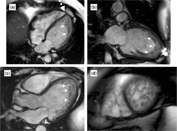
Figure 9. End-diastolic steady state free precession cine images in the horizontal long axis (a), the vertical long axis (b), the left ventricular outflow tract (c) and an apical short axis (d) showing a clear distinction of a thick noncompacted inner layering (white solid arrows) and a thin outer compacted layer. The noncompacted layer can be seen to extend throughout the left ventricular cavity in the apical short axis image (d). Right ventricular shape and involvement can be readily assessed on the horizontal long-axis section (a). Note the extreme angulation of the right ventricular cavity relative to the septum (dashed arrow).

Figure 10. Cine magnetic resonance images in four-chamber (left-hand panel) and two-chamber views show an interrupted layer of tissue (arrows) delineating the noncompacted layer of myocardium from the left ventricular cavity. The trabeculations are seen as fine strands extending from the compacted myocardium towards the thin layer of tissue. Note that the abnormal noncompacted myocardium is thickest in the apex. It extends to the atrioventricular junction along the free wall, while the basal one-third of the septum is not involved.
Clinical findings in patients with isolated left ventricular noncompaction
The clinical features and events facing patients with isolated left ventricular noncompaction include facial dysmorphism, neuromuscular disorders, familial recurrence, association with various syndromes as already discussed, a progressive deterioration in cardiac function resulting in congestive heart failure, supraventricular arrhythmias, ventricular arrhythmias with ventricular tachycardia and fibrillation, sudden death, and thromboembolic events.
The condition has been recognized in the fetus, and also in the octogenarian. Thus, the clinical expression is quite diverse. In the fetus, it may be an incidental finding, or it may be defined in the setting of fetal bradycardia and nonimmune hydrops.1–5, 17, 19–29, 118–127 A comprehensive review of the pertinent literature reveals a ratio of nearly 2 to 1 in favour of males over females.5 Facial dysmorphism has been prominent when the diagnosis is made in childhood, including those patients with Barth syndrome, Toriello–Carey syndrome, and so on.1–5, 17, 19–29, 118–127 Some patients, nonetheless, are seemingly asymptomatic, with left ventricular non-compaction diagnosed during the workup of a heart murmur or similar event. The majority of symptomatic patients present either in congestive heart failure, with supraventricular or ventricular arrhythmias, or because of systemic embolization.1–5, 17–24, 29, 178 The initial clinical descriptions tended to be quite pessimistic in terms of prognosis, with the inference that most patients died early after diagnosis with congestive heart failure, complex arrhythmias, and adverse neurological events secondary to thromboembolic phenomenons.19, 23, 29 While indeed some patients do exhibit a more aggressive and rapid course, eventuating in death or cardiac transplantation, others seem to stabilize with anticongestive and beta-blocker therapy, or at least demonstrate transient recovery of function followed by later deterioration. Some infants have been described with mitral regurgitation and preserved ventricular function.179 Patients may also exhibit a so-called “undulating phenotype”, changing over time from a dilated to a hypertrophic cardiomyopathy.29
A variety of electrocardiographic abnormalities have been identified, including biventricular hypertrophy, right or left bundle branch block, first degree atrioventricular block, various intraventricular conduction defects, complete heart block, abnormalities of repolarization, Wolff–Parkinson–White syndrome, sinus bradycardia, sinus arrest, supraventricular tachycardia, atrial fibrillation, ventricular tachycardia, and ventricular fibrillation.1–5, 17, 19–29, 180–185 There does not appear to be a particular arrhythmia that is characteristic of left ventricular noncompaction. Severe ventricular tachyarrhythmias documented in patients with left ventricular noncompaction are reminiscent of those observed in patients with histiocytoid cardiomyopathy, another uncommon but yet different disorder of cardiac muscle identified more commonly in young female infants.186 Left bundle branch block with significant prolongation of the QRS complex has been identified in some patients. The sensitivity and specificity of this finding is unknown. The aetiology of heart failure and systolic dysfunction in patients with myocardial noncompaction, as well as the severe and frequently life-threatening arrhythmias, is unclear. Myocardial ischaemia has been invoked as possibly a contributory factor, being documented using a variety of methodologies, including Thallium-201 scintigraphy, positron emission tomography, magnetic resonance imaging, and single photon computed tomography.60–63, 172 As we stated earlier, decreased coronary flow reserve is not confined to the noncompacted segments, but extends to most segments of the left ventricle showing abnormal wall motion, possibly contributing to an ischaemic substrate and creating the propensity for severe ventricular arrhythmias.
Ventricular noncompaction and neuromuscular disorders
One of the intriguing findings amongst patients with isolated left ventricular noncompaction is the presence of associated neuromuscular disorders.5, 187–191 In one study, screening showed that more than four-fifths of patients, mostly adults with isolated left ventricular noncompaction also had neuromuscular disorders.190 Thus far, Leber's hereditary optic neuropathy, Becker's muscular dystrophy, myotonic dystrophy, and metabolic myopathy have been recognized, as well as a variety of neuromuscular disorders of unknown etiology. Stöllberger and Finsterer190, 191 recommend that all patients diagnosed with isolated left ventricular noncompaction should be screened for neuromuscular abnormalities. In this respect, it should be noted that mutations of the lamin A/C gene have been associated with the so-called nuclear muscular dystrophies,192, 193 and have also been associated with noncompaction. It is also intriguing that a Cypher knockout mouse develops a severe congenital myopathy and dilated cardiomyopathy.194
Determinants of severity and timing of presentation of patients with left ventricular noncompaction
The factors predictive of the severity and timing of clinical presentation have yet to be completely defined. Amongst Japanese children with left ventricular noncompaction,195, 196 more patients are asymptomatic on presentation and have fewer systemic embolic events and episodes of ventricular tachycardia when compared to the observations of Chin et al.1 Ichida et al.19 suggest that this difference may be accounted for by the fact that left ventricular noncompaction in their patients was detected incidentally during a screening study of an entire population of Japanese children. Whether the extent or degree of hypertrabeculation has a role in determining the timing of presentation, and severity of clinical course, is also uncertain, although this has been suggested.1, 197 Because of the known genetic heterogeneity,42 it is yet to be determined if a specific genetic marker may contribute to either the severity or timing of presentation.
Differential diagnosis
Once the dilated and hypertrophic variants of cardiomyopathy are excluded from consideration, there are relatively few further potential diagnoses for myocardial noncompaction. It is, of course, necessary to exclude the false diagnosis of “normal” left ventricular trabeculations as noncompaction, as we discussed earlier. It is also necessary to distinguish noncompaction from the acquired changes seen in the setting of lesions such as pulmonary atresia with intact ventricular septum. Another potentially misleading condition is the appearance of layered mural thrombus of the left ventricle simulating noncompaction.5 It has also been suggested that mycotic invasion of the heart can produce the appearance of abnormal trabeculations,198, 199 while rarely an intramyocardial haematoma may mimic isolated left ventricular noncompaction.200 The question has also been asked as to whether left ventricular noncompaction can be a cardiac manifestation of Fabry's disease?201 In this respect, while the appearances of hypertrophic cardiomyopathy are well-documented in patients with Fabry's disease, to the best of our knowledge there is no data supporting unequivocally the association of this inborn metabolic disorder with left ventricular noncompaction.
Treatment and outcome
Many of the early clinical reports emphasized the dismal outcomes of patients with isolated left ventricular noncompaction,1–5, 17–29 focusing on the egregious nature of the often malignant accompanying arrhythmias. Increasing clinical experience has modified to some extent this bleak outlook.22, 29 In some asymptomatic patients, isolated left ventricular noncompaction has been found as an incidental finding.202, 203 In others it has been recognized in the sixth and seventh decades of life, and beyond. Yet the disorder certainly has the potential for a poor outcome. Even for those patients presenting in the first year of life with depressed left ventricular contractility, with some recovery of ventricular function, recovery can be transient. A variety of medical therapies have been utilized in those symptomatic patients with congestive heart failure, including cardiac glycosides, diuretics, inhibitors of angiotensin converting enzyme, afterload reducing agents, and for those suspected of having an underlying mitochondrial myopathy, a “metabolic” cocktail.29 Beta-blockade has also been used with some success,204 and some have proceeded to cardiac transplantation.205 Disturbances of rhythm have been treated in standard fashion, while some have implanted defibrillators for severe ventricular arrhythmias recognized as predisposing to sudden death.183, 184 Surprisingly, patients with complex congenital cardiac malformations coexisting with noncompaction seem less troubled with severe disturbances of rhythm. In this regard, Ali and Godman22 remind us that some patients with myocardial noncompaction experience congestive heart failure secondary to associated cardiac lesions, yet with preserved systolic function.
Cerebral vascular accidents certainly contribute to co-morbidity in patients with isolated left ventricular noncompaction, the unusual hypertrabeculations not only reducing ventricular function, but also creating a nidus for formation of mural thrombus.1, 5, 19–29 It is difficult, however, precisely to determine the incidence of adverse neurological events. Oechslin et al.23 reported that a quarter of their adult patients experienced some form of thromboembolic event. Stöllberger and Finisterer, who have written extensively about isolated left ventricular noncompaction, have recently reviewed 22 publications discussing formation of thrombus in this disorder,206 concluding that this is, in fact, a rare event. On this basis, they take a conservative position regarding the role of anticoagulation, despite the fact that others have suggested that patients should at least be placed on a protocol of oral anticoagulation once the diagnosis of isolated left ventricular noncompaction is made. Of course, if a patient has sustained an unequivocal thromboembolic event, then full anticoagulation with low-molecular-weight heparin is advised.1, 19, 23, 29
Conclusions
Ventricular noncompaction, a genetically heterogeneous disorder,41 may affect both ventricles, may be isolated, or may be associated with many diverse forms of congenital cardiac malformations. It has been identified in patients with a variety of syndromes. When confined to the left ventricle, patients may present like those with idiopathic dilated cardiomyopathy, or less frequently with restrictive physiology, this latter physiology perhaps more common in children. The entity has now been recognized in the fetus, and also in the octogenarian, with many patients presenting or identified as adults. The peculiar association of isolated left ventricular noncompaction with many kinds of neuromuscular disorders is well-established, but the reasons for this association have not been clarified. The prognosis for the symptomatic patient is generally poor, with progression to chronic cardiac failure and death. Some will die suddenly, reflecting the predisposition to severe ventricular arrhythmias. These symptomatic patients may benefit from orthotopic cardiac transplantation should medical therapy fail to stabilize the condition.205, 207
In our opinion, the weight of evidence, based on the developmental biology of myocardium in fish, chick, mouse, and man, suggests that isolated left ventricular noncompaction is an abnormality reflecting an arrest in early cardiac morphogenesis. If myocardial noncompaction represents a single endpoint of a complex developmental process, nonetheless, it is still likely that many factors participate or contribute to the molecular and genetic regulation of cardiac trabeculation.
Acknowledgements
We thank Ms Ruth Taylor, of the Division of Cardiology, Hospital for Sick Children, Toronto, for her superb secretarial assistance. Ms Linda Gotlieb, librarian at the Hospital for Sick Children, has proven invaluable in obtaining many of the references cited in this review.
The research on which this review is based was supported in parts by grants from the British Heart Foundation together with the Joseph Levy Foundation. Research at the Institute of Child Health and Great Ormond Street Hospital for Children NHS Trust benefits from R&D funding received from the NHS Executive.
References
Chin TK, Perloff JK, Williams RG, Jue K, Mohrmann R. Isolated noncompaction of left ventricular myocardium: a study of eight cases. Circulation 1990; 82: 507–513.Google Scholar
Jenni R, Oechslin E, Schneider J, Jost CA, Kaufmann PA. Echocardiographic and pathoanatomical characteristics of isolated left ventricular non-compaction: a step towards classification as a distinct cardiomyopathy. Heart 2001; 86: 666–671.Google Scholar
Jenni R, Rojas J, Oechslin E. Isolated noncompaction of the myocardium. N Engl J Med 1999; 340: 966–967.Google Scholar
Weiford BC, Subbarao VD, Mulhern KM. Noncompaction of the ventricular myocardium. Circulation 2004; 109: 2965–2971.Google Scholar
Stöllberger C, Finsterer J. Left ventricular hypertrabeculation/ noncompaction. J Am Soc Echocardiogr 2004; 17: 91–100.Google Scholar
Jenni R, Goebel N, Tartini R, Schneider J, Arbenz U, Oelz O. Persisting myocardial sinusoids of both ventricles as an isolated anomaly: echocardiographic, angiographic, and pathologic anatomic findings. Cardiovasc Interv Radiol 1986; 9: 127–131.Google Scholar
Engberding R, Bender F. Identification of a rare congenital anomaly of the myocardium by two-dimensional echocardiography. Persistence of isolated myocardial sinusoids. Am J Cardiol 1984; 53: 1733–1734.Google Scholar
Chenard J, Samson M, Beaulieu M. Embryonal sinusoids in the myocardium. Report of a case successfully treated surgically. Can Med Assoc J 1965; 92: 1356–1359.Google Scholar
Allenby PA, Gould NS, Schwartz MF, Chiemmongkoltip P. Dysplastic cardiac development presenting as a cardiomyopathy. Arch Pathol Lab Med 1988; 112: 1255–1258.Google Scholar
Amann G, Sherman FS. Myocardial dysgenesis with persistent sinusoids in a neonate with Noonan's syndrome. Pediatr Pathol 1992; 12: 83–92.Google Scholar
Richardson P, McKenna W, Bristow M, et al. Report of the 1995 World Health Organization/International Society and Federation of Cardiology task force on the Definition and Classification of Cardiomyopathies. Circulation 1996; 98: 841–842.Google Scholar
Varnava AM. Isolated left ventricular non-compaction: a distinct cardiomyopathy? Heart 2001; 86: 599–600.Google Scholar
Wong SP, Oldfield M, Ko AP, Kerr AJ. Ventricular non-compaction: a rare cause of heart failure. Intern Med J 2003; 33: 262–263.Google Scholar
Baumhakel M, Kindermann I, Kindermann M, Schneider G, Hennen B, Bohm M. Isolated noncompaction of ventricular myocardium syndrome: a rare structural heart disease. Dtsch Med Wochenschr 2003; 128: 562–567.Google Scholar
Buonanno C, Variola A, Dander B, Gabaldo S, Marafioti V. Isolated noncompaction of the myocardium – an exceedingly rare cardiomyopathy: a case report. Ital Heart J 2000; 1: 301–305.Google Scholar
McCrohon JA, Richmond DR, Pennell DJ, Mohiaddin RH. Images in cardiovascular medicine. Isolated noncompaction of the myocardium: a rarity or missed diagnosis? Circulation 2002; 106: E22–E23.Google Scholar
Zambrano E, Marshalko SJ, Jaffe CC, Hui P. Isolated noncompaction of the ventricular myocardium: clinical and molecular aspects of a rare cardiomyopathy. Lab Invest 2002; 82: 117–122.Google Scholar
Oechslin E, Jenni R. Guest editorial. Isolated left ventricular non-compaction: increasing recognition of the distinct, yet “unclassified” cardiomyopathy. Eur J Echocardiogr 2002; 3: 250–251.Google Scholar
Ichida F, Hamamichi Y, Miyawaki T, et al. Clinical features of isolated noncompaction of the ventricular myocardium: long-term clinical course, hemodynamic properties, and genetic background. J Am Coll Cardiol 1999; 34: 233–240.Google Scholar
Ritter M, Oechslin E, Sutsch G, Attenhofer C, Schneider J, Jenni R. Isolated noncompaction of the myocardium in adults. Mayo Clin Proc 1997; 72: 26–31.Google Scholar
Halbertsma FJ, van't Hek LGEM, Daniels O. Spongy cardiomyopathy in a neonate. Cardiol Young 2001; 11: 458–460.Google Scholar
Ali SKM, Godman MJ. The variable clinical presentation and outcomes for noncompaction of the ventricular myocardium in infants and children, and under-diagnosed cardiomyopathy. Cardiol Young 2004; 14: 409–416.Google Scholar
Oechslin EN, Attenhofer Jost CH, Rojas JR, Kaufmann PA, Jenni R. Long-term follow-up of 34 adults with isolated left ventricular noncompaction: a distinct cardiomyopathy with poor prognosis. J Am Coll Cardiol 2000; 36: 493–500.Google Scholar
Neudorf UE, Hussein A, Trowitzsch E, Schmaltz AA. Clinical features of isolated noncompaction of the myocardium in children. Cardiol Young 2001; 11: 439–442.Google Scholar
Kohl T, Villegas M, Silverman N. Isolated noncompaction of ventricular myocardium detection during fetal life. Cardiol Young 1995; 5: 187–189.Google Scholar
Winer N, Lefevre M, Nomballais MF, et al. Persisting spongy myocardium: a case indicating the difficulty of antenatal diagnosis. Fetal Diagn Ther 1998; 13: 227–232.Google Scholar
Moura C, Hillion Y, Daikha-Dahmane F, et al. Isolated non-compaction of the myocardium diagnosed in the fetus: two sporadic and two familial cases. Cardiol Young 2002; 12: 278–283.Google Scholar
Guntheroth W, Komarniski C, Atkinson W, Fligner CL. Criterion for fetal spongiform cardiomyopathy: restrictive pathophysiology. Obstet Gynecol 2002; 99: 882–885.Google Scholar
Pignatelli RH, McMahon CJ, Dreyer WJ, et al. Clinical characterization of left ventricular noncompaction in children: a relatively common form of cardiomyopathy. Circulation 2003; 108: 2672–2678.Google Scholar
Angelini A, Melacini P, Barbero F, Thiene G. Evolutionary persistence of spongy myocardium in humans. Circulation 1999; 99: 2475.Google Scholar
Victor S, Nayak VM, Rajasingh R. Evolution of the ventricles. Tex Heart Inst J 1999; 26: 168–175.Google Scholar
Steiner I, Hrubecky J, Pleskot J, Kokstejn Z. Persistence of spongy myocardium with embryonic blood supply in an adult. Cardiovasc Pathol 1996; 5: 47–51.Google Scholar
Dusek J, Ostadal B, Duskova M. Postnatal persistence of spongy myocardium with embryonic blood supply. Arch Pathol 1975; 99: 312–317.Google Scholar
Burchell HB. Large vascular sinuses in the myocardium of a dog. Anat Rec 1939; 74: 195–197.Google Scholar
Feldt RH, Rahimtoola H, Davis GD, Swan HJC, Titus JL. Anomalous ventricular myocardial patterns in a child with complex congenital heart disease. Am J Cardiol 1969; 23: 732–734.Google Scholar
Gittenberger-de Groot AC, Tennstedh C, Chaoui R, Lie-Venema H, Sauer U, Poelmann RE. Ventriculo-coronary arterial communications (VCAC) and myocardial sinusoids in hearts with pulmonary atresia with intact ventricular septum: two different diseases. Prog Pediatr Cardiol 2001; 13: 157–164.Google Scholar
Freedom RM, Mawson J, Yoo S-J, Benson LN. Congenital Heart Disease: Textbook of Angiocardiography. Futura Publishing Co., Armonk, NY, 1997, pp 617–672.
Freedom RM, Mawson J, Yoo S-J, Benson LN. Congenital Heart Disease: Textbook of Angiocardiography. Futura Publishing Co., Armonk, NY, 1997, pp 731–765.
Tsang JC-C, Chiu RC-J. The phantom of “myocardial sinusoids”: a historical reappraisal. Ann Thorac Surg 1995; 60: 1831–1835.Google Scholar
Anderson RH. Anatomy. In: Anderson RH, Baker E, Macartney F, Rigby ML, Shinebourne EA, Tynan M (eds). Paediatric Cardiology, 2nd edn. Churchill Livingstone, London, 2002, pp 37–55.
Petersen SE, Selvanayagam JB, Wiesmann F, et al. Left ventricular non-compaction: insights from cardiovascular magnetic resonance imaging. J Am Coll Cardiol; In press.
Digilio MC, Marino B, Bevilacqua M, Musolino AM, Giannotti A, Dallapiccola B. Genetic heterogeneity of isolated noncompaction of the left ventricular myocardium. Am J Med Genet 1999; 85: 90–91.Google Scholar
Boyd MT, Seward JB, Tajik AJ, Edwards WD. Frequency and location of prominent left ventricular trabeculations at autopsy in 474 normal human hearts: implications for evaluation of mural thrombi by two-dimensional echocardiography. J Am Coll Cardiol 1987; 9: 323–326.Google Scholar
Luetmer PH, Edwards WD, Seward JB, Tajik AJ. Incidence and distribution of left ventricular false tendons: an autopsy study of 483 normal human hearts. J Am Coll Cardiol 1986; 8: 179–183.Google Scholar
Malouf J, Gharzuddine W, Kutayli F. A reappraisal of the prevalence and clinical importance of left ventricular false tendons in children and adults. Br Heart J 1986; 55: 587–591.Google Scholar
Abdulla AK, Frustaci A, Martinez JE, Florio RA, Somerville J, Olsen EG. Echocardiography and pathology of left ventricular “false tendons”. Chest 1990; 98: 129–132.Google Scholar
Witter BA, De Cristofaro D. Echocardiography of left ventricular trabeculations, bands and false tendons. Am J Cardiol 1993; 71: 499–500.Google Scholar
Keren A, Billingham ME, Popp RL. Echocardiographic recognition and implications of ventricular hypertrophic trabeculations and aberrant bands. Circulation 1984; 70: 836–842.Google Scholar
Deniz M, Kilinc M, Hatipoglu ES. Morphologic study of left ventricular bands. Surg Radiol Anat 2004; 26: 230–234.Google Scholar
Kervancioglu M, Ozbag D, Kervancioglu P, et al. Echocardiographic and morphologic examination of left ventricular false tendons in human and animal hearts. Clin Anat 2003; 16: 389–395.Google Scholar
Sethuraman KR, Sriram R, Balachandar J. Left ventricular false tendons: echocardiographic incidence in India and clinical importance. Int J Cardiol 1984; 6: 385–387.Google Scholar
Brenner JI, Baker K, Ringel RE, Berman MA. Echocardiographic evidence of left ventricular bands in infants and children. J Am Coll Cardiol 1984; 3: 1515–1520.Google Scholar
Perry LW, Ruckman RN, Shapiro SR, Kuehl KS, Galioto FM, Scott LP. Left ventricular false tendons in children: prevalence as detected by 2-dimensional echocardiography and clinical significance. Am J Cardiol 1983; 52: 1264–1266.Google Scholar
Tamborini G, Pepi M, Celeste F, et al. Incidence and characteristics of left ventricular false tendons and trabeculations in the normal and pathologic heart by second harmonic echocardiography. J Am Soc Echocardiogr 2004; 17: 367–374.Google Scholar
Casta A, Wolf WJ. Left ventricular bands (false tendons): echocardiographic and angiocardiographic delineation in children. Am Heart J 1986; 111: 321–324.Google Scholar
Michel RS, Carpenter MA, Lovell MA. Pathological case of the month. Noncompaction of the left ventricular myocardium. Arch Pediatr Adolesc Med 1998; 152: 709–710.Google Scholar
Finsterer J, Stöllberger C, Feichtinger H. Non-compaction on echocardiography and autopsy. Acta Cardiol 2003; 58: 165–168.Google Scholar
Finsterer J, Stöllberger C, Feichtinger H. Histological appearance of left ventricular hypertrabeculation/noncompaction. Cardiology 2002; 98: 162–164.Google Scholar
Junga G, Kneifel S, Von Smekal A, Steinert H, Bauersfeld U. Myocardial ischaemia in children with isolated ventricular non-compaction. Eur Heart J 1999; 20: 910–916.Google Scholar
Jenni R, Wyss CA, Oechslin EN, Kaufmann PA. Isolated ventricular noncompaction is associated with coronary microcirculatory dysfunction. J Am Coll Cardiol 2002; 39: 450–454.Google Scholar
Soler R, Rodriguez E, Monserrat L, Alvarez N. MRI of subendocardial perfusion deficits in isolated left ventricular noncompaction. J Comput Assist Tomogr 2002; 26: 373–375.Google Scholar
Nil M, Mori K, Yuasa Y, Ichida F. Isolated noncompaction of myocardium associated with calcification in the interventricular septum. Pediatr Cardiol 2003; 24: 591–594.Google Scholar
Icardo JM. Developmental biology of the vertebrate heart. J Exp Zool 1996; 275: 144–161.Google Scholar
Sanchez-Quintana D, Garcia-Martinez V, Climent V, Hurle JM. Morphological changes in the normal pattern of ventricular myoarchitecture in the developing human heart. Anat Rec 1995; 243: 483–495.Google Scholar
Moore KL (ed.). Cardiovascular system. In: The Developing Human: Clinically Oriented Embryology. WB Saunders Co., Philadelphia, 1982, pp 262–276.
Torry TW. Morphogenesis of the Vertebrates. John Wiley & Sons, New York, 1962, pp 407–469.
Hyman LH. Comparative Vertebrate Anatomy. University of Chicago Press, Chicago, 1970, p 544.
Van Mierop LHS, Kutsche LM. Comparative anatomy and embryology of the ventricles and arterial pole of the vertebrate heart. In: Nora JJ, Takao A (eds). Congenital Heart Disease. Causes and Processes. Futura Publishing Co., Mt. Kisco, NY, 1984, pp 459–474.
Kirby ML. Molecular embryogenesis of the heart. Pediatr Develop Pathol 2002; 5: 516–543.Google Scholar
Wessels A, Markwald R. Cardiac morphogenesis and dysmorphogenesis. 1. Normal development. In: Tuan RS, Lo CW (eds). Methods in Molecular Biology, Vol. 136. Developmental Biology Protocols. Humana Press, Totowa, NJ, 2000, pp 239–259.
Davis CL. Development of the human heart from its first appearance to the stage found in embryos of twenty paired somites. Carneg Inst Contrib Embryol 1927; 107: 245–283.Google Scholar
Gittenberger-de Groot AC. Mannheimer Lecture. The quintessence of the making of the heart. Cardiol Young 2003; 13: 175–183.Google Scholar
Manasek FJ. Embryonic development of the heart. I. A light and electron microscopic study of myocardial development in the early chick embryo. J Morphol 1968; 125: 329–365.Google Scholar
Sedmera D, Pexieder T, Vuillemin M, Thompson RP, Anderson RH. Developmental patterning of the myocardium. Anat Rec 2000; 258: 319–337.Google Scholar
Sedmera D, Pexieder T, Hu N, Clark EB. Developmental changes in the myocardial architecture of the chick. Anat Rec 1997; 248: 421–432.Google Scholar
Icardo JM, Fernandez-Teran A. Morphologic study of ventricular trabeculation in the embryonic chick heart. Acta Anat 1987; 130: 264–274.Google Scholar
Ben-Shachar G, Arcilla RA, Lucas RV, Manasek FJ. Ventricular trabeculations in the chick embryo heart and their contribution to ventricular and muscular septal development. Circ Res 1985; 57: 759–766.Google Scholar
Olsen EN, Srivastava D. Molecular pathways controlling heart development. Science 1996; 272: 671–676.Google Scholar
Rychter Z, Ostadal B. Fate of “sinusoidal” intertrabecular spaces of the cardiac wall after development of the coronary vascular bed in chick embryo. Folia Morphol 1971; 19: 31–44.Google Scholar
Ostadal B. Developmental relationships between the structure, blood supply and metabolic pattern of the vertebrate heart. Cor Vasa 1979; 21: 380–386.Google Scholar
Ostadal B, Schiebler TH, Rychter Z. Relations between development of the capillary wall and myoarchitecture of the rat heart. Adv Exp Med Biol 1975; 53: 375–388.Google Scholar
Ostadal B, Rychter Z, Poupa O. Comparative aspects of the development of the terminal vascular bed in the myocardium. Physiol Bohemoslov 1970; 19: 1–7.Google Scholar
Davie PS, Farrell AP. The coronary and luminal circulation of the myocardium of fishes. Can J Zool 1991; 69: 1993–2001.Google Scholar
Sanchez-Quintana D, Hurle JM. Ventricular myocardial architecture in marine fishes. Anat Rec 1987; 217: 263–273.Google Scholar
Agnisola C, Tota B. Structure and function of the fish cardiac ventricle: flexibility and limitations. Cardioscience 1994; 5: 145–153.Google Scholar
Breisch EA, White F, Jones HM, Laurs RM. Ultrastructural morphometry of the myocardium of Thunnus alalunga. Cell Tissue Res 1983; 233: 427–438.Google Scholar
Tota B. Vascular and metabolic zonation in the ventricular myocar-dium of mammals and fishes. Comp Biochem Physiol A 1983; 76: 423–437.Google Scholar
Tota B, Cimini V, Salvatore G, Zummo G. Comparative study of the arterial and lacunary systems of the ventricular myocardium of elasmobranch and teleost fishes. Am J Anat 1983; 167: 15–32.Google Scholar
Simoes K, Vicentini CA, Orsi AM, Gregorio EA, Da Cruz C. Morphological studies on the heart ventricle of African catfish (Clarias gariepinus). Anat Histol Embryol 2002; 31: 247–251.Google Scholar
Simoes K, Vicentini CA, Orsi AM, Cruz C. Myoarchitecture and vasculature of the heart ventricle in some freshwater teleosts. J Anat 2002; 200: 467–475.Google Scholar
Simoes K, Vicentini CA, da Cruz C, Benetti EJ. Structure and vascularization of the ventricular myocardium of Piaractus mesopotamicus and Clarias gariepinus (Teleostei). Anat Histol Embryol 1999; 28: 243–246.Google Scholar
De Andres AV, Munoz-Chapull R, Sans-Coma V. Development of the coronary arteries and cardiac veins in the dogfish (Scyliorhinus caniculus). Anat Rec 1993; 235: 436–442.Google Scholar
Basile C, Goldspink G, Modigh M, Tota B. Morphological and biochemical characterisation of the inner and outer ventricular myocardial layers of adult tuna fish (Thunnus thynnus L.). Comp Biochem Physiol B 1976; 54: 279–283.Google Scholar
Grant RT, Regnier M. The comparative anatomy of the cardiac coronary vessels. Heart 1925–1926; 12–13: 285–310.Google Scholar
O'Brien KM, Xue H, Sidell BD. Quantification of diffusion distance within the spongy myocardium of hearts from antarctic fishes. Respir Physiol 2000; 122: 71–80.Google Scholar
O'Brien KM, Sidell BD. The interplay among cardiac ultrastructure, metabolism and the expression of oxygen-binding proteins in Antarctic fishes. J Exp Biol 2000; 203: 1287–1297.Google Scholar
Santer RM, Greer Walker M. Morphological studies on the ventricle of teleost and elasmobranch hearts. J Zool (Lond) 1980; 190: 259–272.Google Scholar
Ruud JT. Vertebrates without erythrocytes and blood pigment. Nature 1954; 173: 848–850.Google Scholar
Feller G, Goessens G, Gerday Gh, Bassleer R. Heart structure and ventricular ultrastructure of hemoglobin- and myoglobin-free icefish Channichthys rhinoceratus. Cell Tissue Res 1985; 242: 669–676.Google Scholar
Sidell BD. Life at body temperatures below 0 degrees C: the physiology and biochemistry of Antarctic fishes. Gravit Space Biol Bull 2000; 13: 25–34.Google Scholar
Greco G, Lipari D, Tota B, Zummo G. Preliminary observations on the heart of the haemoglobinless Antarctic fish Champsocephalus gunnari, Lonnberg. Boll Soc Ital Biol Sper 1981; 57: 1807–1813.Google Scholar
Zummo G, Acierno R, Agnisola C, Tota B. The heart of the icefish: bioconstruction and adaptation. Braz J Med Biol Res 1995; 28: 1265–1276.Google Scholar
Acierno R, Agnisola C, Tota B, Sidell BD. Myoglobin enhances cardiac performance in antarctic icefish species that express the protein. Am J Physiol 1997; 273: R100–R106.Google Scholar
Tota B, Cerra MC, Mazza R, Pellegrino D, Icardo J. The heart of the Antarctic icefish as paradigm of cold adaptation. J Therm Biol 1997; 22: 409–417.Google Scholar
Moylan TJ, Sidell BD. Concentrations of myoglobin and myoglobin mRNA in heart ventricles from Antarctic fishes. J Exp Biol 2000; 203: 1277–1286.Google Scholar
Di Prisco G, Cocca E, Parker SK, Detrich III HW. Tracking the evolutionary loss of hemoglobin expression by the white-blooded Antarctic icefishes. Gene 2002; 295: 185–191.Google Scholar
Vayda ME, Small DJ, Yuan ML, Costello L, Sidell BD. Conservation of the myoglobin gene among Antarctic notothenioid fishes. Mol Mar Biol Biotechnol 1997; 6: 207–216.Google Scholar
Eastman JT. Antarctic fish biology. Evolution in a unique environment. Academic Press Inc, San Diego, 1993, p 322.
Sedmera D, Pexieder T, Hu N, Clark EB. A quantitative study of the ventricular myoarchitecture in the stage 21–29 chick embryo following decreased loading. Eur J Morphol 1998; 36: 105–119.Google Scholar
Sedmera D, Pexieder T, Rychterova V, Hu N, Clark EB. Remodeling of chick embryonic ventricular myoarchitecture under experimentally changed loading conditions. Anat Rec 1999; 254: 238–252.Google Scholar
Hu N, Sedmera D, Yost HJ, Clark EB. Structure and function of the developing zebrafish heart. Anat Rec 2000; 260: 148–157.Google Scholar
King T, Bland Y, Webb S, Barton S, Brown NA. Expression of Peg1 (Mest) in the developing mouse heart: involvement in trabeculation. Dev Dyn 2002; 225: 212–215.Google Scholar
Sasse-Klaassen S, Probst S, Gerull B, et al. Novel gene locus for autosomal dominant left ventricular noncompaction maps to chromosome 11p15. Circulation 2004; 109: 2720–2723.Google Scholar
Nugent AW, Daubeney PE, Chondros P, et al. The epidemiology of childhood cardiomyopathy in Australia. N Engl J Med 2003; 348: 1639–1646.Google Scholar
Gedeon AK, Wilson MJ, Colley AC, et al. X linked fatal infantile cardiomyopathy maps to Xq28 and is possibly allelic to Barth syndrome. J Med Genet 1995; 32: 383–388.Google Scholar
Matsuda M, Tsukahara M, Kondoh O, Mito H. Familial isolated noncompaction of ventricular myocardium. J Hum Genet 1999; 44: 126–128.Google Scholar
Bleyl SB, Mumford BR, Brown-Harrison MC, et al. Xq28-linked noncompaction of the left ventricular myocardium: prenatal diagnosis and pathologic analysis of affected individuals. Am J Med Genet 1997; 72: 257–265.Google Scholar
Bione S, D'Adamo P, Maestrini E, Gedeon AK, Bolhuis PA, Toniolo D. A novel X-linked gene, G4.5 is responsible for Barth syndrome. Nat Genet 1996; 12: 385–389.Google Scholar
Chen R, Tsuji T, Ichida F, et al. Mutation analysis of the G4.5 gene in patients with isolated left ventricular noncompaction. Mol Genet Metab 2002; 77: 319–325.Google Scholar
Bleyl SB, Mumford BR, Thompson V, et al. Neonatal, lethal noncompaction of the left ventricular myocardium is allelic with Barth syndrome. Am J Hum Genet 1997; 61: 868–872.Google Scholar
Ichida F, Tsubata S, Bowles KR, et al. Novel gene mutations in patients with left ventricular noncompaction or Barth syndrome. Circulation 2001; 103: 1256–1263.Google Scholar
Neustein HB, Lurie PR, Dahma B, Takahashi M. An X-linked recessive cardiomyoapthy with abnormal mitochondria. Pediatrics 1979; 64: 24–29.Google Scholar
Barth PG, Scholte HR, Berden JA, et al. An X-linked mitochondrial disease affecting cardiac muscle, skeletal muscle and neutrophil leucocytes. J Neurol Sci 1983; 62: 327–355.Google Scholar
Pauli RM, Scheib-Wixted S, Cripe L, Izumo S, Sekhon GS. Ventricular noncompaction and distal chromosome 5q deletion. Am J Med Genet 1999; 85: 419–423.Google Scholar
Vatta M, Mohapatra B, Jimenez S, et al. Mutations in Cypher/ZASP in patients with dilated cardiomyopathy and left ventricular non-compaction. J Am Coll Cardiol 2003; 42: 2014–2027.Google Scholar
Shou W, Aghdasi B, Armstrong DL, et al. Cardiac defects and altered ryanodine receptor function in mice lacking FKBP12. Nature 1998; 391: 489–492.Google Scholar
Kenton AB, Sanchez X, Coveler KJ, et al. Isolated left ventricular noncompaction is rarely caused by mutations in G4.5, alpha-dystrobrevin and FK Binding Protein-12. Mol Genet Metab 2004; 82: 162–166.Google Scholar
Sasse-Klaassen S, Gerull B, Oechslin E, Jenni R, Thierfelder L. Isolated noncompaction of the left ventricular myocardium in the adult is an autosomal dominant disorder in the majority of patients. Am J Med Genet 2003; 119A: 162–167.Google Scholar
Hermida-Prieto M, Monserrat L, Castro-Beiras A, et al. Familial dilated cardiomyopathy and isolated left ventricular noncompaction associated with lamin A/C gene mutations. Am J Cardiol 2004; 94: 50–54.Google Scholar
Wong JA, Bofinger MK. Noncompaction of the ventricular myocardium in Melnick–Needles syndrome. Am J Med Genet 1997; 71: 72–75.Google Scholar
Mandel K, Grunebaum E, Benson L. Noncompaction of the myocardium associated with Roifman syndrome. Cardiol Young 2001; 11: 240–243.Google Scholar
Toriello HV, Carey JC. Corpus callosum agenesis, facial anomalies, Robin sequence, and other anomalies: a new autosomal recessive syndrome? Am J Med Genet 1988; 31: 17–23.Google Scholar
Yamatogi Y, Ohtahara S. Early infantile epileptic encephalopathy with suppression-bursts, Ohtahara syndrome; its overview referring to our 16 cases. Brain Dev 2002; 24: 13–23.Google Scholar
Ohtahara S, Yamatogi Y. Epileptic encephalopathies in early infancy with suppression-burst. J Clin Neurophysiol 2003; 20: 398–407.Google Scholar
Finsterer J, Stöllberger C, Kopsa W. Noncompaction on cardiac MRI in a patient with Nail–Patella syndrome and mitochondriopathy. Cardiology 2003; 100: 48–49.Google Scholar
Happle R, Daniels O, Koopman RJJ. MIDAS syndrome (micropthalmia, dermal aplasia, and sclerocornea): an X-linked phenotype distinct from Goltz syndrome. Am J Med Genet 1993; 47: 710–713.Google Scholar
Lacombe D, Creusot G, Battin J. New case of Toriello–Carey syndrome. Am J Med Genet 1992; 42: 374–376.Google Scholar
Grant RT. An unusual anomaly of the coronary vessels in the malformed heart of a child. Heart 1926; 13: 273–283.Google Scholar
Bellet S, Gouley BA. Congenital heart disease with multiple cardiac anomalies. Report of a case showing aortic atresia, fibrous scar in myocardium and embryonal sinusoidal remains. Am J Med Sci 1932; 183: 458–465.Google Scholar
Raghib G, Bloemendaal RD, Kanjuh VI, Edwards JE. Aortic atresia and premature closure of foramen ovale. Myocardial sinusoids and coronary arteriovenous fistula serving as outflow channel. Am Heart J 1965; 70: 476–480.Google Scholar
Mocellin R, Sauer U, Simon B, Comazzi M, Sebening F, Buhlmeyer K. Reduced left ventricular size and endocardial fibroelastosis as correlates of mortality in newborns and young infants with severe aortic valve stenosis. Pediatr Cardiol 1983; 4: 265–272.Google Scholar
Kim SH, Woo H-O, Kim YM, et al. Abnormal morphological patterns of left ventricular myocardium in the critical stenosis of the aortic valve and the intact ventricular septum. JKMS 1997; 12: 49–54.Google Scholar
Freedom RM, Benson L, Wilson GJ. The coronary circulation and myocardium in pulmonary and aortic atresia with an intact ventricular septum. In: Marcelletti C, Anderson RH, Becker AE, Corno A, di Dicarlo D, Mazzera E (eds). Paediatric Cardiology, Vol. 6. Churchill Livingstone, Edinburgh, 1986, pp 78–96.
Freedom RM, Culham JAG, Moes CAF. Angiocardiography of Congenital Heart Disease. Macmillan Publishing Co., New York, 1984, pp 358–362.
Finegold M, Klein KM. Anastomotic coronary vessels in hypoplasia of the right ventricle. Am Heart J 1971; 82: 678–683.Google Scholar
Marin-Garcia J, Roca J, Blieden LC, Lucas Jr RV, Edwards JE. Congenital absence of the pulmonary valve associated with tricuspid atresia and intact ventricular septum. Chest 1973; 64: 658–661.Google Scholar
Cox JN, De Seigneux R, Bolens M, Haenni P, Bopp P, Bruins C. Tricuspid atresia, hypoplastic right ventricle, intact ventricular septum and congenital absence of the pulmonary valve. Helv Paediatr Acta 1975; 30: 389–398.Google Scholar
Freedom RM, Patel RG, Bloom KR, et al. Congenital absence of the pulmonary valve, associated imperforate membrane type of tricuspid atresia, right ventricular tensor apparatus and intact ventricular septum: a curious developmental complex. Eur J Cardiol 1979; 10: 171–196.Google Scholar
Forrest P, Bini RM, Wilkinson JL, et al. Congenital absence of the pulmonic valve and tricuspid atresia with intact ventricular septum. Am J Cardiol 1987; 59: 482–484.Google Scholar
O'Connor WN, Cottrill CM, Marion MT, Noonan JA. Defective regional myocardial development and vascularization in one variant of tricuspid atresia – clinical and necropsy findings in three cases. Cardiol Young 1992; 2: 42–52.Google Scholar
Mori K, Ando M, Satomi M, Nakazawa M, Momma K, Takao A. Imperforate tricuspid valve with dysplasia of the right ventricular myocardium, pulmonary valve, and coronary artery: a clinicopathologic study of nine cases. Pediatr Cardiol 1992; 13: 24–29.Google Scholar
Quero-Jimenez M, Maitre Azcarate MJ, Alvarez Bejarno H, Vasquez Martul E. Tricuspid atresia: an anatomical study of 17 cases. Eur J Cardiol 1975; 3: 337–348.Google Scholar
Litovsky S, Choy M, Park J, et al. Absent pulmonary valve with tricuspid atresia or severe tricuspid stenosis: report of three cases and review of the literature. Pediatr Dev Pathol 2000; 3: 353–366.Google Scholar
Attenhofer CH, Connolly HM, Warnes CA, et al. Noncompacted myocardium in Ebstein's anomaly: initial description in three patients. J Am Soc Echocardiogr 2004; 17: 677–680.Google Scholar
Lamers WH, Viragh S, Wessels A, Moorman AFM, Anderson RH. Formation of the tricuspid valve in the human heart. Circulation 1995; 91: 111–121.Google Scholar
Vijayalakshmi IB, Chitra N, Prabhu Deva AN. Use of an Amplatzer duct occluder for closingan aortico-left ventricular tunnel in a case of noncompaction of the left ventricle. Pediatr Cardiol 2004; 25: 77–79.Google Scholar
Tavli V, Kayhan B, Okur FF, et al. Noncompaction of the right ventricle following Senning repair. Turk J Pediatr 2001; 43: 261–263.Google Scholar
Lemke RP, Giddens NG, Odim JNK. Absent pulmonary valve in tricuspid atresia with left ventricular outflow tract obstruction and patent arterial duct. Cardiol Young 1997; 7: 94–97.Google Scholar
Bax JJ, Lamb HJ, Poldermas D, Schalij MJ, de Roos A, van der Wall EE. Non-compaction cardiomyopathy–echocardiographic diagnosis. Eur J Echocardiogr 2002; 3: 301–302.Google Scholar
Bax JJ, Atsma DE, Lamb HJ, et al. Noninvasive and invasive evaluation of noncompaction cardiomyopathy. J Cardiovasc Magn Reson 2002; 4: 353–357.Google Scholar
Williams RI, Masani ND, Buchalter MB, Fraser AG. Abnormal myocardial strain rate in noncompaction of the left ventricle. J Am Soc Echocardiogr 2003; 16: 293–296.Google Scholar
Borow KM, Lang RM, Neumann A, Carroll JD, Rajfer SI. Physiologic mechanisms governing hemodynamic responses to positive inotropic therapy in patients with dilated cardiomyopathy. Circulation 1988; 77: 625–637.Google Scholar
Sengupta PP, Mohan JC, Mehta V, et al. Comparison of echocardiographic features of noncompaction of the left ventricle in adults versus idiopathic dilated cardiomyopathy in adults. Am J Cardiol 2004; 94: 389–391.Google Scholar
Moon JC, Fisher NG, McKenna WJ, Pennell DJ. Detection of apical hypertrophic cardiomyopathy by cardiovascular magnetic resonance in patients with non-diagnostic echocardiography. Heart 2004; 90: 645–649.Google Scholar
Chung T, Yiannikas J, Lee LC, et al. Isolated noncompaction involving the left ventricular apex in adults. Am J Cardiol 2004; 94: 1214–1216.Google Scholar
Corrado G, Santarone M, Miglierina E, et al. Isolated noncompaction of the ventricular myocardium: a study in an adult male and literature review. Ital Heart J 2000; 1: 372–375.Google Scholar
Koo BK, Choi D, Ha JW, et al. Isolated noncompaction of the ventricular myocardium: contrast echocardiographic findings and review of the literature. Echocardiography 2002; 19: 153–156.Google Scholar
Korcyk D, Edwards CC, Armstrong G, et al. Cardiac-enhanced cardiac magnetic resonance in a patient with familial isolated ventricular non-compaction. J Cardiovasc Magn Reson 2004; 6: 569–576.Google Scholar
Weiss F, Habermann CR, Lilje C, et al. MRI in the diagnosis of non-compacted ventricular myocardium (NCVM) compared to echocardiography. Rofo 2003; 175: 1214–1219.Google Scholar
Hamamichi Y, Ichida F, Hashimoto I, et al. Isolated noncompaction of the ventricular myocardium: ultrafast computed tomography and magnetic resonance imaging. Int J Card Imaging 2001; 17: 305–314.Google Scholar
Gebker R, Paetsch I, Wahl A, Meyer R, Nagel E. Ventricular non-compaction. Eur Heart J 2004; 25: Cover Image.Google Scholar
Barkhausen J, Hunold P, Eggebrecht H, et al. Detection and characterization of intracardiac thrombi on MR imaging. Am J Roentgenol 2002; 179: 1539–1544.Google Scholar
Petersen SE, Timperley J, Neubauer S. Left ventricular thrombi in a patient with left ventricular non-compaction – visualisation of the rationale for anticoagulation. Heart 2005; 91: e4Google Scholar
Rigopoulos A, Rizos IK, Aggeli C, et al. Isolated left ventricular noncompaction: an unclassified cardiomyopathy with severe prognosis in adults. Cardiology 2002; 98: 25–32.Google Scholar
Ali SK, Omran AS, Najm H, Godman MJ. Noncompaction of the ventricular myocardium associated with mitral regurgitation and preserved ventricular systolic function. J Am Soc Echocardiogr 2004; 17: 87–90.Google Scholar
Duru F, Candinas R. Noncompaction of ventricular myocardium and arrhythmias. J Cardiovasc Electrophysiol 2000; 11: 493.Google Scholar
Nihei K, Shinomiya N, Kabayama H, et al. Wolff–Parkinson–White (WPW) syndrome in isolated noncompaction of the ventricular myocardium (INVM). Circ J 2004; 68: 82–84.Google Scholar
Yasukawa K, Terai M, Honda A, Kohno Y. Isolated noncompaction of ventricular myocardium associated with fatal ventricular fibrillation. Pediatr Cardiol 2001; 22: 512–514.Google Scholar
Celiker A, Kafali G, Dogan R. Cardioverter defibrillator implantation in a child with isolated noncompaction of the ventricular myocardium and ventricular fibrillation. Pacing Clin Electrophysiol 2004; 27: 104–108.Google Scholar
Seres L, Lopez J, Larrousse E, Moya A, Pereferrer D, Valle V. Isolated noncompaction left ventricular myocardium and polymorphic ventricular tachycardia. Clin Cardiol 2003; 26: 46–48.Google Scholar
Valdes-Dapena M, Gilbert-Barness E. Cardiovascular causes for sudden infant death. Pediatr Pathol Mol Med 2002; 21: 195–211.Google Scholar
Ferrans VJ, McAllister HA, Haese WH. Infantile cardiomyopathy with histiocytoid change in cardiac muscle cells. Report of six patients. Circulation 1976; 53: 708–719.Google Scholar
Finsterer J, Stöllberger C, Blazek G, Spahits E. Cardiac involvement in myotonic dystrophy, Becker muscular dystrophy and mitochondrial myopathy: a five-year follow-up. Can J Cardiol 2001; 17: 1061–1069.Google Scholar
Stöllberger C, Winkler-Dworak M, Blazek G, Finsterer J. Left ventricular hypertrabeculation/noncompaction with and without neuromuscular disorders. Int J Cardiol 2004; 97: 89–92.Google Scholar
Finsterer J, Stöllberger C, Prainer C, Hochwarter A. Lone noncompaction in Leber's hereditary optic neuropathy. Acta Cardiol 2004; 59: 187–190.Google Scholar
Stöllberger C, Finsterer J, Blazek G. Left ventricular hypertrabeculation/noncompaction and association with additional cardiac abnormalities and neuromuscular disorders. Am J Cardiol 2002; 90: 899–902.Google Scholar
Stöllberger C, Finsterer J, Blazek G. Isolated left ventricular abnormal trabeculation: follow-up and association with neuromuscular disorders. Can J Cardiol 2001; 17: 163–168.Google Scholar
Charniot JC, Pascal C, Bouchier C, et al. Functional consequences of an LMNA mutation associated with a new cardiac and non-cardiac phenotype. Hum Mutat 2003; 21: 473–481.Google Scholar
Raharjo WH, Enarson P, Sullivan T, Stewart CL, Burke B. Nuclear envelope defects associated with LMNA mutations cause dilated cardiomyopathy and Emery-Dreifuss muscular dystrophy. J Cell Sci 2001; 114: 4447–4457.Google Scholar
Zhou Q, Chu PH, Huang C, et al. Ablation of Cypher, a PDZ-LIM domain Z-line protein, causes a severe form of congenital myopathy. J Cell Biol 2001; 155: 605–612.Google Scholar
Hosaki J. The heart disease screening system for school children in Japan and its results. Acta Paediatr Jpn 1985; 27: 360–365.Google Scholar
Ino T, Yabuta K, Yamauchi K. Heart disease screening in Japanese children. BMJ 1993; 306: 1128.Google Scholar
Wald RM, Veldtman GR, Golding F, Kirsh J, McCrindle BW, Benson LN. Determinants of outcome in isolated ventricular noncompaction in childhood. Am J Cardiol 2005; In press.Google Scholar
Stöllberger C, Preiser J, Finsterer J. Histological detection of intramyocardial abscesses in Candida sepsis mimicking left ventricular noncompaction/hypertrabeculation on echocardiography. Mycoses 2004; 47: 72–75.Google Scholar
Stöllberger C, Preiser J, Finsterer J. Candida sepsis with intramyocardial abscesses mimicking left ventricular noncompaction. Eur J Echocardiogr 2004; 5: 76–78.Google Scholar
Stöllberger C, Finsterer J, Waldenberger FR, Hainfellner JA, Ullrich R. Intramyocardial hematoma mimicking abnormal left ventricular trabeculation. J Am Soc Echocardiogr 2001; 14: 1030–1032.Google Scholar
Stöllberger C, Finsterer J, Voigtlander T, Slany J. Is left ventricular hypertrabeculation/noncompaction a cardiac manifestation of Fabry's disease? Z Kardiol 2003; 92: 966–969.Google Scholar
Tsui KL, Chan KK, Leung TC, Lam KH, Li SK. Isolated ventricular non-compaction presenting with ventricular tachycardia. Hong Kong Med J 2003; 9: 137–140.Google Scholar
Elshershari H, Okutan V, Celiker A. Isolated noncompaction of ventricular myocardium. Cardiol Young 2001; 11: 472–475.Google Scholar
Toyono M, Kondo C, Nakajima Y, Nakazawa M, Momma K, Kusakabe K. Effects of carvedilol on left ventricular function, mass, and scintigraphic findings in isolated left ventricular non-compaction. Heart 2001; 86: e4.Google Scholar
Stamou SC, Lefrak EA, Athari FC, Burton NA, Massimiano PS. Heart transplantation in a patient with isolated noncompaction of the left ventricular myocardium. Ann Thorac Surg 2004; 77: 1806–1808.Google Scholar
Stöllberger C, Finsterer J. Thrombi in left ventricular hypertrabeculation/noncompaction: review of the literature. Acta Cardiol 2004; 59: 341–344.Google Scholar
Murphy RT, Thaman R, Gimeno Blanes J, et al. Natural history and familial characteristics of isolated left ventricular non-compaction. Eur Heart J 2005; 24: 187–192.Google Scholar

The normal right ventricle has been opened to show its inlet, apical trabecular, and other components. The coarse apical trabeculations form the luminal surface of the compact myocardium.

The left ventricle is shown from the same heart that contained the right ventricle seen in Figure 3. The apical trabeculations are characteristically fine and criss-crossing when compared to the coarse trabeculations of the right ventricle (Fig. 3). The trabeculations, nonetheless, are no more than the luminal surface markings of the layer of compact myocardium.

This series of sections, from human embryos (A) at Carnegie stage 16, (B) at stage 18, and (C) immediately after the close of the embryonic interventricular foramen, show that, during development, there is an extensive trabecular layer which forms the greater part of the thickness of the ventricular wall when compared to the extent of the compact layer. It is also the trabecular layer which becomes compacted to form the papillary muscles of the atrioventricular valves (asterisks).

The sections shown in panels (a) and (b) are whole mounts of right and left ventricle, respectively, from the heart shown in Figure 1. There are many intertrabecular spaces with many deep recesses. Higher power histological sections, as seen in panels (c) and (d) show deep and numerous intertrabecular recesses that extend almost to the epicardial surfaces. Note the fibroelastic layer that enclosed the trabeculations (Movat pentachrome stain).

This scanning electron micrograph shows the arrangement of the developing myocardium in the mouse heart as the apical components of the right and left ventricles balloon from the primary heart tube. Specimen prepared and reproduced by kind permission of Professor Nigel Brown, St George's Hospital Medical School, London.

Echocardiograms in four-chamber (left-hand panel) and short axis planes show the bilayered arrangement of the myocardium. The inner hyperechogenic layer (arrows) represents the noncompacted layer, and the outer echolucent layer represents the compacted layer.

End-diastolic steady state free precession cine images in the horizontal long axis (a), the vertical long axis (b), the left ventricular outflow tract (c) and an apical short axis (d) showing a clear distinction of a thick noncompacted inner layering (white solid arrows) and a thin outer compacted layer. The noncompacted layer can be seen to extend throughout the left ventricular cavity in the apical short axis image (d). Right ventricular shape and involvement can be readily assessed on the horizontal long-axis section (a). Note the extreme angulation of the right ventricular cavity relative to the septum (dashed arrow).

Cine magnetic resonance images in four-chamber (left-hand panel) and two-chamber views show an interrupted layer of tissue (arrows) delineating the noncompacted layer of myocardium from the left ventricular cavity. The trabeculations are seen as fine strands extending from the compacted myocardium towards the thin layer of tissue. Note that the abnormal noncompacted myocardium is thickest in the apex. It extends to the atrioventricular junction along the free wall, while the basal one-third of the septum is not involved.
 You have
Access
- 147
- Cited by