A 5-year-old girl with a previously undiagnosed anomalous aortic origin of the left coronary artery presented with shortness of breath and experienced a myocardial infarction associated with cardiac arrest. She was resuscitated with extracorporeal cardiopulmonary resuscitation, with institution of femoral extracorporeal membrane oxygenation, followed by transcatheter creation of an atrial septal defect and placement of an atrial stent to decompress the left atrium. She subsequently underwent unroofing of the anomalous coronary artery and placement of a Berlin Heart left ventricular assist device (Berlin EXCOR [Berlin Heart, Inc., Berlin, Germany]). The child is currently 2 weeks status post coronary unroofing and insertion of left ventricular assist device and is stable on mechanical support awaiting either ventricular recovery or cardiac transplantation.
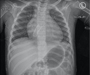
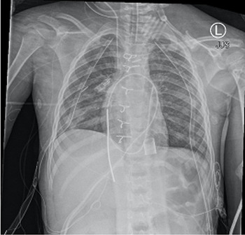
Images of chest radiography compares left ventricular decompression in the same patient supported with extracorporeal membrane oxygenation with atrial septal fenestration (Fig 1) and subsequently supported with left ventricular assist device with apical cannulation (Fig 2). Radiographic images document that direct cannulation of the left ventricle with a left ventricular assist device notably decompressed the left ventricle in comparison to extracorporeal membrane oxygenation with atrial septal fenestration.
An anterior–posterior chest radiograph of a 5-year-old girl with an atrial stent on femoral veno-arterial extracorporeal membrane oxygenation.

An anterior–posterior chest radiograph of the same patient status post-direct cannulation of the left ventricle with a Berlin left ventricular assist device.

Acknowledgements
The authors wish to thank our colleagues from interventional cardiology, James C. Fudge, MD and Himesh V Vyas, MD, for their assistance in caring for this patient and for their clinical support by fenestrating the atrial septum and placing the atrial stent promptly after the initiation of extracorporeal membrane oxygenation.
Financial support
This research received no specific grant from any funding agency, commercial or not-for-profit sectors.
Conflicts of interest
None.