Introduction
Somatic cell nuclear transfer (SCNT) is a technique in which the nucleus of a somatic cell is transferred into an enucleated metaphase-II oocyte. SCNT may be used to produce transgenic animals and to preserve endangered species. It can be also applied in therapeutic cloning procedures. However, the majority of embryos derived from nuclear transfer is lost during different stages of pre-implantation and pregnancy. In this line, there are various issues that need to be addressed. For instance, an appropriate in vitro culture system for recipient and SCNT oocytes is a crucial factor that should be designed and optimized.
All living cells produce reactive oxygen species (ROS), which are essential pre-requisites for their normal functions; however, excessive ROS formation can influence the structure and function of different kinds of biomolecules such as lipids, proteins and nucleic acids, which results in mitochondrial damage, embryo cell block, ATP depletion, and apoptosis (Halliwell & Aruoma, Reference Halliwell and Aruoma1991; Guerin et al., Reference Guerin, El Mouatassim and Menezo2001). Increased oxidative stress appears to be a major factor impairing in vitro mammalian embryo development. Naturally, embryos may be protected against free radicals by oxygen protectors in the oviductal fluid (Legge & Sellens, Reference Legge and Sellens1991), but pre-implantation embryos are susceptible to oxidative stress during in vitro culture (Guerin et al., Reference Guerin, El Mouatassim and Menezo2001).
The protective effect of free radical scavengers in embryo culture medium has been demonstrated by several groups. Melatonin (N-acetyl-5-methoxytryptamine) is mainly synthesized and secreted in the pineal gland, ovary, testes, bone marrow, retina and lens in mammalian species (Reiter et al., Reference Reiter, Tan, Osuna and Gitto2000). It is involved in the detoxification of ROS and protects embryos from oxidative damage. Melatonin acts as a potential free radical scavenger, including peroxyl radical and hydroxyl radical (Poeggeler et al., Reference Poeggeler, Reiter, Tan, Chen and Manchester1993; Reiter et al., Reference Reiter, Tan, Osuna and Gitto2000). In addition, it can stimulate the activity of antioxidant enzymes such as superoxide dismutase and glutathione peroxidase (Mayo et al., Reference Mayo, Sainz, Antoli, Herrera, Martin and Rodriguez2002; Tamura et al., Reference Tamura, Nakamura, Korkmaz, Manchester, Tan, Sugino and Reiter2009).
The beneficial effect of supplementing culture medium with melatonin has been reported previously in species such as mouse, bovine, porcine and buffalo during in vitro embryo development (McElhinny et al., Reference McElhinny, Davis and Warner1996; Ishizuka et al., Reference Ishizuka, Kuribayashi, Murai, Amemiya and Itoh2000; Papis et al., Reference Papis, Poleszczuk, Wenta-Muchalska and Modlinski2007; Rodriguez-Osorio et al., Reference Rodriguez-Osorio, Kim, Wang, Kaya and Memili2007; Choi et al., Reference Choi, Park, Lee, Kim, Jeong, Lee, Park, Kim, Hossein, Jeong, Kim, Hyun and Hwang2008; Tian et al., Reference Tian, Wen, Shi, Liang, Zeng, Tian, Zhou, Zhu and Liu2010).
We have previously examined the effect of melatonin addition to the culture medium on the in vitro maturation of porcine oocytes, and evaluated whether melatonin increased the in vitro development of parthenogenetic and SCNT porcine oocytes (Nakano et al., Reference Nakano, Kato and Tsunoda2012). However, to our knowledge there are no reports on the effect of melatonin on mouse SCNT during in vitro embryonic development under culture medium. Therefore, in the present study melatonin was added to the culture medium and its effect on mouse SCNT embryos was investigated.
Materials and methods
All experiments and protocols were performed in strict accordance with the Guiding Principles for the Care and Use of Research Animals adopted by the Kinki University Committee on Animal Research and Bioethics. All the chemicals used in the present study were purchased from Sigma-Aldrich Chemical Co. (St. Louis, MO, USA) unless otherwise specified.
Production of nuclear-transferred (NT) embryos
Recipient oocytes were recovered from 8- to 10-week-old B6D2F1 female mice (Clea Japan, Inc.) following superovulation with 5 IU of pregnant mare's serum gonadotropin (PMSG) and 5 IU of human chorionic gonadotrophin (hCG), 48 h apart (Rybouchkin et al., Reference Rybouchkin, Kato and Tsunoda2006, Tsuji et al., Reference Tsuji, Kato and Tsunoda2009). Females were sacrificed at 13 to 14 h after injection of hCG and cumulus–oocyte complexes were released into FHM medium with 300 U/ml hyaluronidase. Following 5-min incubation, cumulus-denuded oocytes were collected and washed in FHM. Groups of oocytes were enucleated in FHM medium with 5 μg/ml of cytochalasin B washed in plain FHM, and maintained in KSOM at 37 °C temperature before NT. Cumulus cells released by hyaluronidase treatment were collected into 1 ml of FHM in a 1.5-ml Eppendorf tube, washed by centrifugation. Immediately before NT, cumulus cells were mixed with 10% polyvinylpyrrolidine solution. All micromanipulations were performed at room temperature using a PMM-150FU piezo-actuated micromanipulator unit (Prime Tech Ltd, Tsuchikura, Japan). Following NT and a short 10-min recovery period, oocytes were transferred for pre-activation into KSOM medium with 100 nM trichostatin A (TSA) and placed in a humidified incubator at 37 °C in 5% CO2 in air for 2 h. The oocytes were activated in 10 mM SrCl2 and 5 μg/ml cytochalasin B-supplemented calcium-free KSOM with TSA for 6 h (Kishigami et al., Reference Kishigami, Mizutani, Ohta, Hikichi, Thuan, Wakayama, Bui and Wakayama2006a, Rybouchkin et al., Reference Rybouchkin, Kato and Tsunoda2006, Tsuji et al., Reference Tsuji, Kato and Tsunoda2009). Embryos were cultured to the blastocyst stage (96 h) in KSOM, KSOM containing dimethyl sulphoxide (DMSO) or KSOM containing different concentrations of melatonin.
Melatonin treatment of embryos
To assess melatonin effect on embryo development, embryos were cultured in KSOM, KSOM containing DMSO and three concentrations of melatonin (KSOM containing 10−6, 10−9 or 10−12 M melatonin). The numbers of 2-cell, 4-cell, morula embryos and blastocysts were recorded 1, 2, 3 and 4 days later, respectively.
Differential staining of blastocysts for inner cell mass and trophectoderm cell counting
Nuclear-transferred embryos reaching the blastocyst stage 96 h after activation were denuded with acid Tyrode solution and incubated in rabbit anti-mouse serum in FHM (1:2) solution for 30 min at 37 °C. Afterwards, they were briefly washed in FHM and incubated in 30% guinea pig complement in FHM supplemented with 10 μg/ml propidium iodide (PI) and 10 μg/ml Hoechst H33342 for 30 min. After incubation in complement, blastocysts were fixed in ethanol for 1 min and transferred to glycerol on a glass slides, and flattened with coverslips. They were immediately observed and photographed in order to count nuclei under a fluorescence microscope.
Statistical analysis
All statistical analysis was performed using SPSS 16 software for Windows. The means of rates of cleavage and development to the blastocyst stage were compared by non-parametric analysis test (Kruskal–Wallis). Average number of cells in blastocysts obtained under each culture condition was compared by Student's t-test. Data are expressed as means ± standard error of the mean (SEM). A statistically significant difference was accepted at P< 0.05.
Results
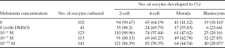
The effects of melatonin supplementation on cleavage rates and blastocyst formation of mouse SCNT were evaluated. After oocyte activation, embryos were monitored for pronucleus (PN) formation. Reconstructed embryos were cultured in media supplemented with varying doses of melatonin, DMSO and also in control medium for 5 days. The numbers of cultured embryos, cleavage rates and blastocyst formations are shown in Table 1.
Effect of melatonin addition to the culture medium on the developmental potential of single cell nuclear transfer (SCNT) oocytes

aSignificantly different (P < 0.05). DMSO, dimethyl sulphoxide.
There was no significant difference observed between the cleavage rates in the groups supplemented with melatonin, DMSO and the control.
The rate of development to blastocyst stage was significantly higher in the group supplemented with 10−12 M melatonin compared with the control group (P < 0.05).
Our findings also demonstrated that the rate of development to blastocyst stage in the 10−9 M group was higher than the control group but the difference was not statistically significant (P > 0.05).
In each group, the blastocysts were double stained in order to determine the cell number of inner cell mass (ICM) and trophectoderm (TE; Fig. 1). The result of cell number for each groups is depicted in Fig. 2. There was no significant difference between cell number in the ICM, TE and total cells from each group.
Fluorescence micrograph of a differentially labelled mouse blastocyst showing trophectoderm nuclei (asterisk, pink) and inner cell mass nuclei (arrow, blue).
Effect of melatonin concentration addition to the culture medium on the cell numbers of blastocyst (mean ± standard error of the mean (SEM)). DMSO, dimethyl sulphoxide; ICM, inner cell mass; TE, trophectoderm.
Discussion
Improving the cultivation systems is essential to achieve major success in nuclear transfer of somatic cells. Embryos derived from nuclear transfer have low developmental potency which may be due to many factors such as in vitro culture parameters, genome re-programming etc. Previous studies have shown that ROS produced during the normal process of embryo culture may damage the lipid membrane of the cell, destroying DNA, accelerating apoptosis and blocking the 2-cell stages and subsequent programmed cell death (Halliwell & Aruoma, Reference Halliwell and Aruoma1991; Legge & Sellens, Reference Legge and Sellens1991; Noda et al., Reference Noda, Matsumoto, Umaoka, Tatsumi, Kishi and Mori1991; Kowaltowski & Vercesi, Reference Kowaltowski and Vercesi1999; Guerin et al., Reference Guerin, El Mouatassim and Menezo2001; Tamura et al., Reference Tamura, Nakamura, Korkmaz, Manchester, Tan, Sugino and Reiter2009). Balance between ROS and antioxidant activity is an important factor for an appropriate embryo development (Tamura et al., Reference Tamura, Nakamura, Korkmaz, Manchester, Tan, Sugino and Reiter2009). Considering melatonin as a potential free radical scavenger, the effects of different concentrations on the development of mouse embryos derived from cumulus cell nuclear transfer was investigated in the present study.
The result of our experiments indicated that cleavage rate and blastocyst cell number were not statistically different between different groups. However, blastocyst formation rate was higher in the group which received 10−12 M melatonin. Transition from the 2- to 4-cell stage, that is the most important stage in the development of mouse embryo, was not significantly different between the groups. Contrary to our findings, Ishizuka has shown a decrease in the 2-cell block following culture of in vitro fertilized embryos in medium containing 10−6 M melatonin (Ishizuka et al., Reference Ishizuka, Kuribayashi, Murai, Amemiya and Itoh2000). They obtained these results during the natural process of embryo development but our data were collected during SCNT. In addition, these researchers cultured fertilized embryos in different concentrations of melatonin for only 24 h. However, in the present study, activated embryos were cultured at different concentrations of melatonin for 96 h. In another study, Tian and colleagues found that 10−9 M melatonin increased blastocyst formation rate, blastocyst hatching rate, and cell number per blastocyst (Tian et al., Reference Tian, Wen, Shi, Liang, Zeng, Tian, Zhou, Zhu and Liu2010). Contrary to Tian's study, McElhinny et al. did not observe any differences between the blastocyst formation and cleavage rates at different concentrations of melatonin in the 2-cell embryo culture (McElhinny et al., Reference McElhinny, Davis and Warner1996). They stated that, except for a few strains of mice, most strains did not have detectable melatonin. However, they employed the above two strains and found that cleavage rate, blastocyst formation and cell number of blastocysts were not affected by melatonin. The effect of melatonin on mouse embryo development has remained a challenge and there exist conflicting data in this field. Rodriguez-Osorio et al. found positive effects of melatonin at a concentration of 10−9 M on the cleavage rate, blastocyst formation and blastocyst cell numbers in pig embryos (Rodriguez-Osorio et al., Reference Rodriguez-Osorio, Kim, Wang, Kaya and Memili2007). There were some positive effects of melatonin observed in Choi's study on parthenogenetic and nuclear transfer embryos in pigs (Choi et al., Reference Choi, Park, Lee, Kim, Jeong, Lee, Park, Kim, Hossein, Jeong, Kim, Hyun and Hwang2008), which were due to its antioxidant properties. They showed that the amount of ROS was significantly reduced in the presence of melatonin.
The increase of cell number in blastocysts was due to decreased levels of apoptosis. Normally, apoptosis occurs during embryo development in order to eliminate abnormal cells. Apoptosis induction may happen for different reasons such as a high level of ROS and problems with the embryonic genome. Culture of parthenogenetic and in vitro fertilized embryos in medium containing melatonin showed an increased expression level of the anti-apoptotic gene Bcl-xl and also reduced the expression level of the proapoptotic gene Bax (Choi et al., Reference Choi, Park, Lee, Kim, Jeong, Lee, Park, Kim, Hossein, Jeong, Kim, Hyun and Hwang2008). However, in the present study, ICM, TE and total cells in different groups were not significantly different. Probably, in all of them, the processes of apoptosis were the same possibly due to re-programming problems that melatonin failed to prevent.
Positive effects of melatonin on the pre-implantation embryos may be due to a variety of reasons, although some of them so far have not been identified. Melatonin can directly scavenge free radicals and up-regulate the expression of several antioxidant genes such as superoxide dismutase (Mayo et al., Reference Mayo, Sainz, Antoli, Herrera, Martin and Rodriguez2002). Melatonin and its metabolites can inhibit DNA methyltransferase enzyme (DNMT) by masking target sequences or by blocking the active site of the enzyme (Korkmaz & Reiter, Reference Korkmaz and Reiter2008). Naturally, during the early stages of embryonic development, rapid demethylation occurs and, after implantation, methylation patterns re-establish (Momparler & Bovenzi, Reference Momparler and Bovenzi2000; Korkmaz & Reiter, Reference Korkmaz and Reiter2008). However, following nuclear transfer, the genome is hypermethylated 10 h after activating the oocyte which is inhibited artificially by adding trichostatin A (TSA) (Kishigami et al., Reference Kishigami, Van Thuan, Hikichi, Ohta, Wakayama, Mizutani and Wakayama2006b). Inhibition of the DNMT may be effective for reorganization of the donor nucleus after nuclear transfer. Melatonin also increases the expression of both class I and class II histone deacetylases (HDAs) such as HDA3, HDA5, and HDA7 at low concentration in neural stem cells (Sharma et al., Reference Sharma, Ottenhof, Rzeczkowska and Niles2008). Moreover, melatonin causes a significant increase in histone H3 acetylation, which is linked to chromatin modification and gene transcription. A similar effect of melatonin was observed in TSA-treated nuclear-transferred oocytes during activation and 10 h post activation. TSA improves the development of nuclear transfer embryo and blastocyst cell number (Kishigami et al., Reference Kishigami, Mizutani, Ohta, Hikichi, Thuan, Wakayama, Bui and Wakayama2006a; Rybouchkin et al., Reference Rybouchkin, Kato and Tsunoda2006). In the present study, we observed an accelerated development to blastocyst stage but we did not observe significant difference in blastocyst cell number. Previous studies have determined that if TSA treatment is given more than 10 h after activation, embryo development will reduce (Kishigami et al., Reference Kishigami, Mizutani, Ohta, Hikichi, Thuan, Wakayama, Bui and Wakayama2006a). Herein, melatonin treatment started after activation and continued to the blastocyst stage. Further studies are needed in order to examine histone acetylation and DNA methylation at all stages from 2-cell to blastocyst in the presence of melatonin. In conclusion, we infer that the effect of melatonin in different organisms can have different effects on embryo development in the natural process of fertilization and in nuclear transfer. In addition, adding melatonin to pre-implantation mouse nuclear transfer embryos can accelerate blastocyst formation.
Acknowledgements
We thank Dr T. Tsuji for his assistance. This work was supported by a grant from the Ministry of Education, Science, and Culture (no. 23013020).