Introduction
Advances in perinatal medicine have increased survival after preterm birth so that the concept of prematurity is shifting from simply a pregnancy complication to a common developmental basis for a whole and new generation of young adults. In Europe alone, there are presently 9 million children and adolescents who have been born preterm and the numbers are increasing.Reference Fellman, Hellstrom-Westas and Norman1 Although this progress is very welcome for women delivering preterm and their families, there is an increasing concern that preterm birth is an emerging risk factor for early arterial hypertensionReference Rotteveel, van Weissenbruch, Twisk and Delemarre-Van de Waal2–Reference Irving, Belton, Elton and Walker6 and diabetes,Reference Hofman, Regan and Jackson7–Reference Hovi, Andersson and Eriksson9 which may predict increased risk for cardiovascular disease and stroke.Reference Koupil, Leon and Lithell10
If and how the abrupt hemodynamic changes after preterm birth affect the cardiovascular system in a life-course perspective are so far poorly understood. Awaiting data on mechanisms, short-term and long-term follow-up studies have contributed to the identification of important cardiovascular differences in subjects born preterm compared with those born at term. Besides possible cardiac re-modeling so far only observed in lambsReference Bensley, Stacy, De Matteo, Harding and Black11 and altered autonomic control characterized by increased sympathoadrenal activity,Reference Cohen, Vella, Jeffery, Lagercrantz and Katz-Salamon12, Reference Johansson, Norman and Legnevall13 several reports point at the fact that the vascular tree is significantly smaller in people born preterm, both at the microvascularReference Bonamy, Martin, Jorneskog and Norman14–Reference Mitchell, Liew and Rochtchina17 and large artery level.Reference Bonamy, Bendito and Martin3, Reference Edstedt Bonamy, Bengtsson, Nagy, De Keyzer and Norman18, Reference Singhal, Kattenhorn, Cole, Deanfield and Lucas19 The underlying causes of such cardiovascular abnormalities are important to explore as they may provide a basis for early intervention. Smaller arteries and arterioles are likely to contribute to increased long-term cardiovascular disease susceptibility, for example, among adult patients referred for coronary arteriography, small coronary diameter was an independent predictor of atherosclerotic lesions in both the right and left coronary arteries.Reference Nwasokwa, Weiss, Gladstone and Bodenheimer20
Pediatric ophthalmologists have previously demonstrated that vascular growth in the retina stops after very preterm birth, mainly as a result of the birth-related drop in circulating insulin-like growth factor 1 (IGF-1).Reference Hellstrom, Perruzzi and Ju21, Reference Hellstrom, Engstrom and Hard22 We hypothesized that the vascular growth arrest of the eye is not restricted to the retina but is a more general phenomenon,Reference Hellstrom, Hard, Niklasson, Svensson and Jacobsson23 also affecting large vessels such as the aorta and carotid artery. As blood flow to the placenta drives fetal aortic growth in utero,Reference Langille, Brownlee and Adamson24 we also suggest that premature uncoupling of the placental circulation makes the aorta a particularly sensitive vessel for growth arrest after preterm birth. Therefore, the primary aim of this study was to compare aortic and carotid artery growth after very preterm birth with normal fetal and infant arterial growth.
Methods
This longitudinal and observational study measured aortic and carotid growth during a 6-month period, two developmental periods corresponding to the last trimester of pregnancy and the first three months of postnatal life after term. The study protocol was approved by the regional ethics committee and parental informed consent was obtained before each examination.
Subjects and study protocol
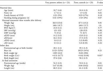
Two groups were compared: the study group comprised 21 (11 boys) very preterm infants born at Karolinska University Hospital between August 2008 and September 2009 at the start of the third trimester (gestational age between 26 and 30 weeks, mean birth weight 1209 g). All very preterm infants were singletons, without any malformations and appropriate for gestational age, defined as a birth weight within ±2 s.d. from the mean for normal fetal weight according to Swedish sex and gestational age-specific reference data.Reference Marsal, Persson and Larsen25 As controls, we selected 29 (10 boys) healthy infants born at term with normal birth weights (mean = 3420 g). Recruitment and enrollment of the controls were performed at a scheduled routine antenatal visit in the second trimester at three primary health-care maternity clinics. Gestational age had been prospectively determined in all pregnancies by fetal ultrasound examination at 17–18 postmenstrual weeks, according to Swedish recommendations for antenatal care. The parents were interviewed about their family history of cardiovascular disease and a positive history was defined as a report of myocardial infarction, stroke, treated hypertension and/or hyperlipidemia among their first-degree relatives. Infant and maternal characteristics are presented in Table 1. There was no case of maternal diabetes and one mother delivering very preterm had a diagnosis of preeclampsia. Before delivery, umbilical artery blood flow velocity measurements had been performed on clinical indication in 7 of 21 pregnancies ending very preterm. Fetal umbilical artery blood flow was found to be normal in all cases (pulsatility index = mean ± 2 s.d. for gestational length). There were no data on fetal umbilical blood flow in controls. Besides one case of preeclampsia (maternal indication for Cesarean section), primary causes of preterm delivery were preterm labor (n = 1), pre-labor premature rupture of membranes (n = 11), vaginal bleeding (n = 7) and one delivery was medically indicated because of intercurrent maternal disease (liver tumor). Once included in the study, there were no dropouts because of pregnancy or postnatal complications, fetal or infant death or withdrawal of parental consent.
Maternal and infant characteristics

CVD, cardiovascular disease; BMI, body mass index; SBP, systolic blood pressure; DBP, diastolic blood pressure; aEDD, aortic end-diastolic diameter; aSD, aortic systolic diameter; cEDD, carotid end-diastolic diameter; cSD, carotid systolic diameter.
Data are presented as mean (s.d.) values or proportions (%).
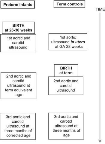
The study protocol consisted of three consecutive assessments. In both groups, ultrasonography of the major arteries was performed at 3 months before term, at term and 3 months after term-equivalent age (Fig. 1). The first ultrasound was performed at a mean age of 206 (s.d. = 10) days of postmenstrual age in very preterm infants and at 198 (s.d. = 6) days of postmenstrual age in the fetal reference group. At the third and last ultrasonographic investigation, the very preterm infants were 380 (s.d. = 16) days and the reference infants born at term 371 (s.d. = 10) days of postmenstrual age. At the final investigation, weight, length and blood pressure (mean of two measurements using an oscillometric device; Omron HEM-907, Omron Healthcare Inc., IL, USA) were measured in all infants. At the same time and using the same protocol (anthropometry, blood pressure and ultrasonography of the large arteries), we also examined the mothers once.
Flow chart of study protocol.

Ultrasonographic assessments
All recordings were performed by one experienced examiner (U.S.) using the same ultrasound machine (GE Vingmed Vivid 7, General Electric, Horten, Norway, http://www.ge.com/no/). For fetal investigations of the aorta, a phased array matrix sector probe (GE M3S 1.5–4.0 MHz) and for recordings in infants, a phased array sector probe (GE 10-S 4.0–10.5 MHz) was used. A complete diagnostic echocardiographic assessment was performed after birth in all participants to rule out malformations. All recordings in infants included electrocardiogram registration for identification of systole and end of diastole. Three consecutive angle-corrected M-mode recordings, each including at least six heart cycles, were performed to determine the diameter of the upper abdominal aorta at the level of the diaphragm, and the diameter of the common carotid artery just before the bifurcation.
All recordings were evaluated off-line by an independent examiner (M.M.) blinded for infant group belonging. Peak-systolic and end-diastolic aortic and carotid artery diameters were measured using commercially available software (Echo-PAC), and mean values from the three consecutive measurements were calculated. In addition, recordings from 12 infants were re-analyzed by another investigator (U.S). The inter-observer coefficient of variation for end-diastolic aortic diameters was found to be 2.3% and the intra-observer coefficient of variation (three consecutive evaluations of the same recording and by same examiner) was 3.0%.
Statistical methods
The results are presented as mean and s.d., median and interquartile range or proportions (%). Student's t-test, Wilcoxon rank-sum test and the χ 2 test were used to investigate group differences. The α-level was chosen to be 0.05.
Aortic and carotid diameters were considered as outcomes. Besides group, sex, weight, maternal smoking in pregnancy, maternal arterial dimensions and family history of cardiovascular disease were considered as covariates. Data with skewed distribution were log transformed before regression analyses to obtain a better approximation of normal distribution. Associations between all covariates and outcomes at the last examination were tested in univariate analyses, and associations with a P < 0.25 were entered into a forward stepwise multivariate regression model. A final multiple regression model was created from the variables surviving stepwise regression. In the final multiple regression model, the infant's current weight was always included.
Associations between outcomes at the last examination and neonatal characteristics and morbidities, such as gestational age, birth weight, standard deviations for birth weight, antenatal steroid exposure, neonatal septicemia, persistent ductus arteriosus and use of umbilical arterial catheter, were investigated within the very preterm group. All data were analyzed using JMP 8.0.1 (SAS Institute Inc., Cary, NC, USA).
Results
Maternal age, parity, smoking in pregnancy and family history of cardiovascular disease did not differ between the two study groups. In addition, there were no significant group differences in maternal height, weight, body mass index, blood pressures, aortic or carotid diameters (Table 1).
At the final follow-up, systolic [mean = 91 (s.d. = 13) v. 94 (s.d. = 9.0) mmHg] and diastolic [mean = 59 (s.d. = 11) v. 63 (s.d. = 12) mmHg] blood pressures did not differ between infants born very preterm and those born at term (P = 0.30 and 0.11, respectively).
Aortic diameters and growth
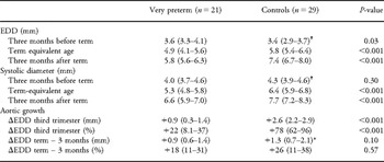
At the first assessment corresponding to an average post-conceptional age of 28 weeks, aortic end-diastolic diameter (aEDD) was somewhat larger in very preterm infants as compared with age-matched controls assessed in fetal life (median = 3.6 v. 3.4 mm, P = 0.03), whereas there was no significant group difference in systolic diameters (aSD; median = 4.0 v. 4.3 mm, P = 0.30). At the next investigation 3 months later, both aEDD (4.9 v. 5.8 mm) and aSD (5.3 v. 6.4 mm) were significantly smaller in infants born very preterm as compared with infants born at term (P < 0.001 for both comparisons). At the final assessment 6 months after the first comparison, these group differences remained highly significant: on average, the aEDD was 1.6 mm or 22% smaller in infants born very preterm as compared with those born at term (5.8 v. 7.4 mm; Table 2).
Aortic diameters (mm) in very preterm infants and controls born at term. Longitudinal assessments during a 6-month period, starting 3 months before and ending 3 months after term

EDD, end-diastolic diameter; ΔEDD, change in EDD.
Data are presented as median and interquartile range.
*P < 0.001 v. growth in fetal life.
†Fetal assessments.
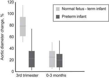
In the fetus, the aEDD increased by +2.6 mm during the third trimester. In contrast, very preterm infants exhibited only +0.9 mm increase in aEDD during the same developmental period (P < 0.001). During the following 3-month period, aortic growth velocity (aGV) continued unchanged (+0.9 mm) in very preterm infants, whereas the aGV in term controls slowed down to +1.3 mm (P < 0.001 v. fetal aortic growth), and the group difference in aGV was no longer statistically significant (Table 2 and Fig. 2).
Relative increase (%) in aortic end-diastolic diameter during a period corresponding to third trimester for term infants (in utero) and the first 3 months of extrauterine life for the very preterm group and in the first 3 months after term. Aortic growth stops prematurely after very preterm birth (P < 0.001). Horizontal lines are median values; boxes are 25–75th percentiles and vertical lines indicate 10th and 90th percentiles.

Within groups, there were no significant associations between increase in weight or length from birth to final follow-up and increase in aEDD or cEDD (carotid EDD).
At the end point of our study, aEDD did not vary in relation to infant sex, postmenstrual age, maternal smoking in pregnancy or a family history of cardiovascular disease. However, we found borderline significant correlations between infant aEDD and infants’ current weight (β = 0.42 mm/kg, r = 0.26, P = 0.07) and between infant and maternal aEDD (β = 0.072 mm/1 mm increase in maternal aEDD, r = 0.27, P = 0.06).
In the final multiple regression model including all three covariates (group, infant weight at last examination and maternal aEDD), only group belonging (very preterm-term) contributed significantly to the variation in infant aEDD (R 2 = 0.54, P < 0.001). The result of this regression analysis was the same after log-transforming infant aEDD. Excluding weight at last examination from the final multiple regression model changed the estimate for the group difference in aEDD at final follow-up from 1.40 to 1.34 mm.
Carotid artery diameters and growth
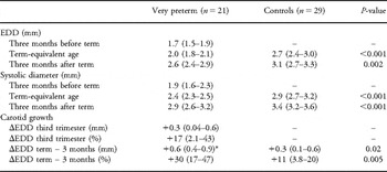
At the first assessment, the cEDD was 1.7 (interquartile range = 1.5–2.0) mm in very preterm infants (no fetal data available). At term, both cEDD (2.0 v. 2.7 mm, P < 0.001) and systolic diameters (cSD; 2.4 v. 2.9 mm, P < 0.001) were significantly smaller in very preterm as compared with term infants. Three months later and at the end point of our study, the average group difference in cEDD was 0.43 mm, corresponding to 14% smaller carotid diameter in infants born very preterm (P = 0.002; Table 3).
Carotid diameters (mm) in very preterm infants and controls born at term. Diameters were assessed during a 6-month period, starting 3 months before term and ending 3 months after term

EDD, end-diastolic diameter; ΔEDD, change in EDD.
Data are presented as median and interquartile range.
*P = 0.01 v. postnatal growth in the first 3 months period corresponding to the third trimester.
In very preterm infants, the cEDD increased on average by +0.3 mm during the first 3 months of postnatal life. During the next 3 months period, carotid growth velocity (cGV) increased in very preterm infants to +0.58 mm (P = 0.01 for group comparison), and it was higher in preterm infants as compared with controls born at term (+0.31 mm, P = 0.02 for group difference; Table 3).
At the last examination, infant cEDD did not vary in relation to infant sex, infants’ current weight maternal smoking, maternal cEDD or a family history of cardiovascular disease.
Neonatal morbidity among very preterm infants and associations to arterial size and growth
Within the very preterm group, there was no case of necrotizing enterocolitis and no infant underwent surgical ligation of a patent ductus arteriosus. Antenatal steroids for induction of lung maturation had been administered to 17 out of 21 (81%) infants, and seven (33%) infants needed ventilator support during their initial hospitalization. Sixteen (76%) had an umbilical artery catheter inserted after birth, with the catheter tip located in the lower thoracic aorta (Th6-10). All umbilical catheters had been removed before the first ultrasonographic assessment of aortic dimensions. In addition, four infants (19%) were pharmacologically treated with ibuprofen for a hemodynamically significant patent ductus arteriosus, and eight infants (38%) suffered from septicemia. Two infants had a diagnosis of a mild intraventricular hemorrhage (grade 1–2), one had chronic lung disease (defined as need of supplemental oxygen at 36 weeks of postmenstrual age) and one infant had retinopathy of prematurity stage 1.
We found no significant associations between gestational age, s.d. scores of birth weight and birth weight and the corresponding aEDD and cEDD at final follow-up. In addition, there were no statistically significant associations between neonatal treatments (antenatal steroids, ventilator treatment or umbilical artery catheterization) or morbidity (septicemia and patent ductus arteriosus) and aEDD and cEDD.
Discussion
This study presents three major and novel findings: first, the data confirm our hypothesis that the aortic narrowing previously found in children and adolescents born very pretermReference Bonamy, Bendito and Martin3, Reference Edstedt Bonamy, Bengtsson, Nagy, De Keyzer and Norman18 has a perinatal origin, occurring during a developmental period corresponding to the third trimester of pregnancy. Second, we also found smaller carotid arteries in infants born very preterm, suggesting that impaired growth of the arterial vascular tree may be a generalized phenomenon induced by very preterm birth. Finally, fetal aGV was found to be high and the aortic diameter increased by 60% during the third trimester of pregnancy. This rapid and physiological aortic growth decelerated dramatically after birth.
The strengths of this study include the longitudinal and controlled design, as well as blinded analyses of the ultrasonography images. We also excluded small-for-gestational-age subjects as a proxy for fetal growth restriction and controlled and adjusted for several potential confounders such as maternal smoking, family history of cardiovascular disease, maternal aortic size, neonatal exposures and morbidities, as well as infant body size. One limitation was that the very preterm group was on average 1 week older than the control group. If anything, this difference would introduce conservative bias and would contribute to a small underestimation of the true difference in aortic and carotid diameters between the two groups. Accordingly, including postmenstrual age in the models the group difference for aEDD was even more pronounced (it increased from 1.40 to 1.46 mm), whereas cEDD did not change. Another limitation may be that the cohort represents a convenience sample and not all consecutive births within the study period. However, using the same inclusion criteria, there is no reason to believe that vascular growth and development in this sample would differ from that in another random sample from the same population.
In utero, almost half of the combined fetal cardiac output is distributed through the aorta to the placenta. Accordingly, placental blood flow seems to drive aortic growth in the third trimester. The birth-related termination of the placental circulation and closure of fetal shunts lead to a drop in aortic blood flow velocity, which is associated with a reduction in aortic growth postpartum.Reference Langille, Brownlee and Adamson24 If this circulatory transition – including a raise in cardiac output and in systemic blood pressure – occurs well before term, the arterial system is exposed to an increased mechanical stress. Adequate postnatal adaptation and normal aortic growth do not seem to be compatible, that is, the full growth potential of the aorta during the third trimester will not be reached.
Another role of the placenta is that of an endocrine organ. The placenta produces vascular growth factors such as IGF-1. Several studies have shown that human aortic smooth muscle cells are sensitive to IGF-1 at the receptor level and that a normal concentration of IGF-1 is necessary for normal vascular development.Reference Chisalita, Johansson, Liefvendahl, Back and Arnqvist26, Reference Colao27 IGF-1 also regulates synthesis and accumulation of elastin in the arterial wall, processes which in animal models have been shown to be particularly active in the immediate perinatal period.Reference Bendeck and Langille28, Reference Wagenseil, Ciliberto and Knutsen29 In humans, downregulation of IGF-1 caused by placental insufficiency has been associated with thinner and stiffer umbilical arteries in newborn infants who had suffered from intrauterine growth restriction.Reference Burkhardt, Matter and Lohmann30 Moreover, children born small for gestational age continue to exhibit low levels of IGF-1 and vascular dysfunction.Reference Sesso and Franco31 Taken together, these findings could explain why fetal growth restriction is a developmental risk factor for lasting arterial narrowing.Reference Brodszki, Lanne, Marsal and Ley32, Reference Ley, Stale and Marsal33 After preterm birth and cessation of placental circulation IGF-1 levels drop, and persistent low serum concentrations of IGF-I in very preterm infants have been associated with growth arrest of the microvasculature in the eye, preceding retinopathy of prematurity.Reference Hellstrom, Perruzzi and Ju21, Reference Hellstrom, Engstrom and Hard22 Accordingly, insufficient IGF-1 may also be responsible for the growth arrest of the great arteries seen in the very preterm infants of our study. In fact, the great arteries may be even more sensitive to insufficiency of IGF-1 levels than the microvascular system of the eye, because only one of the very preterm infants in our study suffered from mild retinopathy of prematurity, whereas almost all showed great artery involvement. As IGF-1 levels in very preterm infants are modifiable and related to infant nutrition,Reference Engstrom, Niklasson, Wikland, Ewald and Hellstrom34 this is an important area for further research.
In the first 1–2 weeks of postnatal life, ductal systemic-to-pulmonary shunting may completely abolish or even reverse diastolic blood flow in the distal aorta of preterm newborns (4 out of 21 infants in our study).Reference Groves, Kuschel, Knight and Skinner35 In addition, arterial catheterization of the umbilical artery and distal aorta (16 out of 21 infants in our study) may also contribute to a reduced aortic blood flow in very preterm infants. Interestingly, we found no contribution of these factors to the observed aortic growth arrest after very preterm birth.
The aortic diameters observed herein correspond to those previously reported in normal fetuses.Reference Gardiner, Brodszki, Eriksson and Marsal36 However, the aortic growth retardation found after very preterm birth in our study exceeded that associated with poor fetal growth,Reference Gardiner, Brodszki, Eriksson and Marsal36 indicating that very preterm birth may be a stronger risk factor for vascular narrowing than intrauterine growth restriction.
In 6-month-old infants born very preterm, the aortic diameter was 22% and the carotid artery diameter 14% shorter as compared with age-matched controls born at term. As indicated by the growth acceleration of the carotid artery seen in very preterm infants at the final assessment, later catch-up growth may occur. In previous studies of healthy adolescents, we found 16–19% narrower aortic cross-sectional area in subjects born very preterm as compared with matched controls born at term,Reference Edstedt Bonamy, Bengtsson, Nagy, De Keyzer and Norman18 suggesting that no catch-up had occurred. Others studies have found that the brachial artery is narrower in young people born preterm as compared with those born at term.Reference Singhal, Kattenhorn, Cole, Deanfield and Lucas19, Reference Hovi, Turanlahti and Strang-Karlsson37 In contrast, carotid diameters were found to be similar in a cohort of healthy schoolchildren born very preterm and at termReference Bonamy, Andolf, Martin and Norman38 although a borderline significant narrowing of the carotid artery has recently been shown in young adults born preterm.Reference Hovi, Turanlahti and Strang-Karlsson37 Accordingly, childhood growth of the large arteries after very preterm birth may be non-uniform, with the aorta being more vulnerable for long-lasting effects after growth arrest in early life.
The significance of aortic narrowing for cardiac function and development is so far largely unknown and represents areas for further research. The significance of luminal narrowing for mechanical properties of the aorta is also an important issue, as EDD is one determinant of arterial elasticity and compliance. In spite of a narrower vessel, we have previously reported that the relative pulsatile diameter change of the abdominal aorta was significantly larger in teenagers born very preterm as compared with age-matched controls born at term.Reference Bonamy, Bendito and Martin3 However, in another cohort with older subjects, aortic elasticity was found to be decreased in adults born preterm as compared with those born at term.Reference Oren, Vos and Bos39 These findings could signal early mechanical exhaustion followed by accelerated aging of the aorta in subjects born preterm. Future studies should therefore rule out if preterm birth is a risk factor for aortic aneurysm development.
In conclusion, recent observations indicate that the growing number of people born preterm are at increased risk for hypertension,Reference Norman40 diabetesReference Hofman, Regan and Jackson7–Reference Hovi, Andersson and Eriksson9 and stroke.Reference Koupil, Leon and Lithell10 Although the underlying causes are still poorly defined, there are convincing experimental, epidemiological and clinical data for a perinatal contribution from preterm birth, rather than a common genetic trait.Reference Bonamy, Bendito and Martin3–Reference Kaijser, Bonamy and Akre8, Reference Bensley, Stacy, De Matteo, Harding and Black11, Reference Johansson, Norman and Legnevall13, Reference Edstedt Bonamy, Bengtsson, Nagy, De Keyzer and Norman18, Reference Lawlor, Hubinette and Tynelius41–Reference Singhal, Cole and Lucas43 Our study shows growth failure of the major arteries after very preterm birth, a result that is likely to have implications for follow-up and health in the new and growing population of adult people born preterm.
Acknowledgments
We acknowledge valuable assistance from midwives and nurses at Karolinska University Hospital in recruiting the mothers and infants participating in this study. This study was supported by unrestricted grants from Swedish Heart Lung Foundation, Stockholm County Council and the Swedish Order of Freemasons. The authors had full access to the data and were responsible for its integrity. All authors have read and agreed to the article as written. The authors report no conflicts of interest and have nothing to disclose.