INTRODUCTION
In some eutrophic coastal waters, successions of dominant primary producers from perennial macrophytes to opportunistic epiphytes, free-floating macroalgae, and then microalgae with increasing phases of eutrophication have been reported (Schramm, Reference Schramm1999). The excessive proliferation of free-floating green algae is generally termed as ‘green tide’ (Fletcher, Reference Fletcher, Schramm and Nienhuis1996), while that of microalgae is known as ‘red tide’. The successions of major primary producers, particularly the occurrence of green and red tides in eutrophic coastal waters, have been attributed to complex interactions among biotic and abiotic factors (Schramm, Reference Schramm1999), and those interactions between microalgae and seaweed have received much attention in ecological studies of harmful algal blooms (Smith & Horne, Reference Smith and Horne1988; Sfrifo & Pavoni, Reference Sfrifo and Pavoni1994; Alamsjah et al., Reference Alamsjah, Hirao, Ishibashi and Fujita2005).
Large-scale green tides of Ulva prolifera occurred consecutively after 2007 in the Yellow Sea (YS), China, and attracted extensive attention (Ding & Luan, Reference Ding and Luan2009; Leliaert et al., Reference Leliaert, Zhang and Ye2009; Shimada et al., Reference Shimada, Nagano, Hiraoka, Ichihara, Mineur and Zhu2010; Liu et al., Reference Liu, Keesing, He, Wang, Shi and Wang2013). Ulva prolifera has multiple reproduction modes and complex life histories (Hiraoka et al., Reference Hiraoka, Dan, Shimada, Hagihira, Migita and Ohno2003; Liu et al., Reference Liu, Yu, Yan, Zhang and Zhou2015b), with both haploid and diploid macroscopic, as well as microscopic phases occuring in its life cycles. The microscopic forms of U. prolifera, such as spores and gametes produced by thalli, are collectively known as propagules (Chapman, Reference Chapman, Blaxter and Southwood1986; Hoffmann & Santelices, Reference Hoffmann and Santelices1991; Santelices et al., Reference Santelices, Aedo and Hoffmann2002; Zhang et al., Reference Zhang, Wang, Mao, Liang, Zhuang, Wang and Ye2010). These propagules will undergo two basic processes, settlement and germination, before developing into multi-cellular thalli (Clayton, Reference Clayton1992; Fletcher & Callow, Reference Fletcher and Callow1992; Schories & Reise, Reference Schories and Reise1993; Gao et al., Reference Gao, Chen, Yi, Wang, Pan, Lin and Peng2010). Therefore, the microscopic propagules are important in controlling the population dynamics of macroalgae (Lotze et al., Reference Lotze, Schramm, Schories and Worm1999).
The amount of microscopic propagules of bloom-forming Ulva prolifera in the YS is huge and cannot be ignored. Zhang et al. (Reference Zhang, Ma and Hu2011) reported that one floating haploid thallus of U. prolifera could produce about 2.31 × 10⁷ gametes during a bloom season. Wang et al. (unpublished data) utilized fluorescence quantitative PCR to analyse the temporal-spatial distribution of Ulva microscopic propagules during the green tide in 2012. They found that the abundance of propagules increased rapidly from 0.30 to 102 cells ml–1 in May, and the maximum density of propagules could reach 104 cells · ml–1 in mid-May. The rapid increase of microscopic propagules in the YS, as addressed by Zhang et al. (Reference Zhang, Liu and Yu2015) and Liu et al. (Reference Liu, Yu, Yan, Zhang and Zhou2015b), was mainly due to the proliferation of free-floating green algae. In Subei Shoal, where green tides initially developed, microscopic propagules of green algae presented in seawater throughout the year and could serve as seed stock for the formation of green tides (Song et al., Reference Song, Li and Fang2015). These investigations suggest that the propagules play a critical role in regulating the occurrence and development of green tides in the YS (Liu et al., Reference Liu, Pang, Zhao and Hu2012; Li et al., Reference Li, Song, Xiao, Wang, Fu, Zhu, Li, Zhang and Wang2014; Song et al., Reference Song, Li and Fang2015).
Microscopic propagules of Ulva spp. and microalgae floating in seawater have a similar habitat, which gives them a chance to interact with each other. It is well known that thalli of green algae can inhibit the growth of unicellular microalgae (Jin & Dong, Reference Jin and Dong2003; Wang et al., Reference Wang, Wang, Zhou, Sun and Tang2013). Many previous studies have indicated that green algae can adversely affect phytoplankton in general, and lead to changes in composition and primary productivity of the phytoplankton community, even the occurrences of microalgal blooms (Smith & Horne, Reference Smith and Horne1988; Jin & Dong, Reference Jin and Dong2003; Nan et al., Reference Nan, Zhang and Zhao2004; Alamsjah et al., Reference Alamsjah, Hirao, Ishibashi and Fujita2005, Reference Alamsjah, Hirao, Ishibashi, Oda and Fujita2008; Wang et al., Reference Wang, Yu, Song and Zhang2006; Tang & Gobler, Reference Tang and Gobler2011; Wang et al., Reference Wang, Yu and Zhou2012). There is little recent research on the affects of microalgae on germlings of macroalgae (Schonbeck & Norton, Reference Schonbeck and Norton1979; Huang & Boney, Reference Huang and Boney1983). Since microscopic propagules are critical for the population dynamics of U. prolifera, any variation may eventually influence the subsequent population development and the magnitude of green tides. Therefore, it is necessary to look into the interactions between microalgae and microscopic propagules of macroalgae to examine all potential factors affecting the formation and consequences of green tides in detail.
In this study, we selected nine species of microalgae, including some toxic or harmful species commonly occurring in the coastal waters of China, to study the potential interactions between these microalgae and the gametes of bloom-forming Ulva prolifera isolated from the YS, through two co-culture experiments under laboratory conditions.
MATERIALS AND METHODS
Sample identification and preparation
Thalli of Ulva prolifera were collected from Huiquan Bay, Qingdao in July 2011. All samples were identified as U. prolifera using morphological and molecular methods as described in Zhang et al. (Reference Zhang, Liu and Yu2015). Thalli were washed several times with sterile seawater, gently cleaned with soft brushes, and checked under a microscope to ensure that they were free of epiphytes. The treated thalli were maintained with 1 mg l–1 GeO2 to control the growth of diatoms and other silicified algae in the laboratory.
Nine species of microalgae representing five different phyla were selected to study the interactions with gametes of Ulva prolifera. They were provided by the Algal Culture Center of the Institute of Oceanology, Chinese Academy of Sciences (IOCAS). Detailed information concerning the microalgae is listed in Table 1.
List of microalgal species used in the experiment.

PST, Paralytic shellfish toxins; DST, Diarrhetic shellfish toxins; CCMP, Provasoli-Guillard National Center for Marine Algae and Microbiota; IOCAS, Institute of Oceanology, Chinese Academy of Sciences.
Microalgae and Ulva prolifera were cultured in flasks containing specific medium (Guillard, Reference Guillard, Smith and Chanley1975; Guillard & Hargraves, Reference Guillard and Hargraves1993) (Table 1), and maintained at 19 ± 1°C. Irradiance was 50 µmol photons m−2 s−1, and the light:dark cycle was 14:10. Natural seawater for preparation of culture medium was collected from Huiquan Bay, Qingdao, and sand-filtered prior to use. The seawater was further filtered through a 0.45 µm membrane and autoclaved to prepare the culture medium. Salinity of the seawater was 30 ± 1.
Obtaining gametes of Ulva prolifera
Thalli of Ulva prolifera cultured in the laboratory showed alternative phases of parthenosporophytes and gametophytes as described in Liu et al. (Reference Liu, Yu, Yan, Zhang and Zhou2015b). The gametophytes produced biflagellate unfertilized gametes, which could develop parthenogenetically into sporophytes. The sporophytes then produced tetraflagellate spores, which would develop into gametophytes. Gametes and spores could be easily distinguished by their size, numbers of flagella and phototactic responses. The gametes with two flagella were obviously smaller than spores that had four flagella. Besides, the gametes showed positive phototaxis, while the spores showed negative phototaxis.
To prepare gametes for the experiments, spores produced by a single thallus were cultured until they developed into gametophytes. A modified punching method was then adopted to induce the formation of gametes (Dan et al., Reference Dan, Hiraoka, Ohno and Critchley2002). The thalli of gametophytes were cut into small fragments (1–2 cm long), and incubated in a glass Petri dish (95 mm in diameter, 10 mm high) containing 20 ml f/2 medium. The medium was replaced every day for 2–3 days until the gametes were released.
Design of experiments
Two experiments were designed to study the interactions between microalgae and propagules of Ulva prolifera. Based on the abundance of these microalgae in natural seawater, the microalgal cell density in the experiments was set to be 1.0 × 10²–1.0 × 10⁴ cells ml–1. For all the experiments, microalgae at the exponential phase were collected and used. Cell density of gametes used in the experiments was set to be 2.0 × 10⁴ cells ml–1, which is similar to the abundance of microscopic propagules (2.0 × 10⁴ cells ml–1) in the YS determined by the qPCR assay (Wang et al., unpublished data).
EXPERIMENT I: INTERACTIONS BETWEEN MICROALGAE AND GAMETES OF ULVA PROLIFERA AT THE SETTLEMENT STAGE
To study the interactions between microalgae and gametes of Ulva prolifera at the settlement stage, the newly released gametes were collected and put into small beakers containing 40 ml of sterile seawater (final cell density at 2.0 × 10⁴ cells ml–1). For each species of microalgae, three levels of cell density were set up (1.0 × 10², 1.0 × 10³ and 1.0 × 10⁴ cells ml–1). Monocultures of U. prolifera gametes and nine species of microalgae (cell density at 1.0 × 10², 1.0 × 10³ and 1.0 × 10⁴ cells ml–1 for each species, respectively) were used as controls. Every treatment or control had three replicates. To avoid nutrient limitation, 40 µl of f/2 or L1 stock solution was added at the beginning of the experiments, the concentrations of NO3-N, PO4-P in the f/2 or L1 stock solution were 882 and 36 mmol l−1.
Part 1: Effects of microalgae on settlement of gametes. To test the effects of microalgae on gametes of Ulva prolifera, a coverslip (10 × 10 mm) was placed at the bottom of each beaker to facilitate the counting of attached gametes. The experiments were conducted under darkness in a stable environment for one day to ensure gametes randomly attached to the coverslip. After 24 h, the coverslips were removed and placed in sterilized seawater several times to remove unattached gametes. Attached gametes were then counted in 20 fields under a microscope (400 × magnification), and the average value of the 20 fields was used to represent the number of settled gametes.
Part 2: Effects of gametes on the growth of microalgae. To test the effects of gametes on the growth of microalgae, 200 µl of culture medium was collected from each group of treatments and controls after the experiments, and fixed with Lugol's solution. The cell density of microalgae was counted under a microscope, and the significance of variation was compared between each treatment and the control.
EXPERIMENT II: INTERACTIONS BETWEEN MICROALGAE AND GAMETES OF ULVA PROLIFERA AT THE GERMINATION STAGE
For this experiment, gametes of Ulva prolifera (2.0 × 10⁴ cells ml–1) were incubated in small beakers containing 40 ml of sterile seawater under darkness for one day to ensure settlement of gametes. Then the beakers were put back to the light condition for the germination experiment. Nine species of microalgae were then inoculated into the medium, and the cell density was adjusted to 1.0 × 10⁴ cells ml–1. Monocultures of U. prolifera gametes and nine species of microalgae were used as controls. Each group of treatments and controls was performed in three beakers. Every day, 200 µl of f/2 or L1 stock solution was added to avoid nutrient limitation.
Part 1: Effects of microalgae on germination of gametes. To test the effects of microalgae on germination of gametes, a coverslip (10 × 10 mm) was placed at the bottom of each beaker prior to the experiment to observe the germination of gametes (a gamete that divided into at least two cells was considered as the standard of germination). Every day, the coverslip in each beaker was taken out to count the germination rates and then put back. Germinated gametes were counted for 20 fields under a microscope (400 × magnification), and the average germination rate was calculated according to the following formula: GR (germination rate, %) = N/N 0 × 100%, where N 0 is the total number of gametes observed in one field, and N is the number of germinated gametes in one field. The experiments lasted for 7 days. All beakers were shaken twice a day during the experiments to avoid adherence of microalgae to the beakers.
Part 2: Effects of germinated gametes on the growth of microalgae. To test the effects of germinated gametes on the growth of microalgae, 200 µl of seawater was collected from each beaker every day, and 200 µl of f/2 or L1 stock solution was added to ensure a constant volume and to avoid nutrient limitation. All samples were fixed with Lugol's solution prior to cell counting. The experiment lasted for 7 days and the growth inhibition rates on the 7th day were calculated with the following formula: GIR (growth inhibition rate, %) = (1–N/N 0) × 100%, where N 0 is the cell density of the control groups on the 7th day; and N is the cell density of the treatment groups on the 7th day.
Statistical analysis
Data were calculated as mean ± standard deviation from the three replicates (N = 3). Statistical differences were analysed with Origin 8.5 and SPSS 16.0, and the differences were considered significant when P < 0.05.
Two statistical methods were used in the experiments. First, the significance of difference among nine species of microalgae in affecting the settlement and germination of gametes were tested with one-way ANOVA, after testing for normal distribution and homogeneity of variance. For the data with heterogeneity of variances, non-parametric tests (Kruskal–Wallis one-way ANOVA) were used. If the difference was significant, a post-hoc test (Tukey's test or Dunn's test) was performed to test the difference among the experimental groups. For the effects of gametes on the growth of microalgae, t-test for independent samples was used to test the significance between the treatment and the control.
RESULTS
Effects of microalgae on the settlement and germination of Ulva prolifera gametes
EFFECTS OF MICROALGAE ON THE SETTLEMENT OF ULVA PROLIFERA GAMETES
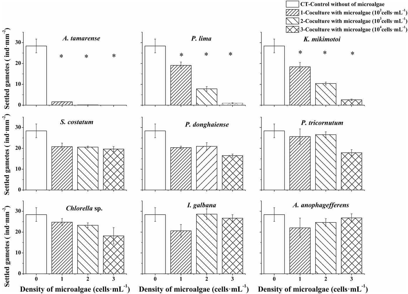
Effects of nine microalgal species on the settlement of Ulva prolifera gametes were shown in Figure 1. The tested microalgal species displayed significant differences in affecting the settlement of gametes (one-way ANOVA, P < 0.05). Six microalgal species, i.e. Skeletonema costatum, Prorocentrum donghaiense, Phaeodactylum tricornutum, Chlorella sp., Isochrysis galbana and Aureococcus anophagefferens, had no significant effects on the settlement of gametes at the three levels of cell density (Figure 1D–H). The other three microalgal species, i.e. Alexandrium tamarense, Prorocentrum lima and Karenia mikimotoi, significantly decreased the settled number of gametes compared with the control, and the inhibitory effect increased with cell density (Figure 1A–C). These microalgae will lead to adverse effects on the settlement of gametes of U. prolifera at the bloom density (Table 1).
Effects of microalgae on the settlement of Ulva prolifera gametes. Cell densities of microalgae were set at three levels: 1.0 × 10², 1.0 × 10³, 1.0 × 10⁴ cells ml–1, respectively, and the concentration of U. prolifera gametes was 2.0 × 10⁴ cells ml–1. Data points are means ± SD (N = 3). *P < 0.05 as compared with the control.

EFFECTS OF MICROALGAE ON THE GERMINATION OF ULVA PROLIFERA GAMETES
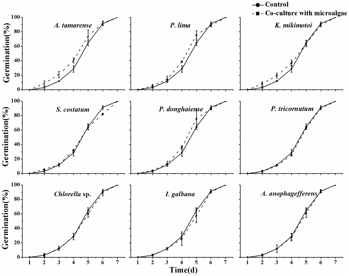
Gametes in all tested groups of Experiment II germinated completely within 7 days, and there was no significant difference in the final germination rate of gametes co-cultured with nine species of microalgae (Figure 2). Although the germination process of gametes seems slightly accelerated in the groups co-cultured with A. tamarense, P. lima, K. mikimotoi, S. costatum and P. donghaiense from 2–6 day, the differences between control and treatments were not significant (one-way ANOVA, P > 0.05).
Effects of microalgae on the germination of Ulva prolifera gametes. Cell density of microalgae was set at 1.0 × 10³ cells ml–1, and the concentration of U. prolifera gametes was 2.0 × 10⁴ cells ml–1. Data points are means ± SD (N = 3). *P < 0.05 as compared with the control.

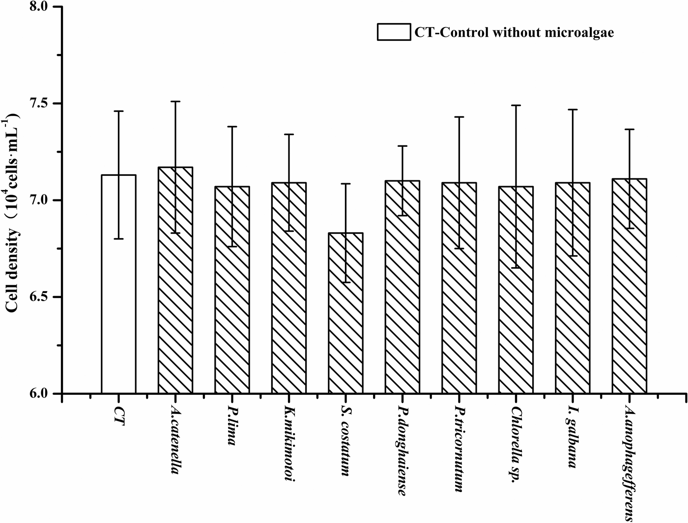
In addition, the total number of cells of germinated and un-germinated gametes were counted every day to reflect the increase of green algal biomass during the experiment. A rapid increase of Ulva prolifera biomass was observed over the 7-day experiment. The total biomass of U. prolifera increased about 3–4 times as reflected from the total cell number recorded every day. There was no significant difference among the treatments of germinated gametes co-cultured with nine species of microalgae (Figure 3) (one-way ANOVA, P > 0.05).
The total number of germinated and un-germinated gametes of Ulva prolifera recorded on the 7th day of Experiment II. Data points are means ± SD (N = 3). * P < 0.05 as compared with the control.

Effects of Ulva prolifera gametes on the growth of microalgae
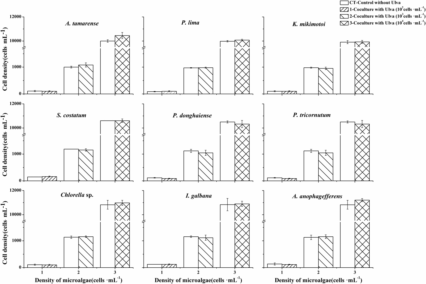
The growth of microalgae co-cultured with gametes of Ulva prolifera at the settlement stage is shown in Figure 4. Compared with the controls, no significant inhibitory or lethal effects of gametes were observed on the microalgae at the three different levels of cell density (t-test, P > 0.05). The results suggest that gametes of U. prolifera had no significant inhibitory effects on microalgae.
Effects of gametes on the growth of microalgae at the settlement stage. The experiment lasted for 1 day. Cell density of microalgae were set at three levels of 1.0 × 10², 1.0 × 10³, 1.0 × 10⁴ cells ml–1, respectively, and the concentration of Ulva prolifera gametes was 2.0 × 10⁴ cells ml–1. The data are presented as the means ± SD (N = 3). * P < 0.05 as compared with the control.

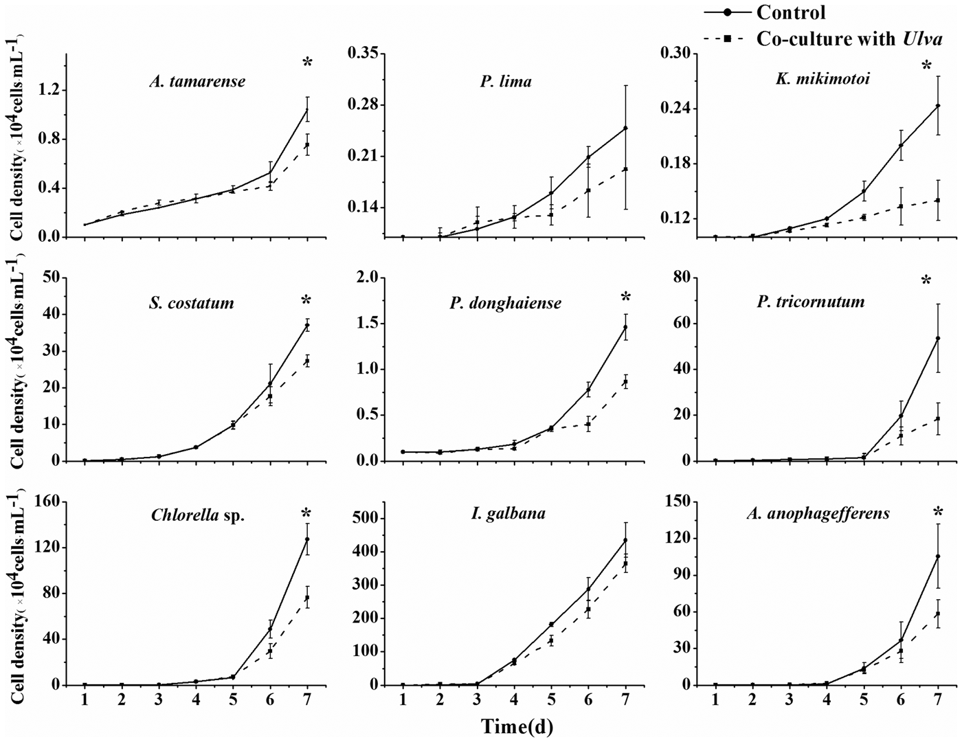
The effects of germlings germinated from gametes on the growth of microalgae are shown in Figure 5. Compared with the controls, the growths of microalgae co-cultured with germlings of U. prolifera were significantly inhibited. On the 7th day, the growth inhibition rates of the nine species of microalgae were 28%, 43%, 27%, 41%, 66%, 40% and 45% for A. tamarense, K. mikimotoi, S. costatum, P. donghaiense, P. tricornutum, Chlorella sp. and A. anophagefferens, respectively (t-test, P < 0.05).
Effects of germinated gametes on the growth of microalgae. The cell density of microalgae was set at 1.0 × 10³ cells ml–1, and the concentration of Ulva prolifera gametes was set as 2.0 × 10⁴ cells ml–1. Data points are means ± SD (N = 3). * P < 0.05 as compared with the control.

DISCUSSION
Complex interactions between microalgae and propagules of green algae
In the current study, the complex interactions between propagules of bloom-forming Ulva prolifera in the YS and microalgae were demonstrated for the first time through two co-culture experiments. Three species, i.e. Alexandrium tamarense, Prorocentrum lima and Karenia mikimotoi, showed significant inhibitory effects on the settlement of gametes, while the gametes had no apparent effects on the growth of microalgae. At the germination stage, however, the microalgae did not affect the germination of gametes, while the germlings showed significant inhibitory effects on the growth of microalgae. It seems that the complex interactions between propagules of U. prolifera and microalgae do not result from resource competition. During the experiments, enough nutrients were supplemented into the culture medium so that nutrients would not be a limiting factor. The impacts were unlikely to be caused by other environmental factors such as light, because the settlement experiment (Experiment I) was performed in darkness. During the germination experiment (Experiment II), the density of microalgae (1.0 × 10⁴ cells ml–1) was not high, and there would be little shading effect. Amsler et al. (Reference Amsler, Reed and Neushul1992) pointed out that propagules of macroalgae would hardly reach a high enough density to compete for light. Other abiotic factors (e.g. temperature and pH) were also measured throughout the experiments and were unlikely to become limiting factors. As mentioned above, it seems that resource competition is unlikely to account for the interactions in our study.
An interesting finding in this study is that three species with significant inhibitory effects on the settlement of Ulva prolifera gametes were species which can produce toxins, such as paralytic shellfish toxins (PST), diarrhetic shellfish toxins (DST) and haemolytic toxins (Table 1). Among the three species, A. tamarense showed most significant inhibition on the settlement of gametes. However, we compared the inhibitory effects of the PST-producing A. tamarense with a non-PST-producing dinoflagellate Alexandrium affine under the same experimental conditions, and found this non-toxic Alexandrium strain also inhibited the settlement of U. prolifera. When A. affine (at the density of 1.0 × 10² cells ml–1) was co-cultured with the gametes, the settled number of gametes was only 2 cells·mm−2, very close to the co-culture with toxic A. tamarense (7 cells mm−2) (Liu et al., Reference Liu, Yan, Zhou, Zhang and Lin2015a). Therefore, it can be implied that the PST produced by Alexandrium did not play a major role in affecting gametes. Other bioactive compounds may be involved in the interactions. However, we did not further test the toxins of P. lima and K. mikimotoi in inhibiting the gametes, for which further elucidation is needed. Recent research has shown that the settlement of Ulva propagules was impacted by some bacteria (Joint et al., Reference Joint, Tait, Callow, Callow, Milton, Williams and Cámara2002; Wheeler et al., Reference Wheeler, Tait, Taylor, Brownlee and Joint2006; Tait et al., Reference Tait, Williamson, Atkinson, Williams, Cámara and Joint2009). Joint et al. (Reference Joint, Tait, Callow, Callow, Milton, Williams and Cámara2002) pointed to the bacterial quorum sensing molecules N-acylhomoserine lactone (AHL) as a cue for the site selection of Ulva zoospore settlement. So bacteria may be an important factor in the settlement mechanisms of U. prolifera propagules; this will need to be carefully evaluated.
Through the germination experiment, we found that the inhibitory effects of Ulva prolifera on microalgae started from the early germination stage of gametes. During the 7-day experiment, the germinated gametes significantly reduced the density of the microalgae, even the toxic microalgal species. The impacts of macroalgae are generally not mediated by toxic substances; some metabolites like polyunsaturated fatty acids were thought to cause negative effects on microalgae. Jensen (Reference Jensen, Faulkner and Fenical1977) examined whether propagules could affect their own microenvironment via the release of certain metabolites during settlement and germination stages. Recent studies also showed that mature thalli of U. prolifera in YS could produce chemicals that inhibit the growth of microalgae (Nan et al., Reference Nan, Zhang and Zhao2004; Alamsjah et al., Reference Alamsjah, Hirao, Ishibashi, Oda and Fujita2008; Huo et al., Reference Huo, Tian, Xu, Wang, Feng, Fang and He2010; Wang et al., Reference Wang, Wang, Zhou, Sun and Tang2013). However, whether the germlings of U. prolifera produce the same compounds as the mature thalli is not clear so far. Some studies have emphasized that certain development stages of macroalgae would associate with their unique characteristics, particularly at the microscopic stage (Amsler et al., Reference Amsler, Reed and Neushul1992).
Ecological implications of the interactions between microalgae and propagules of green algae in the YS
In many eutrophic waters, apparent succession of primary producers has been reported (Schramm, Reference Schramm1999). To study the interactions between opportunistic macroalgae and microalgae is an important approach to understand the succession process and the formation of harmful algal blooms. In this study, we found that some bloom-forming microalgal species could inhibit the settlement of gametes. Based on the studies of green tides in the YS, the Subei Shoal is a key region for the early development of green tides (Keesing et al., Reference Keesing, Liu, Fearns and Garcia2011; Liu et al., Reference Liu, Keesing, He, Wang, Shi and Wang2013). Microscopic propagules are present in this region throughout the year, which offer important seed stock for the recurrent green tides (Li et al., Reference Li, Song, Xiao, Wang, Fu, Zhu, Li, Zhang and Wang2014; Song et al., Reference Song, Li and Fang2015). Besides, the intensive Porphyra culture rafts deployed in this region offer important substrates for the settlement of microscopic propagules, in particular the large amounts of gametes produced in spring (Liu et al., Reference Liu, Yu, Yan, Zhang and Zhou2015b; Song et al., Reference Song, Li and Fang2015). In Subei Shoal, however, there are few reports of microalgal blooms and the biomass of phytoplankton is generally low (Kang et al., Reference Kang, Sun, Sun, Xu and Que2013), probably due to the highly turbid seawater and the strong mixing. Besides, the presence of large amounts of green algae and cultured Porphyra could also inhibit the growth of microalgae (Huo et al., Reference Huo, Tian, Xu, Wang, Feng, Fang and He2010; Sun et al., Reference Sun, Liu, Yan and Ma2010; Wang et al., Reference Wang, Wang, Zhou, Sun and Tang2013). Therefore, the settlement of microscopic propagules is not likely to be inhibited by microalgae in this region.
The occurrences of harmful algal blooms caused by Alexandrium tamarense, Prorocentrum lima and Karenia mikemotoi, however, have been recorded widely in the coastal waters of China (China Marine Environmental Bulletin, 2001–2012). Due to their production of toxins such as paralytic shellfish toxins, diarrhetic shellfish toxins and haemolytic toxins, these bloom-forming species have led to considerable concern regarding their potential ecological consequence. The outbreak stage of these harmful algal blooms coincides well with the green tides in the YS (from May to August). The occurrence of such algal blooms could impair the settlement of coexisting gametes of Ulva prolifera, and the subsequent population dynamics of U. prolifera. The green tides of U. prolifera occurred consecutively in the YS for at least 8 years (2007–2014), but there is no record of an associated population of this bloom-forming U. prolifera in the coastal region of China, except for the Subei Shoal. And the floating population of bloom-forming U. prolifera still remains a unique ecotype in the YS (Zhao et al., Reference Zhao, Jiang and Qin2015). Whether this phenomenon is related to the interactions between microalgae and bloom-forming U. prolifera is not clear but worth further study.
In this study, it was also found that bloom-forming Ulva prolifera exhibited negative effects on the growth of microalgae from the germination stage of gametes. This result, together with some previous studies (Huo et al., Reference Huo, Tian, Xu, Wang, Feng, Fang and He2010; Wang et al., Reference Wang, Yu and Zhou2012, Reference Wang, Wang, Zhou, Sun and Tang2013), reflected the impacts of U. prolifera on the phytoplankton community. During the green tides in the YS, there were huge amounts of floating green algae, which required large amount of nutrients to support such a high biomass. Macroalgae generally showed a higher efficiency in absorption of nutrients than microalgae. Hence, macroalgal blooms could reduce the abundance of phytoplankton, and even the occurrence of HABs through competition on nutrients in the sea. Besides, the floating thalli of U. prolifera could lead to shading effects on phytoplankton, and the high rate of photosynthesis of U. prolifera may draw down CO2 concentration and lead to the variation of pH level in seawater (Xu et al., Reference Xu, Fan and Zhang2012). Our results regarding the inhibition of U. prolifera on some harmful algae fitted with the situation in YS. Through comparing the records of harmful algal blooms before and after the green tides occurred in YS, we found the HAB events decreased by nearly a half over the last five years, from nine times in 2001–2005 to four times in 2008–2012 (China Marine Environmental Bulletin, 2001–2012). Similar situations have been reported in other sea areas. Investigations by Smith & Horne (Reference Smith and Horne1988) showed a dramatic decline of phytoplankton blooms with the introduction of Ulva in San Francisco Bay-Estuary, California. Sfrifo & Pavoni (Reference Sfrifo and Pavoni1994) also found that phytoplankton blooms were relatively scarce when macroalgae were found in the central Venice lagoon.
From the current study, it can be seen that Ulva prolifera inhibit not only the HAB-forming species of microalgae, but also microalgae that could serve as food for zooplankton. Therefore, the inhibition on phytoplankton will decrease marine primary productivity, and influence the energy transfer through the food chains. Qin et al. (Reference Qin, Ji, Song and Xu2011) proved that the diversity and abundance of dominant species in the phytoplankton community changed significantly during the green tides in 2008, compared with those reported prior to the year 2002. Zhang et al. (Reference Zhang, Luan, Sun and Wang2013) also pointed out that green tides of U. prolifera simplified the structure of phytoplankton community in the YS. Therefore, the potential ecological consequences of green tides are closely related with the interactions between U. prolifera and microalgae.
CONCLUSION
In this study, we demonstrated the complex interactions between the propagules of bloom-forming Ulva prolifera and microalgae through two co-culture experiments. Some microalgae could inhibit the settlement of gametes, although the mechanism is not yet clearly understood. The bloom-forming U. prolifera, however, showed strong and non-selective inhibitory effects on the growth of microalgae early from the germination stage of gametes. A better understanding of the interactions between microalgae and the propagules of bloom-forming U. prolifera is necessary in order to understand the mechanisms and ecological consequences of green tides in the Yellow Sea.
ACKNOWLEDGEMENTS
We thank Professor Ian Jenkinson for his kind help in linguistic advice.
FINANCIAL SUPPORT
This study was supported by the National Basic Research Priority Program of the Ministry of Science and Technology (2010CB428705), the Strategic Priority Research Program (XDA01020304) of the Chinese Academy of Sciences, and the joint program of NSFC (41476102) and Jiangsu province (17KJB170019).