Pentalogy of Cantrell, first described in 1958,Reference Cantrell, Haller and Ravitch1 is a rare congenital syndrome that affects roughly one in every 100,000 individuals. Findings include defects in the supra-umbilical abdominal wall and the lower sternum, deficiencies in both the anterior portion of the diaphragm and the diaphragmatic aspect of the pericardium, as well as intracardiac defects.
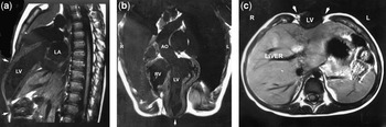
We saw recently an infant with an anterior abdominal and sternal defect, and a prominent left ventricular diverticulum protruding through the anterior chest wall, covered only by skin. There was also a moderately-sized ventricular septal defect. Magnetic resonance imaging (Fig. 1) was performed at 6 months of age to assess the extent of the diverticulum. Cardiac-gated T1-weighted spin-echo images of the chest were obtained, with the patient sedated. Imaging in the oblique coronal plane (a) demonstrated the left ventricular diverticulum (vertical arrow). Imaging in the saggital plane (b) showed the shortened sternum and truncated subcutaneous tissue (top arrow), as well as the upper boundary of the abdominal wall (bottom arrow). Taken together, the arrows demonstrate the extent of the defect in the midline. Imaging in the transverse plane (c) demonstrated the degree of protrusion of the left ventricular (LV) diverticulum through the anterior abdominal wall. The arrows delineate the lateral boundaries of the subcutaneous tissue and rectus muscles (Ao = aorta; LA = left atrium, R = right, RV = right ventricle).
Figure 1

At surgical closure of the ventricular septal defect, the diverticulum was not excised. The vulnerable myocardium was covered with pectoral and abdominal muscle flaps to protect the area from trauma. Six years later the child continues to do well.