I. INTRODUCTION
Over the past decade, the need to create a sustainable energy future has resulted in the increasing global interest in research and development on thermoelectric (TE) materials. The efficiency and performance of TE energy conversion is related to the dimensionless figure of merit (ZT) of the TE materials, given by ZT = S2σT/κ, where T is the absolute temperature, S is the Seebeck coefficient or TE power, σ is the electrical conductivity, and k is the thermal conductivity (Nolas et al., Reference Nolas, Sharp and Goldsmid2001). Only a small number of materials have been found to have practical industrial applications and all of those exhibit ZT values around or below 1.0. Optimization of the ZT values is not straightforward because S, σ, and κ are interrelated. Reports that relatively high ZT values are possible in both thin film and bulk forms have revitalized interest in development of TE materials (Tritt, Reference Tritt1996; Ghamaty and Eisner, Reference Ghamaty and Eisner1999; Venkatasubramanian et al., Reference Venkatasubramanian, Siivola, Colpitts and O'Quinn2001; Hsu et al., Reference Hsu, Loo, Guo, Chen, Dyck, Uher, Hogan, Polychroniadis and Kanatzidis2004; Dresselhaus et al., Reference Dresselhaus, Chen, Tang, Yang, Lee, Wang, Ren, Fleurial and Gogna2007).
The stability of metal oxides at high temperature has made these materials highly relevant to waste heat conversion applications. For example, cobaltates that include La1−x Ax CoO3 (A = Pb, Na) (He et al., Reference He, Chen, Calvarese and Subramanian2006), NaCoO x (Terasaki et al., Reference Terasaki, Sasago and Uchinokura1997), Ca2Co3O6 (Mikami et al., Reference Mikami, Funahashi, Yoshimura, Mori and Sasaki2003; Mikami and Funahashi, Reference Mikami and Funahashi2005), Ca3Co4O9 (Masset et al., Reference Masset, Michel, Maignan, Hervieu, Toulemonde, Studer and Raveau2000; Mikami et al., Reference Mikami, Itaka, Kawaji, Wang, Koinuma and Lippmaa2002; Grebille et al., Reference Grebille, Lambert, Bourée and Petricek2004; Hu et al., Reference Hu, Si, Sutter and Li2005), Bi2Sr2Co2O x (Shin and Murayama, Reference Shin and Murayama2000; Wang et al., Reference Wang, Venimadhav, Guo, Chen, Li, Soukiassian, Schlom, Pan, Wong-Ng, Vaudin, Cahill and Xi2009), and Ba2Ho(Cu3−x Co x )O6+x (Wong-Ng et al., Reference Wong-Ng, Yang, Hu, Huang, Lowhorn, Otani, Kaduk, Green and Li2009) combine large Seebeck coefficients and relatively low thermal conductivities. Consequently in recent years, considerable research has been conducted on oxide TEs, including phase diagram studies of ternary oxide systems using CaO and Co3O4 as two of the end members (Wong-Ng et al., Reference Wong-Ng, Liu, Martin, Thomas, Lowhorn and Otani2010, Reference Wong-Ng, Luo, Tang, Xie, Kaduk, Huang, Yang, Tang and Tritt2011, Reference Wong-Ng, Laws and Yan2013, Reference Wong-Ng, Laws, Talley, Huang, Yan and Kaduk2014).
The lanthanide-containing double perovskite iron cobaltates may also offer desirable TE properties. This paper describes the structure and X-ray reference pattern determination of the R(Fe0.5Co0.5)O3 (R = Pr, Nd, Sm, Eu, and Gd) system. We used density functional theory (DFT) calculations to investigate the structural stability of Pr2(FeCo)O6 in comparison with experimental data, followed by electronic density of states (DOS) calculations to reveal its electrical properties. Since X-ray diffraction (XRD) is a non-destructive technique for phase identification, XRD patterns are especially important for phase characterization; therefore another goal of this investigation was to determine the experimental patterns for R(Fe0.5Co0.5)O3, and to make them widely available through submission to the Powder Diffraction File (PDF, 2016).
II. EXPERIMENTAL
A. Sample preparation
Samples were prepared from stoichiometric amounts of R 2O3 Co3O4, and Fe2O3 (all raw materials are 99.9% pure) using solid-state high-temperature techniques. The starting samples were mixed, pelletized, and heat treated in air at 750 °C for 1 day and subsequently annealed at 950 °C for 15 h, 980 °C for 80 h with intermediate grindings, and 1050 °C for 60 h with intermediate grindings. During each heat treatment, the samples were furnace cooled. The heat treatment process was repeated until no further changes could be detected in the X-ray powder diffraction patterns.
B. X-ray Rietveld refinements and powder reference patterns
X-ray powder diffraction patterns of R(Fe0.5Co0.5)O3 powders were measured using a Bruker D2 Phaser diffractometer equipped with a LynxEye position-sensitive detector (certain commercial equipment, instruments, or materials are identified in this paper in order to specify the experimental procedure adequately. Such identification is not intended to imply recommendation or endorsement by the National Institute of Standards and Technology, nor is it intended to imply that the materials or equipment identified are necessarily the best available for the purpose). Patterns were measured (CuKα radiation, 30 kV, 10 mA, 0.6 mm divergence slit, 2.5° Soller slits, 3 mm scatter screen height) from 5° to 130° 2θ in 0.020 214 4° steps, counting for 1 s step−1. To minimize fluorescence, the lower window of the MCA was changed from its default value of 0.11–0.19 V.
The Rietveld refinement technique (Rietveld, Reference Rietveld1969) with the software package GSAS (Larson and von Dreele, Reference Larson and von Dreele2004) was used to analyze the diffraction patterns of R(Fe0.5Co0.5)O3 (R = Pr, Nd, Sm, Eu, and Gd). The structure of La2FeCoO6 (PDF 4-13-6526, Karpinsky et al., Reference Karpinsky, Troyanchuk, Dobryanskii, Szymczak and Tovar2006) reported previously, was used as a starting model for the refinement process. All Fe/Co sites were refined assuming equiatomic occupancies. The constant wavelength Pseudo-Voigt profile function (function #4 with 18 terms) was used for all refinements (Thompson et al., Reference Thompson, Cox and Hastings1987; Finger et al., Reference Finger, Cox and Jephcoat1994; Stephens, Reference Stephens1999). GSAS offers a number of different background functions. For each of these functions, the number of terms to be used is adjustable. The more number of terms, the more complex is the shape that can be fit. In theory, each function will have advantages under different circumstances. In general, the Shifted Chebyschev (type #1) is preferable to the others for the vast majority of Rietveld refinements. In this work, background correction was performed using the GSAS background function #1 with six terms, and an absorption/surface roughness correction was applied using function #3.
Reference patterns were obtained with a Rietveld pattern decomposition technique. Using this technique, the reported peak positions were derived from the extracted integrated intensities, and positions calculated from the lattice parameters. When peaks are not resolved at the resolution function, the intensities are summed, and an intensity-weighted d-spacing is reported.
C. Transmission electron microscopy (TEM)
Samples of Pr2 (Fe0.5Co0.5)O3 for TEM were prepared by dispersing the crushed powder on a lacey-carbon-coated copper grid. Electron diffraction and imaging were performed using a TEM operated at 300 kV.
D. Bond valence sum (V b) calculations
The bond valence sum values, V
b, for the R, Fe, and Co sites were calculated using the Brown-Altermatt empirical expression (Brown and Altermatt, Reference Brown and Altermatt1985; Breese and O'Keeffe, Reference Brese and O'Keeffe1991). The V
b of an atom i is defined as the sum of the bond valences v
ij
of all the bonds from atoms i to atoms j. The most commonly adopted empirical expression for the bond valence v
ij
as a function of the interatomic distance d
ij
is
 $v_{ij} = {\rm exp}^{{[({R_0}^-{d_{ij}})} {/B}]}$
. The parameter, B, is commonly taken to be a “universal” constant equal to 0.37 Å. The values for the reference distance R
0 for Fe–O, Co–O, Pr–O, Nd–O, Sm–O, Eu–O, and Gd–O are 1.759, 1.70, 2.135, 2.117, 2.088, 2.076, and 2.065, respectively (Brown and Altermatt, Reference Brown and Altermatt1985; Breese and O'Keeffe, Reference Brese and O'Keeffe1991). For sites shared by several species, the resulting bond-valence sum (BVS) is the weighted sum using the site occupancy.
E. Computation methods
Structural optimization and electronic structure calculations of Pr2(FeCo)O6 were performed using Vienna ab initio simulation package (VASP) (Kresse and Furthmuller, Reference Kresse and Furthmuller1996), based on DFT. The generalized gradient approximation with the Perdew–Becke–Erzenhof parameterization was used to estimate the exchange–correlation functional (Perdew et al., Reference Perdew, Burke and Ernzerhof1996). DFT + U calculations were also implemented using a cutoff energy of 400 eV for the expansion of the plane-wave basis set within the projector augmented wave formalism (Blőchl, Reference Blőchl1994; Kresse and Joubert, Reference Kresse and Joubert1999). Perovskite structure relaxation, according to the conjugate-gradient algorithm, was converged with the residual Hellman–Feynman forces of <0.01 eV Å−1. For the electronic structure calculations, we applied the GGA + U scheme of Liechtenstein et al. (Reference Liechtenstein, Anisimov and Zaanen1995) with a Hubbard U effective on-site coulomb interaction to the f-orbitals of Pr and d-orbitals of Fe and Co: U Pr = 18 eV, U Fe = 18 eV, and U Co = 12 eV. The DFT + U method could overcome the band gap underestimation by a traditional DFT method. We also optimized the U values for Pr, Fe, and Co and the k-mesh sampling in the Brillouin zone with DOS convergence tests. The DFT + U method is the common and reliable method to calculate electronic structure for a wide range of materials with highly-correlated elements.
In order to model disorder structures and accommodate 0.5 occupancy factor of Fe and Co and their randomness on the B sites, we used 1 × 1 × 1, 2 × 1 × 1, and 2 × 2 × 1 cells of Pr2(FeCo)O6 with different atomic arrangements of Fe and Co for comparison. Note that the occupancies of Fe and Co atoms in different cells remain equivalent. To further study the electrical properties of Pr2(FeCo)O6 compound, we applied the DFT + U approach to calculate DOS with more dense k-mesh sampling.
III. RESULTS AND DISCUSSION
A. XRD and TEM studies
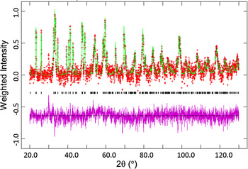
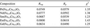
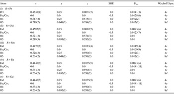
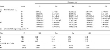
Figure 1 displays the results of Rietveld refinements for Nd2(Fe0.5Co0.5)O3. The observed (crosses), calculated (solid line), and difference XRD patterns (bottom) as determined by the Rietveld analysis, are shown. The difference pattern is plotted on the same scale as other patterns. The tick marks indicate the calculated peak positions. Since Fe and Co fluoresce when illuminated with Cu radiation, even with attempts to reduce the fluorescence by adjusting the analyzer electronics settings, some fluorescence still remains. Figure 1 is a weighted least-squares plot, to highlight the weak reflections (Larson and von Dreele, Reference Larson and von Dreele2004). Table I gives the refinement results. Table II tabulates the lattice parameters and calculated density D x . As expected, the D x values increase as the size of ionic radius, r(R 3+) (Shannon, Reference Shannon1976), decreases. The atomic coordinates and displacement parameters are given in Table III. Table IV summarizes the bond distances, selected bond angles, and BVS values.
Observed (crosses), calculated (solid line), and difference XRD patterns (bottom) for Nd(Fe0.5Co0.5)O3 by the Rietveld analysis technique. The difference pattern is plotted at the same scale as the other patterns. The vertical scale is not the raw intensity, but the weighted intensity; this plot emphasizes the fit to the weaker peaks. The peak at 29.6° results from CuK β contamination.

Rietveld refinement results (R p, R wp, and χ 2) of R(Fe0.5Co0.5)O3 (R = Pr, Nd, Sm, Eu, and Gd), Pnma (No. 62), Z = 2.

Unit-cell parameters of R(Fe0.5Co0.5)O3 (R = Pr, Nd, Sm, Eu, and Gd) [Pnma (No. 62), Z = 2, D x refers to calculated density]. Uncertainties refer to standard deviation. r(R 3+) (Å) is the ionic radius for R (VIII coordination).

Atomic coordinates and isotropic displacement factors for R(Fe0.5Co0.5)O3; [Pnma (No. 62), Z = 2, D x refers to calculated density]. SOF stands for site occupancy factor. Uncertainties refer to standard deviation.

Bond distances and bond-valence sum (BVS) values for R(Fe0.5Co0.5)O3 (R = Pr, Nd, Sm, Eu, and Gd), M = Co/Fe. If more than one atom types occupy the same site, the resulting BVS is the weighted sum of each site occupancy. Uncertainties refer to standard deviation.

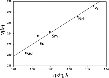
R(Fe0.5Co0.5)O3 phases reportedly acquire orthorhombic Pnma (No. 62) symmetry (Karpinsky et al., Reference Karpinsky, Troyanchuk, Dobryanskii, Szymczak and Tovar2006), as determined by the combined anti-phase and in-phase octahedral rotations, which can be described as a − a − c + using Glazer's notation. The unit-cell volume (Figure 2) for R(Fe0.5Co0.5)O3 increases monotonically with the increasing size of r(R 3+), from Gd to Pr, respectively. The volume increase is mainly due to the larger b and c lattice parameters, although the magnitude of the a-parameter maintains an opposite trend (Table II). This trend implies that the twisting of the [(Fe/Co)O6] octahedra in R(Fe0.5Co0.5)O3 has a larger influence on the magnitude of the a-cell parameter, while that of the size of eight-coordination polyhedra, [RO8], has a stronger effect on b and c.
Plot of unit-cell volume, V, of R(Fe0.5Co0.5)O3 (R = Pr, Nd, Sm, Eu, and Gd) vs. r(R 3+). A monotonic decrease of V is observed which follows the trend of lanthanide contraction.

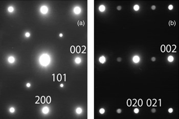
Electron diffraction patterns (Figure 3) feature sharp superlattice reflections at ½ooo and ½ooe locations (“o” and “e” refer to the odd and even reflection indexes, respectively, for a perovskite unit cell with a 0 ≈ 4 Å). These superlattice reflections are consistent with the presence of both anti-phase and in-phase octahedral rotations. All the reflections are indexable according to the unit cell √2a 0 × 2a 0 × √2a 0 and reflection conditions hk0 h = 2n agree with the Pnma (No. 62) space group, which is expected for the a − a − c + octahedral tilting. No hints of Fe/Co ordering could be observed. For example, a common rock-salt-type ordering of these cations would yield 110 reflections, which, however, are notably absent in the present diffraction patterns. These observations agree with Galasso and Pyle (Reference Galasso and Pyle1963a, Reference Galasso and Pyle1963b) and Blasse (Reference Blasse1965) which suggest weakening of long-range B-site order with decreasing mismatch between the ionic radii of B-cations sharing the same crystallographic site, as encountered for Fe and Co.
Electron diffraction patterns for Pr(Fe0.5Co0.5)O3 in [100] (a) and [001] (b) zone axis orientations of the orthorhombic √2a 0 × 2a 0 × √2a 0 unit cell. The superlattice reflections 011 (½ooo in the cubic perovskite indexes) and 210 (½ooe) arise because of octahedral rotations; the intensities of the latter are dominated by contributions from the antiparallel Pr displacements that accompany the octahedral tilting. The 110 reflections are absent consistent with the Pnma space group and disordered Co and Fe.

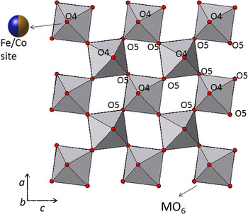
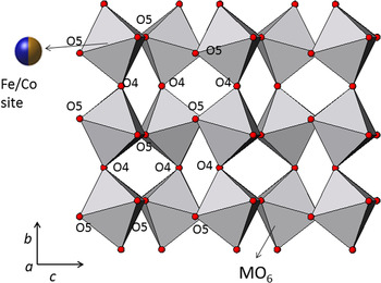
Figures 4 and 5 illustrate the structure of R(Fe0.5Co0.5)O3. In orthorhombic R 2M2O6 (“M” is used to represent the mixed site Co/Fe), there are two distinct M–O–M angles (M–O4–M and M–O5–M) that relate neighboring octahedra. The M–O4–M angle (Figure 5) and the M–O5–M angles (Figure 4) which both deviate from the ideal value of 180° reflect the degree of octahedral tilting. The three tilt angles θ, ϕ, and ω which follow the notation of Zhao et al. (Reference Zhao, Weider, Parise and Cox1993a, Reference Zhao, Weider, Parise and Cox1993b) to represent rotations of octahedral about the pseudocubic [110]p, [001]p, and [111]p axes, respectively, are shown in Table IVb. All the three tilt angles increase for R = Pr to R = Gd: θ from 12.268° to 15.148°, ϕ from 7.522° to 15.776°, and ω from 14.360° to 21.737°. This increase across the lanthanide series indicates a greater octahedral distortion as the size of R decreases.
Crystal structure of R(Fe0.5Co0.5)O3 (R = Pr, Nd, Sm, Eu, and Gd) based on X-ray powder data, showing the unit-cell outline, labeling of various atoms, and the coordination environment of the distorted [(Co, Fe)O6] octahedra view along the b-axis. R atoms are not shown for clarity.

Crystal structure of R(Fe0.5Co0.5)O3 (R = Pr, Nd, Sm, Eu, and Gd) based on X-ray powder data, showing the unit-cell outline, labeling of various atoms, and the coordination environment of the distorted [(Co, Fe)O6] octahedra view along the a-axis. R atoms are not shown for clarity.

The Goldschmidt geometrical tolerance factor (t) is a criterion for the stability and distortion of perovskite structures. It can also be used to calculate the compatibility of an ion with a crystal structure. The tolerance factor t is defined as t = 〈r A + r O〉/√2〈r B + r O〉 (Goldschmidt, Reference Goldschmidt1926), where r A, r B, and r O represent the ionic radii of A-cations, B-cations, and oxygen, respectively. In the ideal cubic structure with t = 1, the unit-cell parameter a can be described as a = √2〈r A + r O〉 = 2〈r B + r O〉. As t approaches 1, the structure is expected to become less distorted; with t < 1 the distortion is accommodated by the cooperative rotations of the [BO6] octahedra, resulting in a lower symmetry. In R(Fe0.5Co0.5)O3, the coordination number (c.n.) is 8 for R 3+ sites and 6 for the Fe3+/Co3+ sites. The experimental R–O and Fe/Co–O values are used to compute the t values. As r(R 3+) decreases across the lanthanide series, so do the t values. The t values for R(Fe0.5Co0.5)O3, as R = Pr, Nd, Sm, Eu, and Gd are 0.889, 0.885, 0.869, 0.863, and 0.857, respectively. These t values fall in the range of Goldschmidt “orthorhombic/rhombohedral” structure type, 0.71–0.90 (Goldschmidt, Reference Goldschmidt1926).
The BVS values (Table IVc) for the R 3+ site (ranging from 2.99 to 3.162) and for the (Fe/Co)–O site (3.019–3.111) in R(Fe0.5Co0.5)O3 imply near strain-free situation (V b > 3.0, the ideal valence value). Indeed, octahedral tilting in these structures relieves bond strain.
B. Structural stability and electrical properties
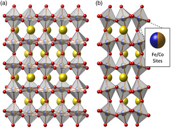
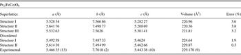
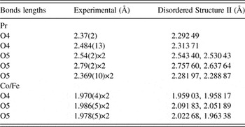
Table V shows the structural data derived for different superlattices and disordered structures for Pr(Fe0.5Co0.5)O3. Through the total energy minimization approach a disordered Pr(Fe0.5Co0.5)O3 structure is more stable than its ordered variants. Disordered Structure II has lattice parameters a = 5.61, b = 7.50, and c = 5.46 Å. Its unit-cell volume is 229.87 Å3. We found that the calculated lattice parameter c and volume agree well with the experimental data, while the lattice parameters a and b are slightly different from the experimental values by <3%. This difference could be attributed to the fact that the experimental lattice parameters are measured as the average values of the unit cells on a macro scale, whereas DFT calculations present the local structure features on a much smaller atomic scale. Table VI compares the calculated and experimental Pr–O and Fe(Co)–O bond distances. In general, the Pr–O bonds are longer than the Fe(Co)–O bonds. DFT calculations indicate that the disorder of Fe and Co leads to larger lattice parameters and unit-cell volume. Expanding the cell size provides more options for the ordered atomic arrangement of Fe and Co on the B sites. Starting from a 1 × 1 × 1 unit cell, we double the lattice parameters a and b, and estimated total energy/atom for each supercell. Disordered Structure II has a 2 × 2 × 1 cell and completely random arrangement of Fe and Co atoms on the B sites (Figure 6). Its total energy/atom is slightly lower (by ~0.42%, i.e. more negative) than that for Disordered Structure I with a 2 × 1 × 1 cell. This result implies that the random occupancy of the B sites is energetically favorable in the Pr(Fe0.5Co0.5)O3 compound.
(a) Front view and (b) side view of the 2 × 2 × 1 supercell of Pr(Fe0.5Co0.5)O3, including disordered B-sites with 50–50 occupation of Fe and Co.

Calculated structure parameters by DFT for Pr(Fe0.5Co0.5)O3 compound. Error % refers to the difference between calculated and experimental volume data.

Calculated bond distances by DFT for Pr(Fe0.5Co0.5)O3 compound.

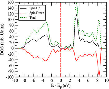
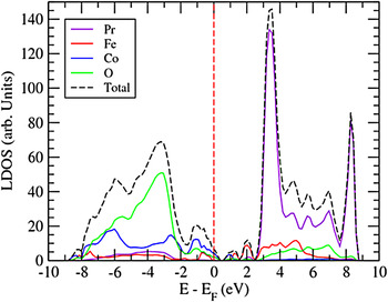
Figure 7 shows a small band gap near the Fermi energy E F. Pr2(FeCo)O6 is a semiconductor with a band gap of E g ≈ 0.43 eV (As mentioned earlier, DFT + U method was applied to overcome the band gap underestimation by traditional DFT method. We also optimized the U values for Pr, Fe, and Co and the k-mesh sampling in the Brillouin zone with DOS convergence tests.) The spin-up and spin-down densities are slightly asymmetric and favor the spin-down configuration below the Fermi energy E f, causing a small net magnetism. As shown in Figure 8, the local DOS is projected onto individual Pr, Fe, Co, and O atoms to reveal their contributions to the electrical properties of the compound. O and Co provide the main contributions to the total DOS below E f, while Pr and Fe dominate above E f between 0 and 2.5 eV. Intense orbital hybridization around E f also gives evidence that covalency plays an important role in the perovskite bonding characteristics.
DOS for Pr(Fe0.5Co0.5)O3 with a band gap of E g ≈ 0.43 eV. E f refers to Fermi energy.

Local DOS for Pr(Fe0.5Co0.5)O3 projected on Pr, Fe, Co, and O atoms in comparison with total DOS.

C. Reference XRD patterns
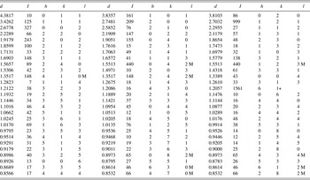
An example of the reference pattern of Sm(Fe0.5Co0.5)O3 is shown in Table VII. In this pattern, the symbols “M” and “+” refer to the peaks that contain contributions of 2 and more than 2 reflections, respectively. The strongest peak in the pattern is assigned an intensity of 999 and other lines are scaled relative to this value. The d-spacing values are calculated values from refined lattice parameters. The intensity values reported are integrated intensities (rather than peak heights) calculated from corresponding profile parameters as reported in Table VII. For peaks containing more than one reflection that can still be resolved , intensity-weighted calculated d-spacing, along with the observed integrated intensity and the hkl indices of both peaks (for “M”), or the hkl indices of the strongest peak (for “+”) are used. For peaks that are not resolved within the instrumental resolution, the intensity-weighted average d-spacing and the summed integrated intensity value are used. In the case of a cluster, unconstrained profile fits often reveal the presence of multiple peaks, even when they are closer than the instrumental resolution. In this situation, both d-spacing and intensity values are reported independently. All patterns have been submitted for inclusion in the Powder Diffraction File (PDF).
X-ray powder pattern for Sm(Fe0.5Co0.5)O3 [Pnma (No. 62), Z = 2, a = 5.4963(2) Å, b = 7.6205(2) Å, c = 5.355 56(10) Å, V = 224.313(11) Å3, D x = 7.574 g cm−3]. The symbols “M” and “+” refer to peaks containing contributions from two and more than two reflections, respectively. The particular peak that has the strongest intensity in the entire pattern is assigned an intensity of 999 and other lines are scaled relative to this value. The d-spacing values are calculated values from refined lattice parameters, and “I” represents integrated intensity values.

IV. SUMMARY
Crystal structure and powder diffraction reference patterns of the perovskite R(Fe0.5Co0.5)O3 (R = Pr, Nd, Sm, Eu, and Gd) compounds have been determined and the latter have been submitted to the PDF. The Co/Fe sites in this double perovskite series are disordered, as supported by both TEM and DFT calculations. Octahedral tilting was observed in these compounds with the tilt angles (distortion) increasing as r(R 3+) decreases; the tilting relieves the R–O bond strain. DFT + U calculations for Pr(Fe0.5Co0.5)O3 indicated it to be a narrow band gap semiconductor, which will be validated by experiment in the future.
SUPPLEMENTARY MATERIAL
To view supplementary material for this article, please visit http://dx.doi.org/10.1017/S088571561600049X.
ACKNOWLEDGEMENT
ICDD is acknowledged for Grant-in-Aid assistance for the project.