Introduction
Mcgovernite was described as a new species from Sterling Hill, New Jersey, by Palache and Bauer (Reference Palache and Bauer1927), its nature as a very large structure was described by Wuensch (Reference Wuensch1960), and a preliminary description of its crystal structure was given by Cooper and Hawthorne (Reference Cooper and Hawthorne2001). Dunn et al. (Reference Dunn, Francis and Innes1988) reported a mcgovernite-like mineral from the Kombat mine, Otavi Valley, Namibia, and Hawthorne et al. (Reference Hawthorne, Abdu, Ball and Pinch2013) recently described this mcgovernite-like mineral as the new species carlfrancisite. Brugger et al. (Reference Brugger, Armbruster, Meisser, Hejny and Grobety2001) described turtmannite from manganoan ores filling paleokarst pockets in the Triassic marbles of the Barrhorn Unit in the Central Alps, Switzerland, and reported its structure. Mcgovernite (Wuensch, Reference Wuensch1960; Cooper and Hawthorne, Reference Cooper and Hawthorne2001), ideally Zn3(Mn2+,Mg,Fe3+,Al)42(As3+O3)2(As5+O4)4[(Si,As5+)O4]8(OH)42 is isostructural with carlfrancisite, ideally Mn32+(Mn2+,Mg,Fe3+,Al)42(As3+O3)2(As5+O4)4[(Si,As5+)O4]8(OH)42, and both are related to turtmannite, ideally (Mn,Mg)22.5Mg3–3x(V,AsO4)3(SiO4)3(AsO3)xO5–5x(OH)20+x (Brugger et al., Reference Brugger, Armbruster, Meisser, Hejny and Grobety2001). All three structures have similar cell dimensions and consist of close-packed (and not-so-close-packed) layers of octahedra and tetrahedra stacked along the c direction. These structures are the most complicated of a family of minerals based on dominantly close-packed layers (Wuensch, Reference Wuensch1968), including arakiite (Cooper and Hawthorne, Reference Cooper and Hawthorne1999), dixenite (Araki and Moore, Reference Araki and Moore1981), franciscanite (Pertlik, Reference Pertlik1986), hematolite (Moore and Araki, Reference Moore and Araki1978), kraisslite (Moore and Ito, Reference Moore and Ito1978; Cooper and Hawthorne, Reference Cooper and Hawthorne2012), örebroite (Dunn et al., Reference Dunn, Peacor, Erd and Ramik1986) and welinite (Moore, Reference Moore1967). Here, I report the structures of mcgovernite and carlfrancisite and examine the similarities and differences between mcgovernite, carlfrancisite and turtmannite.
Sample provenance
The carlfrancisite crystals used in the present work were taken from the holotype sample. A sample of mcgovernite from Sterling Hill, New Jersey, was purchased from a mineral dealer and its identity was confirmed by X-ray diffraction.
Chemical composition
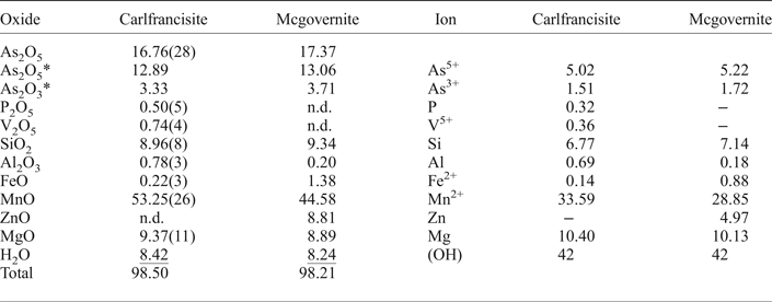
Crystals were mounted in epoxy, polished, and analysed using a Cameca SX-100 electron microprobe operating in wavelength-dispersion mode with an accelerating voltage of 15 kV, a specimen current of 10 nA, and a beam diameter of 10 µm. The following standards were used: cobaltite: As; apatite: P; synthetic VP2O7: V; diopside: Si; andalusite: Al; fayalite: Fe; spessartine: Mn; gahnite Zn; forsterite: Mg. The data were reduced and corrected by the PAP method of Pouchou and Pichoir (Reference Pouchou, Pichoir and Armstrong1985) and the presence and quantity of (OH) groups were established by crystal-structure solution and refinement. Table 1 gives the resultant chemical compositions (each the mean of ten determinations).
The chemical composition (wt.%) and unit formulae (apfu) of mcgovernite and carlfrancisite.
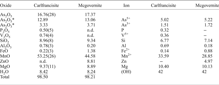
*The As3+/(As3+ + As5+) ratio was derived from the crystal structure; n.d. not detected.
Calculation of the chemical formulae
The empirical formulae were calculated on the basis of 96 anions with (OH) = 42 apfu (anions per formula unit) as indicated by crystal-structure solution and refinement. Interatomic distances show that all Mn and Fe are in the divalent state. Both minerals contain significant As. The refined site-scattering and long <T–O> bond lengths indicate the presence of both trivalent and pentavalent As in mcgovernite and carlfrancisite. These valence states were used in the calculation of the empirical formulae, giving the following: carlfrancisite: Mn2+33.59Mg10.40Fe2+0.14Al0.69As3+1.51Si6.77P0.32V5+0.36As5+5.02O54(OH)42; mcgovernite: Mn2+28.85Mg10.13 Fe2+0.88Al0.18Zn4.97Si7.14As5+5.22O54(OH)42.
X-ray data collection, crystal-structure solution and structure refinement
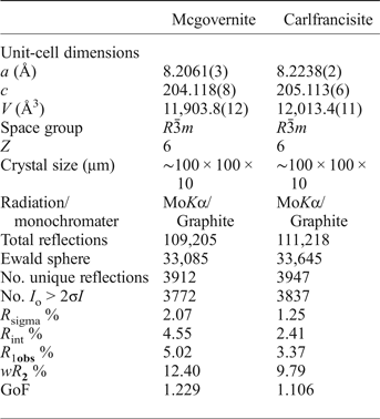
Each crystal was attached to a tapered glass fibre. X-ray-diffraction data for carlfrancisite were collected with MoKα X-radiation on a Bruker D8 three-circle diffractometer equipped with a rotating-anode generator, multilayer optics and an APEX-II CCD detector. Intensities were collected to 60°2θ using 5 s per 0.3° frame with a crystal-to-detector distance of 8 cm. An empirical absorption correction (SADABS; Sheldrick, Reference Sheldrick2008) was applied, and the data were corrected for Lorentz, polarization and background effects. X-ray-diffraction data for mcgovernite were collected with MoKα X-radiation on a Siemens P4 four-circle diffractometer. Cell dimensions were determined from the setting angles of 30 automatically aligned reflections in the range 14 < 2θ < 30°. The refined unit-cell parameters of both structures are given in Table 2, together with other information pertaining to data collection and structure refinement.
Miscellaneous information for mcgovernite and carlfrancisite.
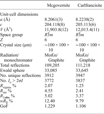
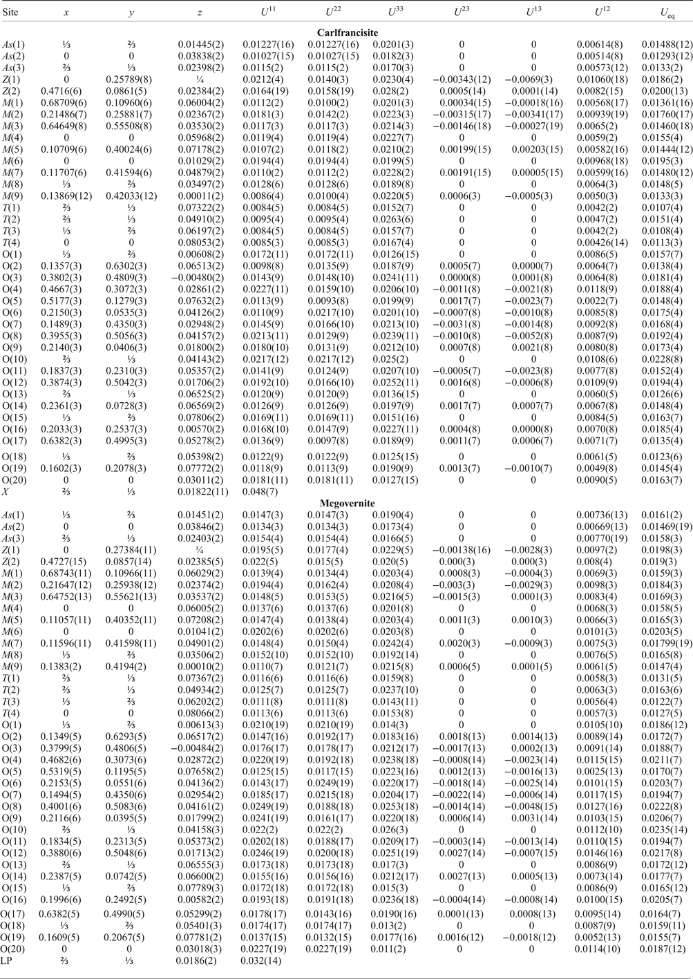
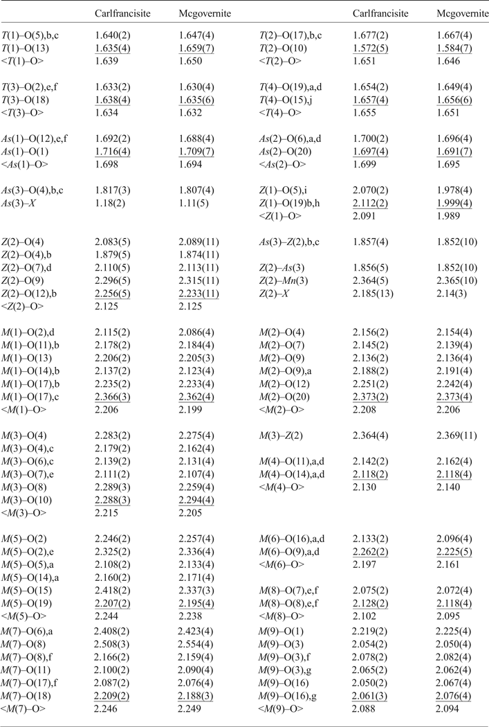
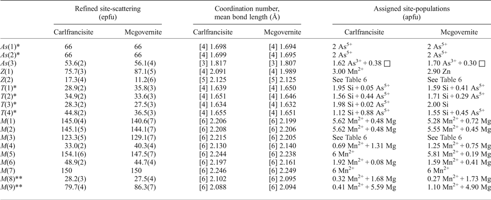
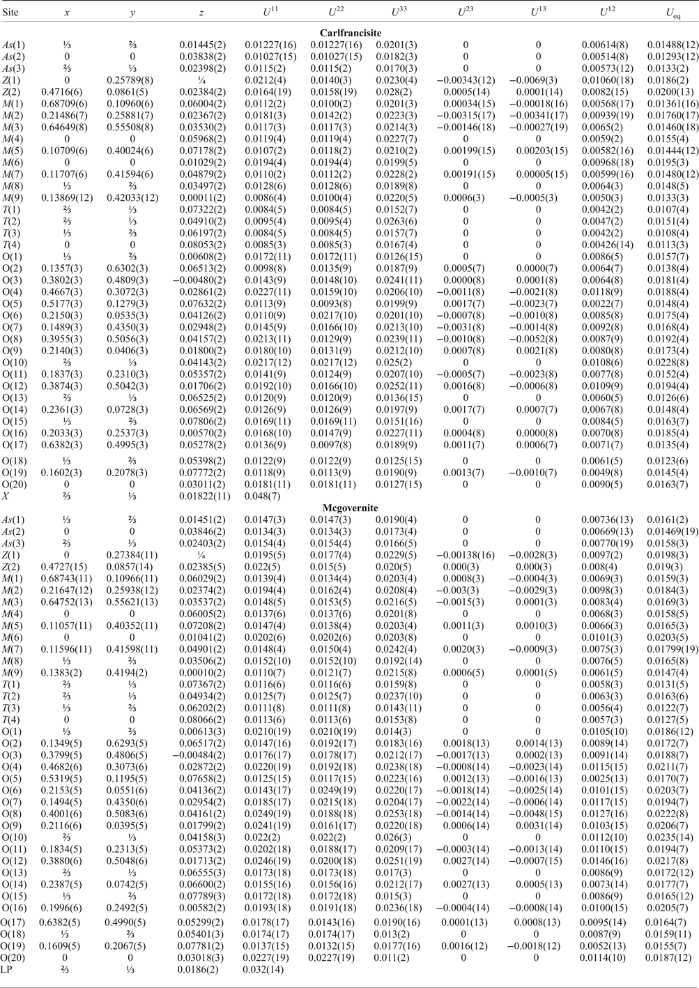
The structures were solved by direct methods and refined in the space groups R3̄m with the SHELXTL version 5.1 system of programs (Bruker, Reference Bruker1997). There are several scattering species to be distributed over a comparatively large number of sites, but this cannot be done unambiguously during refinement (Hawthorne, Reference Hawthorne1983). Hence I assigned what I considered to be the dominant scattering species at each site and refined the occupancies unconstrained. If the absorption corrections are accurate, the resulting site-scattering values (Hawthorne et al., Reference Hawthorne, Ungaretti and Oberti1995) should also be accurate, and one can combine these values with mean bond lengths and constituent-cation radii to derive site populations. The final models refined to R 1 indices of 3.56% for carlfrancisite and 5.02% for mcgovernite. Final atom parameters are given in Table 3, selected interatomic distances in Table 4, and refined site-scattering values and site populations in Tables 5 and 6; a bond-valence table for carlfrancisite was calculated using the parameters of Gagné and Hawthorne (Reference Gagné and Hawthorne2015) and is given in Table 7; crystallographic information files have been deposited with the Principal Editor of Mineralogical Magazine, and are available as Supplementary material (see below).
Atom coordinates and displacement parameters for carlfrancisite and mcgovernite.
Selected interatomic distances (Å) in carlfrancisite and mcgovernite.
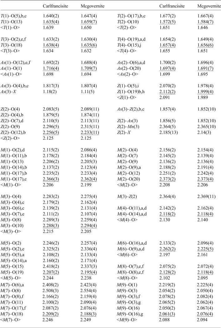
a: –y, x–y, z; b: 1–y, x–y, z; c: 1–x + y, 1–x, z; d: y–x, –x, z; e: 1–y, 1 + x–y, z; f: y–x, 1–x, z; g: x–y, x, –z; h: y + 1/3, x–1/3, –z + 1/6; i: 4/3–x, 2/3–x + y, 1/6–z; j: y–2/3, x–1/3, –z + 1/6.
Site-scattering values and assigned site-populations in carlfrancisite and mcgovernite.
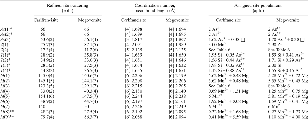
*includes minor V5+ and P;
**includes minor Al.
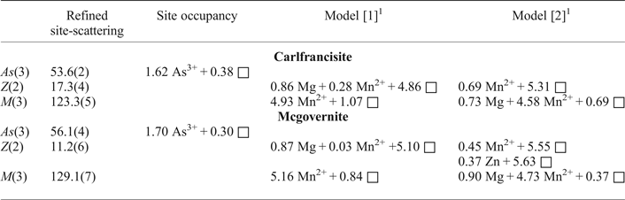
Site occupancies for the As(3), Z(2) and M(3) sites.
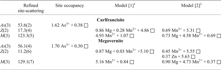
1 see text
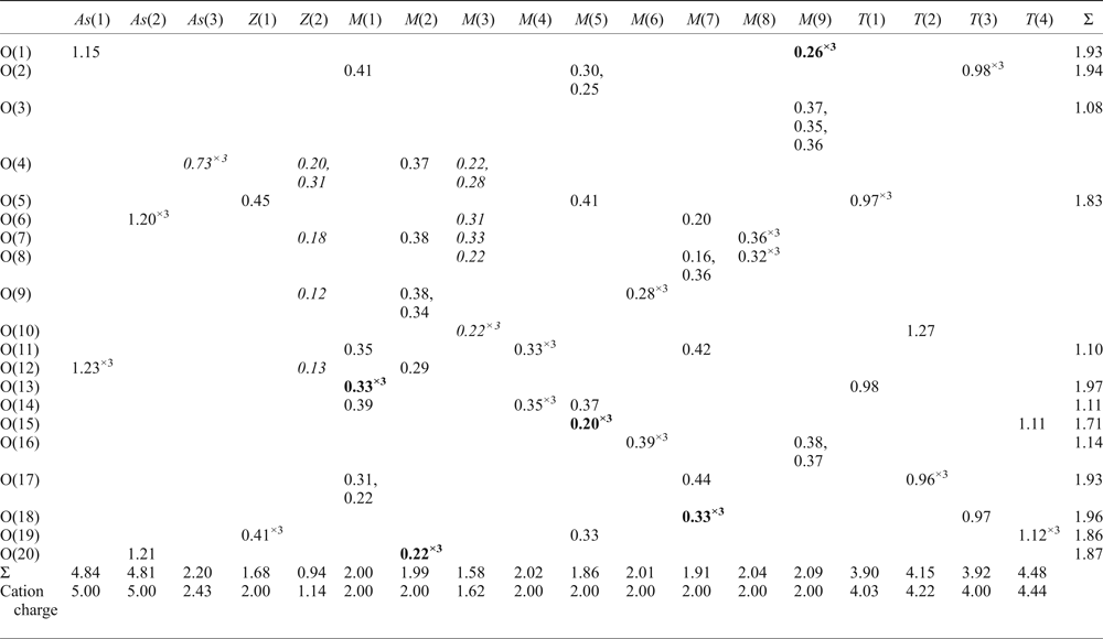
Bond-valence table for carlfrancisite.
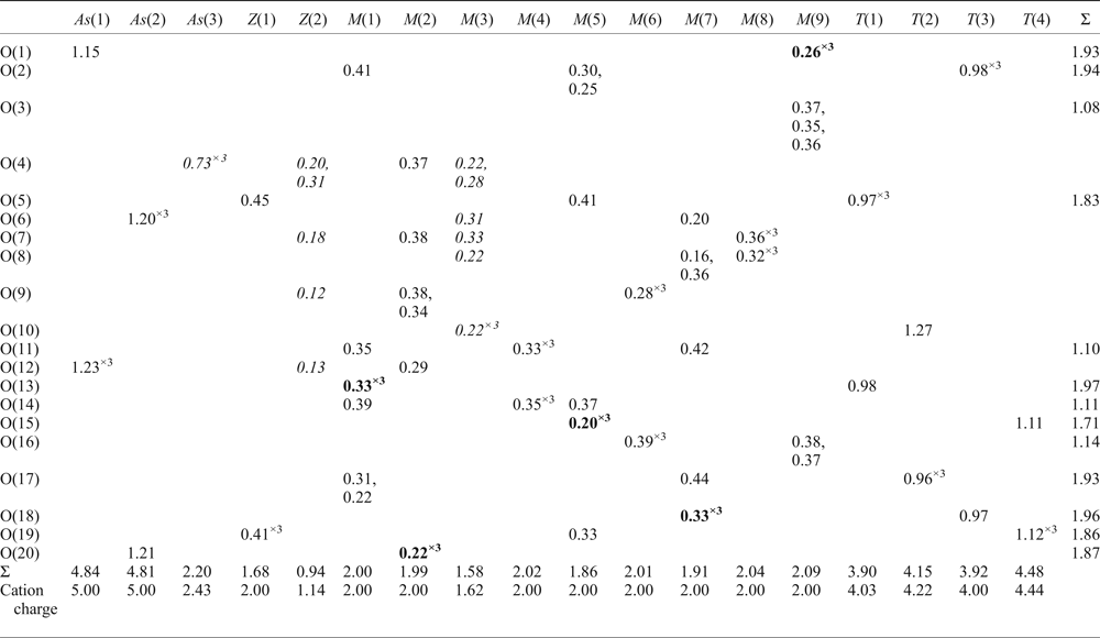
Bold font indicates that the bond-valence sum is included in the sum to the right; italics indicates vacancies.
Assignment of site populations
There are 18 crystallographically distinct cation sites, and the site occupancies were assigned using the refined site-scattering values (Table 5), the observed local stereochemistry, the unit formula derived by electron-microprobe analysis (Table 1), and the constraints exerted by cation sites that are too close for simultaneous local occupancy. Inspection of the interatomic distances in both structures (Table 4) shows that three sites are involved in short (long-range) interatomic separations such that both sites in each local pair of sites cannot each be occupied, and this forces the occurrence of vacancies at each of these sites. This is the case for the As(3), Z(2) and M(3) sites. I will first consider the site populations for the sites not involved in such short separations.
Sites not involving vacancies
There are two As sites, As(1) and As(2), that are [4]-coordinated with site-scattering values of 66 epfu (electrons per formula unit) and grand <As–O> distances of 1.694 and 1.698 Å, in carlfrancisite and mcgovernite, respectively. These values are slightly larger than the grand <[4]As 5+–φ> distances (φ = O2–, (OH)–) in well-refined arsenate structures (1.687 Å, Majzlan et al., Reference Majzlan, Drahota, Filippi, Bowell, Alpers, Jamieson, Nordstrom and Majzlan2014), which is in accord with the presence of a small amount of V5+ in the chemical analysis (Table 1). The observed equivalent isotropic-displacement parameters U eq in each refinement (Table 3) are in accord with these site-scattering values which match with the presence of As (plus minor V5+ and P) in the chemical compositions determined by electron-microprobe analysis (Table 1).
There is one Z(1) site that is [4]-coordinated with refined site-scattering values of 75.7 and 87.1 epfu. From the refined site-scattering values, the possibilities for occupancy of this site are Mn2+ in carlfrancisite and (Mn2+,Zn) in mcgovernite (Table 1). The <Z(1)–O> distances are 2.091 Å in carlfrancisite and 1.989 Å in mcgovernite, in accord with occupancy by Mn2+ and Zn, respectively.
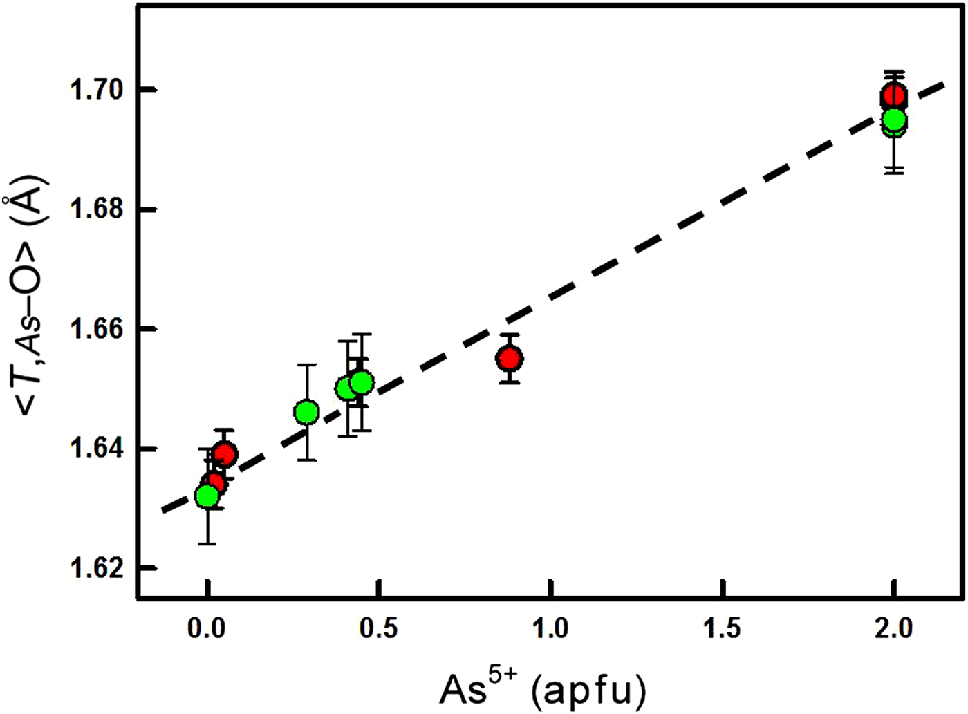
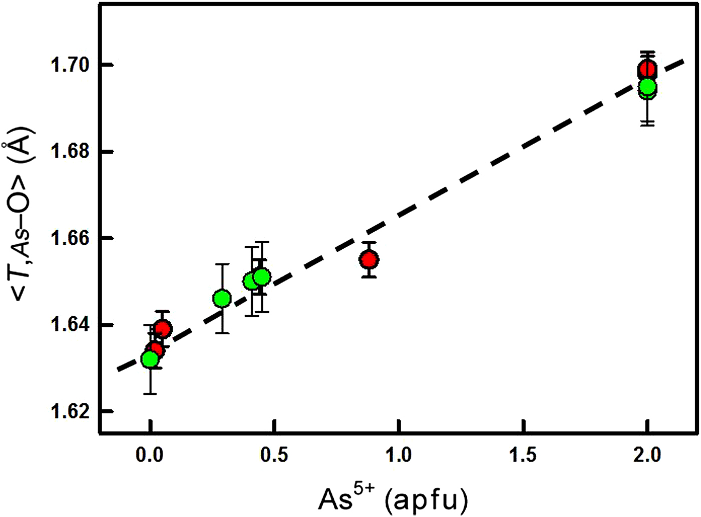
The T(1), T(2), T(3) and T(4) sites in both structures are [4]-coordinated and the <T–O> distances range from ~1.632 to 1.656 Å (Table 4). The refined site-scattering values range from 27.1 to 49.6 epfu (Table 5) which, in combination with the chemical formulae (Table 1), indicate that these sites contain Si in solid solution with a heavier cation. The refined site-scattering values constrain the second cation to be As, and the coordination number and observed <T–O> distances indicate that it is As5+. This being the case, the Si and As site-populations were derived from the refined site-scattering values and the resultant values are given in Table 5. The mean bond-lengths should be related to the site populations, and this is shown to be the case for the T and As sites in both structures in Fig. 1.
Variation in <T,[4]As–O> as a function of the site population of As5+ in mcgovernite (green) and carlfrancisite (red).
There are nine M sites, all of which are octahedrally coordinated and contain dominantly Mn2+ and Mg as indicated by the unit formulae calculated from the electron-microprobe analyses (Table 1). Site-scattering refinement shows that that six M sites have dominant Mn2+, and the remaining M sites have dominant Mg (Table 5). As indicated by the unit formulae (Table 5), the M sites also contain minor Al and Fe. The <M–O> distances range from ~2.09 to 2.25 Å (Table 4), and comparison of the observed mean incident bond-valences suggests that the minor Al and Fe may occur at the M(8) and M(9) sites. The Mg and Mn site-populations were initially assigned from the refined site-scattering values.
Sites involving vacancies
As noted above, the As(3), Z(2) and M(3) sites are involved in short interatomic separations such that both sites in each local pair of sites cannot each be occupied, forcing the occurrence of vacancies at each of these sites.
As(3) site
The As(3) site has triangular pyramidal coordination with <As(3)–O> distances of 1.817 and 1.808 Å typical of As3+ (Majzlan et al., Reference Majzlan, Drahota, Filippi, Bowell, Alpers, Jamieson, Nordstrom and Majzlan2014). The only constituent in either structure that is compatible with the observed stereochemistry is As3+. Moreover, a weak peak is observed in each structure (labelled X in Tables 3 and 4) ~1.1–1.2 Å from the As(3) site. One may interpret X in two ways: [1] X lies on the opposite side of the As(3) site to the anions bonded to the As3+ cation, and is characteristic of the stereoactive lone-pair of electrons expected for As3+. The refined site-scattering values (Table 5) for both structures indicate only partial occupancy by As3+ and the rest of the site must be vacant as there is no other constituent in the structure that is compatible with the local stereochemistry. [2] X lies 2.14–2.18 Å from the Z(2) site and its occupancy when considered as an O anion equals that of the Z(2) cation, with a Z(2)–X distance of ~2.16 Å. Whichever interpretation is correct, it does not affect the cation order in the rest of the structure. Hence the site populations of Table 5 were so assigned.
Z(2) site
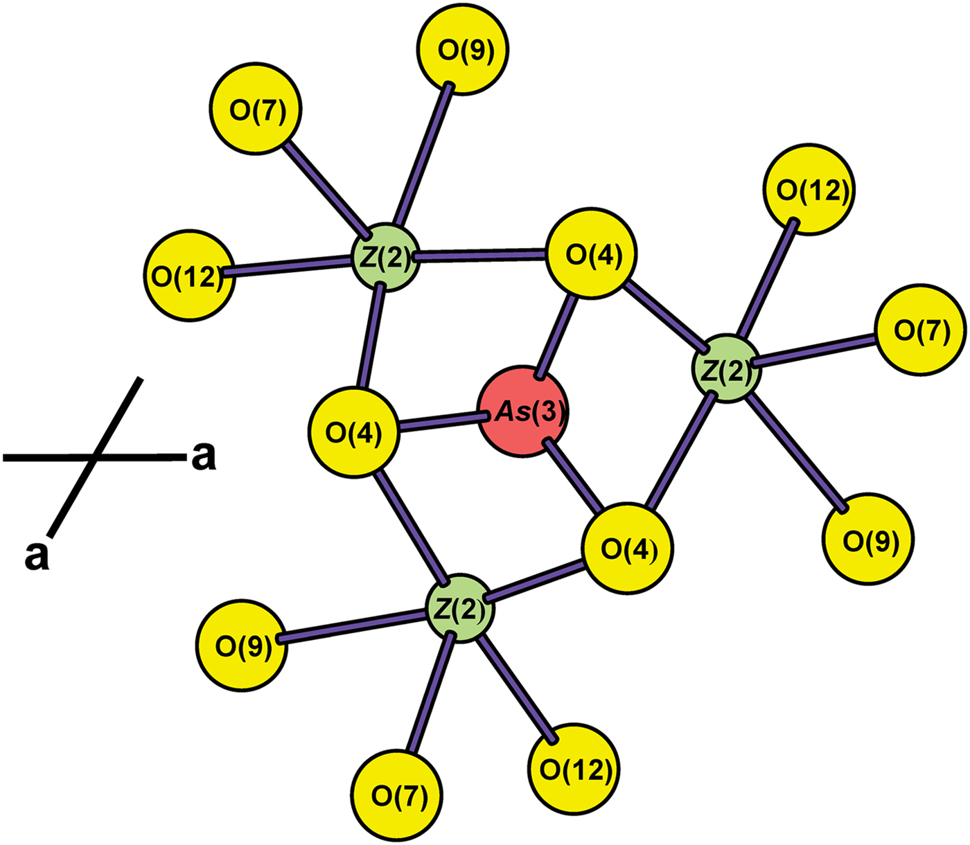
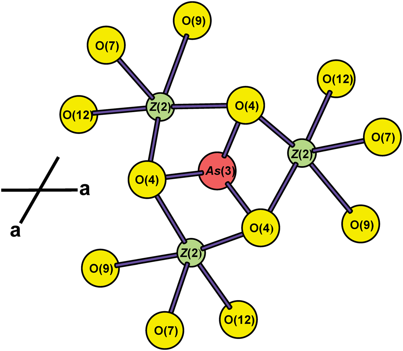
Additional scattering ~2.3 Å from the As(3) site was located in the difference-Fourier maps of both structures. This was designated the Z(2) site, and the site-scattering values at both the As(3) and Z(2) sites were refined independently, initially using As and Mn scattering factors. The arrangement of the Z(2) and As(3) sites in adjacent layers of the structure are shown in Fig. 2. The (As3+O3) group shares three O–O edges with adjacent Z(2) octahedra, with As(3)–Z(2) distances of ~1.85 Å, precluding simultaneous local occupancy of one As(3) site and three Z(2) sites. The site populations of the As(3) site in carlfrancisite and mcgovernite are 1.62 As3+ + 0.38 □ and 1.70 As3+ + 0.30 □, respectively. Thus the Z(2) sites in carlfrancisite and mcgovernite must have a minimum of 1.62 × 3 = 4.86 □ and 1.70 × 3 = 5.10 □ pfu, respectively.
The arrangement of Z(2) and As(3) sites in adjacent layers of the mcgovernite structure; these involve a Z(2)–As(3) approach of ~1.86 Å across common polyhedron edges, and polyhedra sharing these edges cannot both be locally occupied. Red circle: As(3); green circle: Z(2); yellow circles: anions.
There are two possibilities as to the occupancy of the Z(2) site in each structure: [1] there are 1.14 and 0.90 pfu of cations occupying Z(2); in this case, the cations give scattering corresponding to 15.2 and 12.4 electrons per cation, i.e. dominantly Mg in each case, with a small amount of Mn2+. [2] There are additional vacancies at the Z(2) site in each structure, and Z(2) is occupied by cations of a higher scattering power than Mg. The possible cations occupying Z(2) in this case are Mn2+ in carlfrancisite and (Mn2+, Zn) in mcgovernite. In this case, the refined scattering values at Z(2) correspond to 0.69 Mn2+ + 0.45 □ in carlfrancisite and 0.45 Mn2+ + 0.45 □ or 0.37 Zn + 0.53 □ pfu in mcgovernite (Table 6). The Z(2) site is surrounded by five O anions in a square-pyramidal arrangement, with <Z(2)–O> distances of 2.126 and 2.125 Å (Table 4) in carlfrancisite and mcgovernite, respectively. As the Z(2) site is dominantly vacant in each structure, these distances are dictated primarily by the arrangement where Z(2) is vacant rather than where it is occupied by cations. The site populations for the two models are given in Table 6.
M(3) site
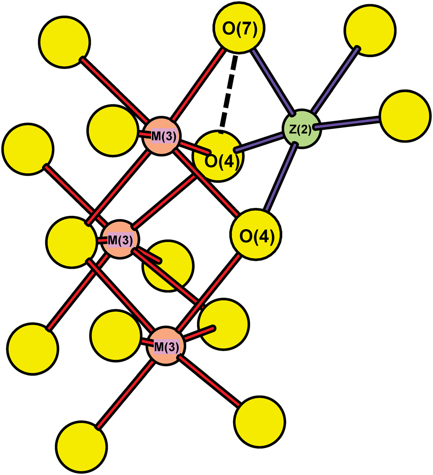
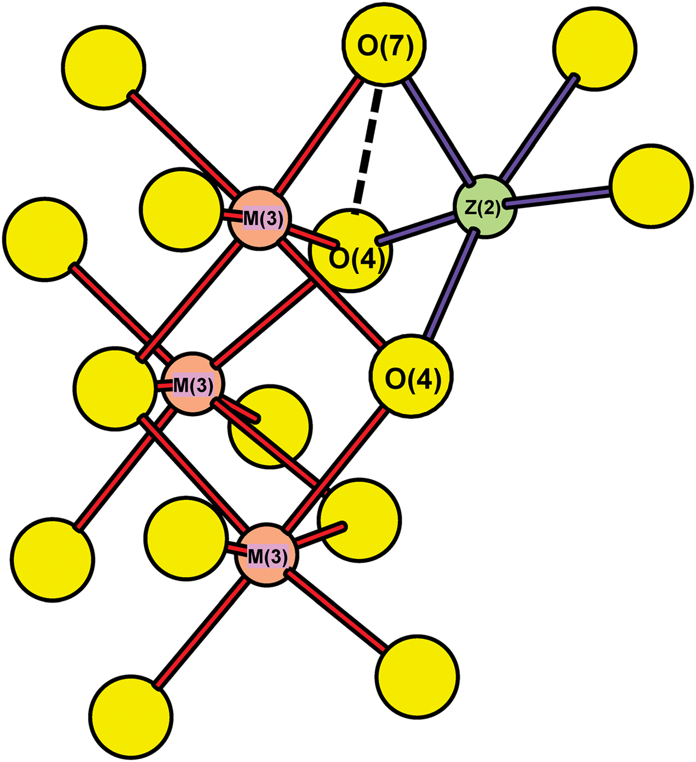
The Z(2) square pyramid links to three M(3) octahedra (Fig. 3). It shares an edge with one M(3) octahedron (shown as a thick dashed line in Fig. 3) and a vertex with two other M(3) octahedra. The Z(2)–M(3) distance across this shared edge is ~2.365 Å in both structures and precludes simultaneous local occupancy of these two sites. Thus each occupied Z(2) must be locally associated with a vacant M(3) site across the edge shared between the constituent polyhedra. The ranges of cation content at Z(2) from the two models described above are as follows: 0.69–1.14 for carlfrancisite and 0.37–0.90 apfu for mcgovernite (Table 6). As each cation at Z(2) must be locally associated with a vacancy at M(3), the ranges of vacancy content at M(3) for carlfrancisite and mcgovernite are 0.69–1.14 and 0.37–0.90 pfu, respectively.
The arrangement of Z(2) and M(3) sites in the mcgovernite structure: these involve a Z(2)–M(3) approach of 2.36 Å across a shared edge (one of which is shown by a thick dashed line) and the polyhedra across these edges cannot both be locally occupied. Pink circles: M(3), legend as in Fig. 2.
There are two ways to assign cations to the M(3) site: [1] we may calculate the Mn2+ content from the refined site-scattering value and compare the resulting vacancy content with the range dictated by the occupancy of the Z(2) site; [2] we may fix the vacancy content at the minimum value allowed by the occupancy of the associated Z(2) site and calculate the Mn2+ and Mg contents from the refined site-scattering values. These two sets of values are given in Table 6. The chemical composition of the two models are the same, they just have a different distribution of cations and vacancies over the Z(2) and M(3) sites.
Anion sites
For the long-range structure, all anions coordinating the cations at the As and T sites are oxygen (O2–) and all anions not coordinating the cations at the As and T sites are (OH)– groups, as indicated by the bond valences incident at each anion (Table 7).
Short-range order
Short-Range Order (SRO) involves the local occupancies of individual (i.e. non-averaged) sites over a scale of a few Å in a crystal structure. Information on disorder and local structure can, in principle, be derived from diffuse (non-Bragg) scattering or via several spectroscopic techniques. However, this information is not easy to extract from the diffuse scattering as the weak intensities involved may not easily be distinguished from background noise, particularly where the disorder is complicated (Hawthorne, Reference Hawthorne2016). An alternative approach is to apply bond-valence theory (Brown, Reference Brown2002, Reference Brown2016) to local atomic-arrangements in minerals (Hawthorne Reference Hawthorne1996, Reference Hawthorne1997, Reference Hawthorne2016; Hawthorne et al., Reference Hawthorne, Ungaretti, Oberti, Caucia and Callegari1993, Reference Hawthorne, Della Ventura and Robert1996), and that is what I shall do now.
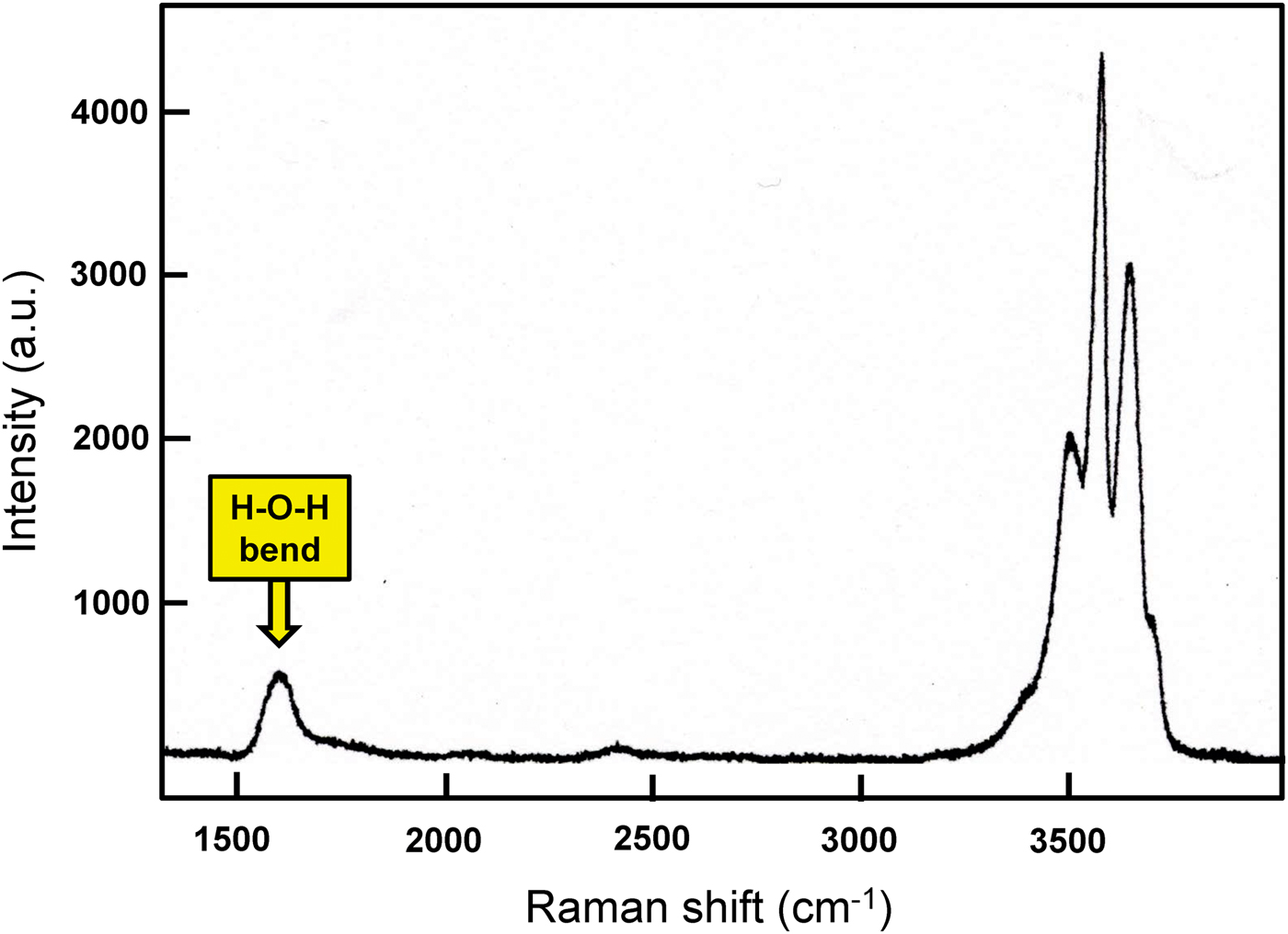
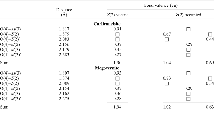
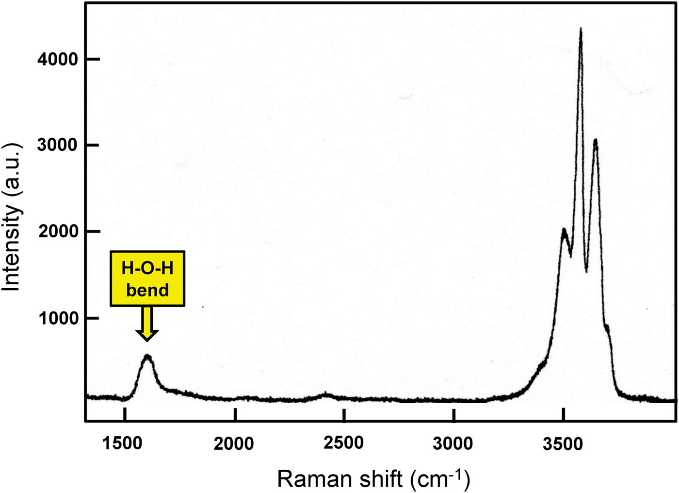
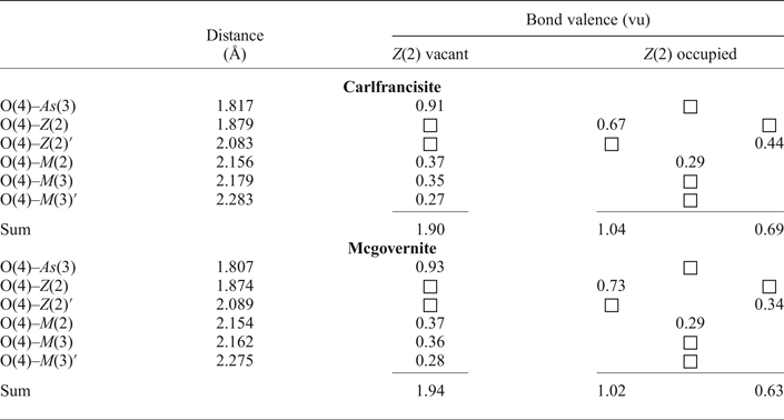
There are three sites involved in SRO of cations: As(3), Z(2), M(3), and their coordinating anions will be greatly affected by the presence of vacancies and/or cations at these three sites. Inspection of Table 6 shows that there are two distinct arrangements in the local structure around these three sites: [1] Z(3) occupied, As(3) and M(3) vacant; [2] Z(3) vacant, As(3) and M(3) occupied. The O(4) anion is of particular importance with regard to incident bond-valence as it is close to the As(3), Z(2), M(3) ×2 and M(2) sites. The environment of the O(4) anion is shown in Table 8. Where the Z(2) site is vacant, the bond valence incident at O(4) sums to 1.90 and 1.94 valence units (vu), in carlfrancisite and mcgovernite respectively, indicating that the O(4) site is occupied by O2– in this arrangement. Where the Z(2) site is occupied, there are two Z(2) sites adjacent to the O(4) site. The site population at the (long-range) Z(2) site is ~1(Mn2+,Mg) + 5 □ pfu, and hence the two Z(2) sites locally adjacent to O(4) are unlikely to both be occupied if they show a random distribution. In this case, the incident bond valence at O(4) is 1.04 and 1.02 vu. if the closer Z(2) site is occupied, and O(4) is an (OH) group. If the closer Z(2) site is vacant, the further Z(2) site is occupied by Mg and the M(2) site is occupied by Mg, the bond valence incident at O(4) is 0.69 and 0.63 vu in carlfrancisite and mcgovernite, respectively. In this case, O(4) must be (locally) an (H2O) group. If both Z(2) sites are occupied, the incident bond valence at O(4) is ~1.5 vu, indicating that it is unlikely that both these sites are locally occupied. Thus the O(4) anion may be O2– where the Z site is vacant and As(3) and M(3) are occupied, and either (OH) or (H2O) or both where the Z site is occupied. The presence of (H2O) is not apparent when considering only the long-range structure, but local bond-valence theory (Hawthorne, Reference Hawthorne1997) shows that (H2O) is present. The Raman spectrum of carlfrancisite (Hawthorne et al., Reference Hawthorne, Abdu, Ball and Pinch2013) has an intense envelope of absorption centred on ~3550 cm–1 (Fig. 4) that has extensive fine-structure with numerous sharp bands characteristic of O–H stretching modes, in accord with the presence of numerous (OH) groups in the structure. However, there is a peak at ~1600 cm–1 that is characteristic of the H–O–H bend of an (H2O) group, in accord with the short-range arrangement of one Z(2) cation and one M(2) cation around (H2O) at O(4) in carlfrancisite. The Raman spectrum of mcgovernite from RRUFF (Lafuente, Reference Lafuente, Downs, Yang, Stone, Armbruster and Danisi2015, http://rruff.info/mcgovernite/) contains only the faintest suggestion of intensity at ~1600 cm–1, suggesting that the local order around O(4) in mcgovernite is somewhat different to that in carlfrancisite. Possibly the small amount of Fe in mcgovernite is trivalent rather than divalent (as I have assumed); in this case, possible ordering of Fe3+ at Z(2) [or even M(8) where Fe3+ would provide increased bond valence to O(7), allowing the Z(2) cation to move toward O(4)] would provide sufficient incident bond valence to obviate the need for H2O at O(4).
The Raman spectrum of carlfrancisite; after Hawthorne et al. (Reference Hawthorne, Abdu, Ball and Pinch2013).
The short-range environment around O(4) in carlfrancisite and mcgovernite.
Bond topology
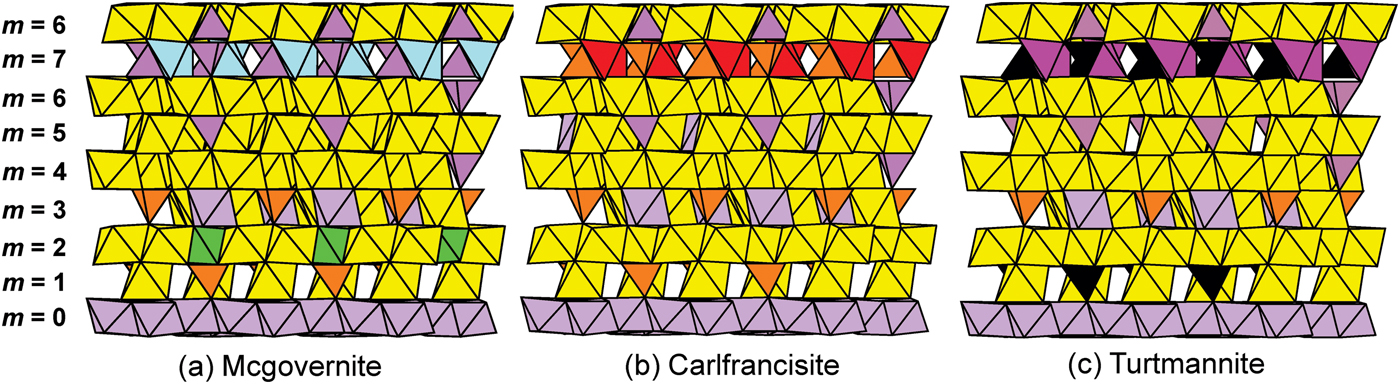
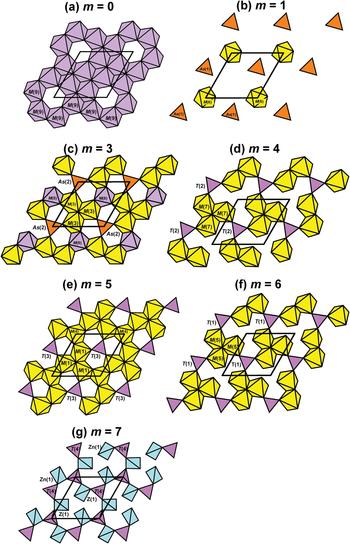
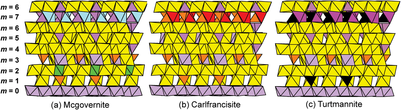
The carlfrancisite–mcgovernite structure contains 84 layers of approximately close-packed polyhedra that comprise one translation along c, giving this structure type the distinction of possessing the largest unit-cell parameter of any mineral. The anions in the structure are arranged in (generally close-packed) layers orthogonal to the c-axis. Fourteen layers stack along the c-axis in the sequence |**hhchch•hchchh| (* denotes a layer of anions displaced from close packed). The first eight anion layers (Fig. 5) repeat via a centre of symmetry (•) at the origin (within the layer m = 0). There are eight distinct layers of cation-centred polyhedra labelled m = 0–7 in Fig. 5, and these layers are shown in plan in Figs 6 and 7.
Part of the crystal structures of mcgovernite, carlfrancisite and turtmannite projected along [100], showing the first eight distinct layers of cation-centred polyhedra labelled m = 0–7; note that the stacking sequence reverses at layer 7 (via a centre of symmetry) and the resulting 14-layer stacking comprises ⅙ of the c-axis repeat. The following cations and cation-centred coordination polyhedra correspond to the following colours in this and the following figures: Mn2+: yellow, red at Z(2) in carlfrancisite; As5+: orange; Si4+: purple; As3+: dull green; Zn2+: pale blue at Z(1), bright green at Z(2) in mcgovernite; Mg2+: pale mauve; and V5+: black. Layers are designated by the letter m where m = 0–7.
The layers of polyhedra in mcgovernite. Legend as in Fig. 5; the unit cell is shown by the black lines.
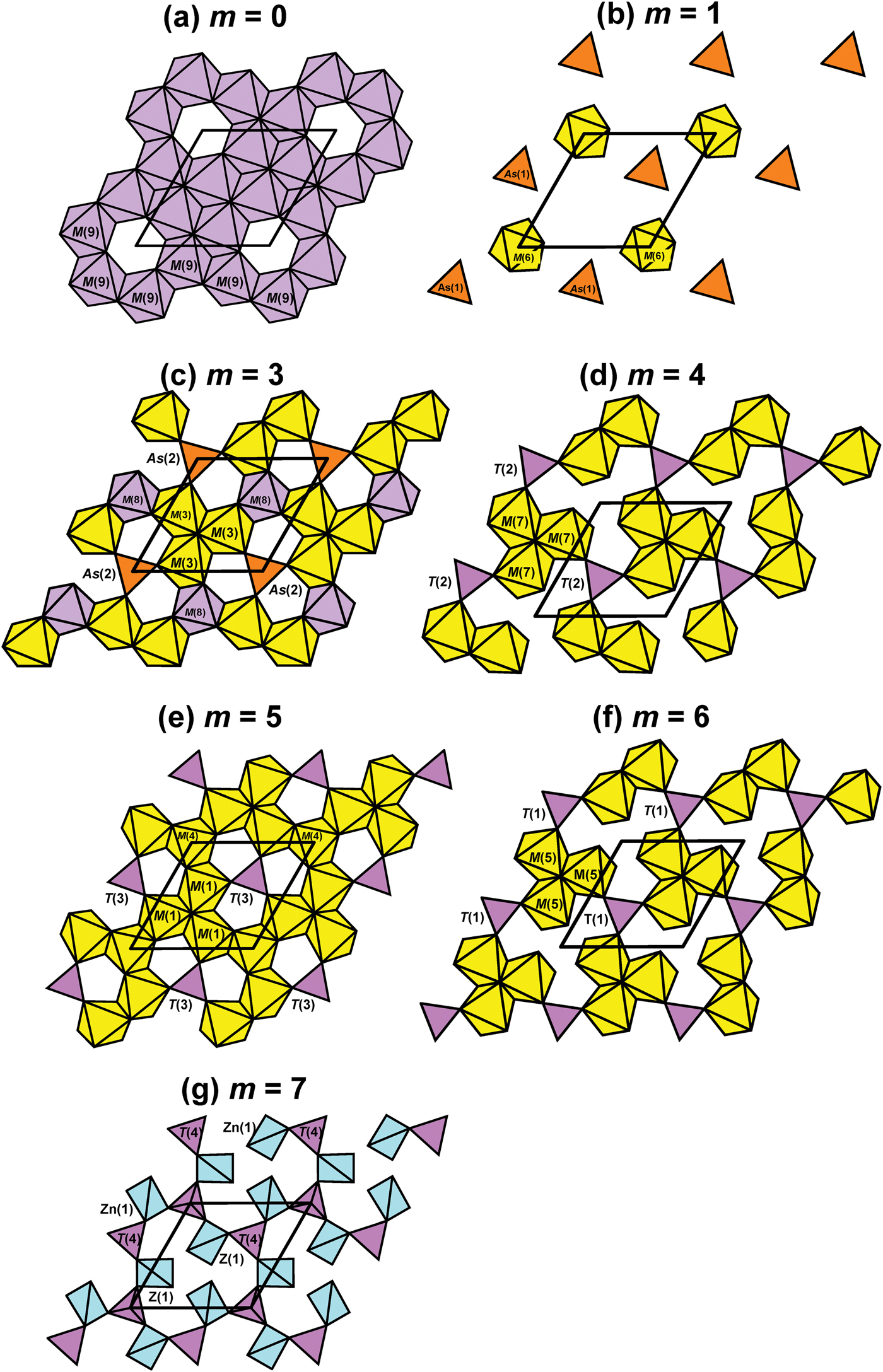
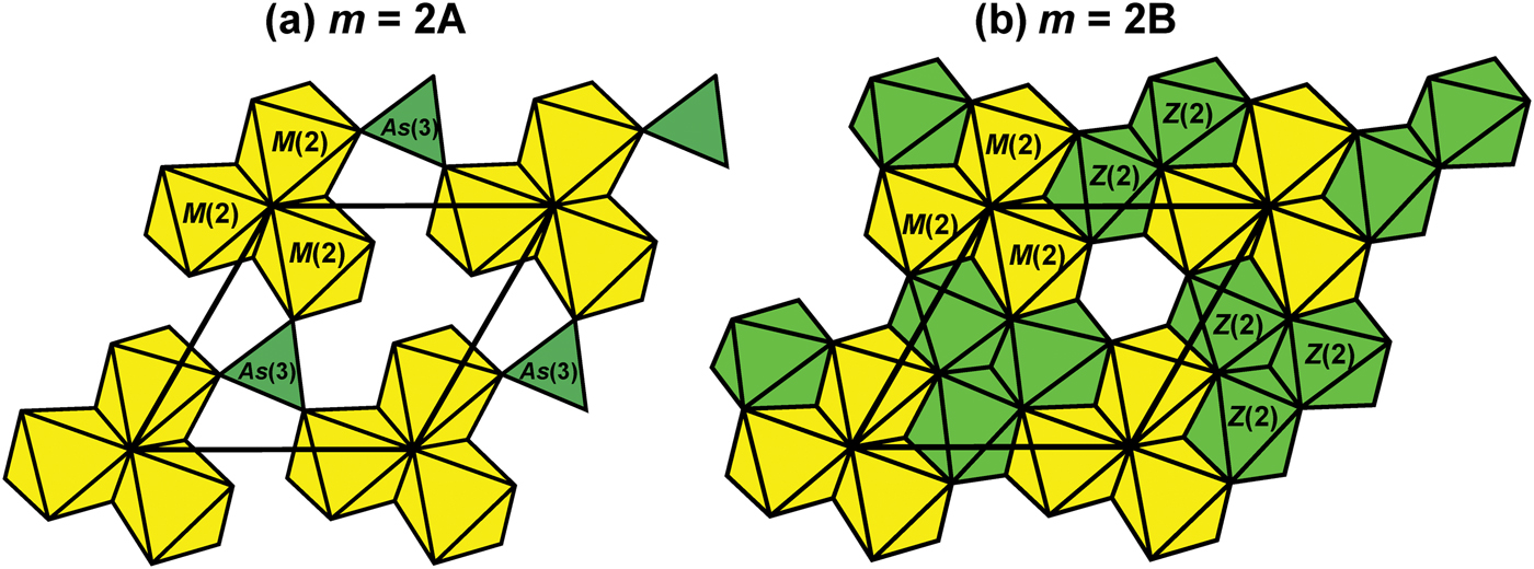
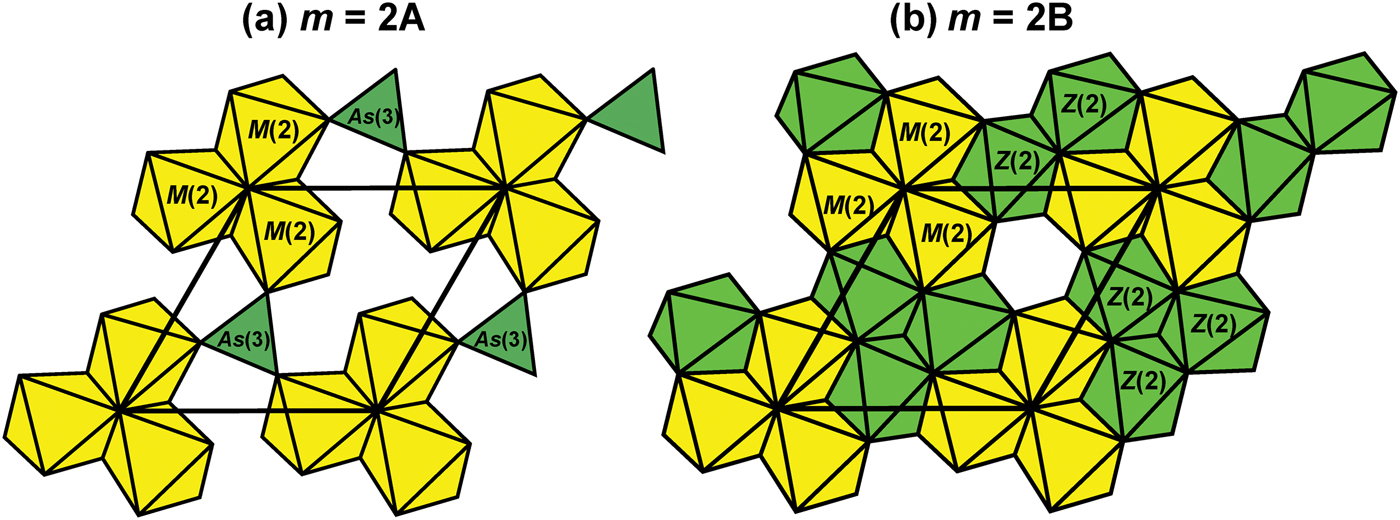
At m = 0 (Fig. 6a), the layer consists of an interrupted sheet of edge-sharing M(9) octahedra that are occupied dominantly by Mg with significant (~0.4–1.1 apfu) Mn2+. The vacant octahedra in the sheet lie on the c axis. At m = 1 (Fig. 6b), the layer consists of a rather sparsely occupied sheet of unconnected (within the m = 1 sheet) As(1) tetrahedra and M(6) octahedra where M(6) is occupied predominantly by Mn2+ but also contains significant (0.1–0.4 apfu) Mg. The m = 2 layer contains significant disorder (Fig. 7). There is a trimer of edge-sharing M(2) octahedra occupied predominantly by Mn2+ but also containing significant (0.46 apfu) Mg, and this trimer is centred on the c axis in both arrangements of the m = 2 layer. In layer m = 2A (Fig. 7a), the trimers are linked into a sheet by As(3) trigonal pyramids that are present 1.66 / 2 × 100 = 83%, whereas in layer m = 2B (Fig. 7b), the trimers are linked into a sheet by sharing edges with trimers of edge-sharing Z(2) octahedra occupied by Mg, Mn2+ and Zn. At m = 3 (Fig. 6c), the layer consists of trimers of edge-sharing M(3) octahedra occupied predominantly by Mn2+ but also containing significant (~2 apfu) Mg, linked into a sheet by sharing edges with M(8) octahedra, occupied predominantly by Mg but also containing some (~0.30 apfu) Mn2+, and sharing corners with As(2) tetrahedra completely occupied by As5+ in both carlfrancisite and mcgovernite. At m = 4 (Fig. 6d), the layer consists of trimers of edge-sharing M(7) octahedra occupied by Mn2+ and linked by sharing corners with T(2) tetrahedra occupied predominantly by Si with minor (~0.29–0.44 apfu) As5+. At m = 5 (Fig. 6e), the layer consists of trimers of edge-sharing M(1) octahedra occupied by Mn2+ (with significant Mg in mcgovernite) and linked by sharing edges with M(4) octahedra that lie on the c axis and T(3) tetrahedra that are dominated by Si; the M(4) octahedron contains substantial Mg and Mn2+, but is dominated by Mg in carlfrancisite and Mn2+ in mcgovernite. At m = 6 (Fig. 6f), the layer consists of trimers of edge-sharing M(5) octahedra occupied by Mn2+ (with minor Mg in mcgovernite) that are linked by sharing corners with T(1) tetrahedra that are dominated by Si (with significant As5+ in mcgovernite). The m = 7 layer (Fig. 6g) is substantially different from the other layers, consisting of 12-membered rings of 3-connected T(4) tetrahedra and 2-connected Z(1) tetrahedra, where T(4) is occupied by dominant Si and substantial As5+ (0.45–0.88 apfu).
Principal chemical differences between mcgovernite, carlfrancisite and turtmannite
The principal compositional differences between each mineral are summarized in Table 9 and may be seen in Fig. 5. In layer m = 1, the As(1) site is occupied by As5+ in carlfrancisite and mcgovernite, and by V5+ in turtmannite. In layer m = 5, the M(4) site is occupied by dominant Mn2+ with minor Mg in mcgovernite and turtmannite, and by dominant Mg with minor Mn2+ in carlfrancisite. In layer m = 7, the Z(1) site is occupied by Zn in mcgovernite, and by Mn2+ in carlfrancisite and turtmannite, and the T(4) site is occupied by dominant Si with minor As5+ in carlfrancisite and mcgovernite, and V5+ in turtmannite.
Principal compositional differences between mcgovernite, carlfrancisite and turtmannite.

Related minerals
We will discuss the detailed structural relations between the minerals of the mcgovernite family following the formal description of two additional (new) minerals. However, it is notable that of the eight distinct layers of polyhedra in this structure type, four contiguous layers form a slab the topology of which is identical to that in arakiite (Cooper and Hawthorne, Reference Cooper and Hawthorne1999) and hematolite (Moore and Araki, Reference Moore and Araki1978). This four-layer unit interleaves with a unit of three [6]mangano-[4]silicate layers and a [4]zinco-[4]silicate layer in mcgovernite, a unit of four [6]mangano-[4]silicate layers in carlfrancisite.
Acknowledgements
I thank Mark Cooper for his help with this work. Financial support was provided by a Canada Research Chair in Crystallography and Mineralogy, and Discovery Grants from the Natural Sciences and Engineering Research Council of Canada, and by Canada Foundation for Innovation Grants, to FCH.
Supplementary material
To view supplementary material for this article, please visit https://doi.org/10.1180/minmag.2017.081.098