Introduction
Global warming and desertification make the camel a promising livestock species for animal production under extreme environmental conditions as a result of its higher adaptability to life in the desert (Moulavi and Hosseini, Reference Moulavi and Hosseini2018; Tibary, Reference Tibary2018). Improvement of reproductive efficiency of she-camels is limited by their short breeding season, longer calving interval and older age of puberty, and the continuous use of traditional reproductive management systems (Basiouni, Reference Basiouni2007). Assisted reproductive technologies (ART) such as in vitro production (IVP) of embryos have been performed in camels, but with varying degree of success (Kafi et al., Reference Kafi, Mesbah, Davoodian and Kadivar2014). For IVP of embryos, oocytes have to be retrieved from ovaries of slaughtered pregnant and non-pregnant females (Moore and Thatcher, Reference Moore and Thatcher2006) or live females using the ovum pick-up (OPU) technique (Pieterse et al., Reference Pieterse, Kappen, Kruip and Taverne1988). Follicles from the abattoir can be used as potential oocyte resources to support IVP development (Adam et al., Reference Adam, Gholib, Hafizuddin, Zamzami and Bahi2019). Oocyte quality and quantity are fundamental for increasing the effectiveness of ART, but oocytes need to acquire a series of competence during follicular development to make them ready to be fertilized (Silva et al., Reference Silva, Van Den Hurk and Figueiredo2016).
Within the dominant follicle and near the ovulation time, the cow oocyte goes through some changes that are crucial for the oocyte to acquire developmental capacity. More oocytes recovered during this period underwent development compared with oocytes recovered at the earlier follicular development stages (Humblot et al., Reference Humblot, Holm, Lonergan, Wrenzycki, Lequarré, Joly, Herrmann, Lopes, Rizos, Niemann and Callesen2005). Similarly, dromedary oocytes developmental competence is acquired late during the final phase of follicular development and this developmental ability translates into greater pregnancy rates after transfer of IVP hatched blastocysts (Khatir et al., Reference Khatir, Anouassi and Tibary2007). During the early stages of pregnancy, there are a clear changes in the endocrine profiles of ovarian tissues (Gong et al., Reference Gong, Chen, Xu, Xie, Yu, Yang and Kuang2017; Quan et al., Reference Quan, Zheng, Ling, Fang, Chu, Zhang, Liu and Li2019), therefore pregnancy could affect ovarian function and subsequently follicular development (Zi et al., Reference Zi, Chen and Wang2012).
Ovarian activity and endocrine function are quite different in pregnant and non-pregnant animals. Non-pregnant animals can normally ovulate and have a higher level of oestrogen to sustain normal oestrous. However, the ovaries of pregnant animals contain corpora lutea that hinder ovulation and allow gradual increases in progesterone levels to maintain the pregnancy (Ling et al., Reference Ling, Guo, Chen, Ding, Ma, Chu, Di, Zhang and Zhang2016). These biological variations between pregnant and non-pregnant animals could be attributed to altered expression of genes that regulate follicular development (such as IGF1, FSHR, and BMP15), follicular survival (PTEN and BCL2), steroid hormone secretion (STAR, IGF1, FSHR, and BMP15), and oocyte development and maturation (BMP6 and BMP15) (Dube et al., Reference Dube, Wang, Elvin, Lyons, Celeste and Matzuk1998; Shimasaki et al., Reference Shimasaki, Moore, Otsuka and Erickson2004; Braw-Tal and Roth, Reference Braw-Tal and Roth2005; Assidi et al., Reference Assidi, Dufort, Ali, Hamel, Algriany, Dielemann and Sirard2008; Orisaka et al., Reference Orisaka, Jiang, Orisaka, Kotsuji and Tsang2009; George et al., Reference George, Dille and Heckert2011; Yamashita et al., Reference Yamashita, Okamoto, Kawashima, Okazaki, Nishimura, Gunji, Hishinuma and Shimada2011; Li et al., Reference Li, Li, Ou, Zhang, Ren, Wei, Zhang and Yang2014; Boruszewska et al., Reference Boruszewska, Sinderewicz, Kowalczyk-Zieba, Grycmacher and Woclawek-Potocka2015; Sargent et al., Reference Sargent, Lu, Clopton, Pohlmeier, Brauer, Ferrara, Silversides and Cupp2015; de Figueiredo et al., Reference de Figueiredo, de Lima, Silva and Santos2018; Ipsa et al., Reference Ipsa, Cruzat, Kagize, Yovich and Keane2019; Quan et al., Reference Quan, Zheng, Ling, Fang, Chu, Zhang, Liu and Li2019). The physiological intimacy between oocytes and the surrounding cumulus cells (CCs) enables gene expression profiling of granulosa cells (GCs; Ola and Sun, Reference Ola and Sun2012) and oocytes (Lonergan et al., Reference Lonergan, Rizos, Gutierrez-Adan, Fair and Boland2003; Emori and Sugiura, Reference Emori and Sugiura2014; Melo et al., Reference Melo, Cordeiro, Pellegrino, Wei, Daye, Nishimura and Dode2017). Moreover, the pregnancy status in camel affects oocyte quality and the ability to resume meiosis in vitro (Torner et al., Reference Torner, Heleil, Alm, Ghoneim, Srsen, Kanitz, Tuchscherer and Fattouh2003; Moawad et al., Reference Moawad, Ghoneim, Darwish and Badr2020).
Little information is known regarding the effect of pregnancy on the expression of growth factor genes (BMP6, BMP15, and IGF1), steroid hormones secretion regulatory genes (STAR and FSHR), and cell cycle regulatory genes (PTEN and BCL2) in camel oocytes and GCs. Therefore, this study aimed to compare expression of these genes in oocytes and GCs and their correlation with FF levels of steroid hormones between pregnant and non-pregnant camels.
Materials and methods
Collection of ovaries
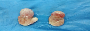
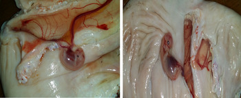
Due to the seasonality of follicular activity and lesser numbers of follicles growing during the non-breeding season in she-camels (Abdoon, Reference Abdoon2001), oocytes had to be retrieved from pregnant and non-pregnant slaughtered animals to maximize the number of collected oocytes. In total, 138 pairs of ovaries were collected from mature (5–10 years old) healthy 138 dromedary camels slaughtered at El-Warak abattoir, Giza, Egypt during the breeding season (November to April) of 2017–2019. The collected ovaries included 92 pairs from non-pregnant and 46 pairs from pregnant camels. The camel is an induced ovulatory and so the corpus luteum can only be detected during pregnancy. Subsequently, early pregnancy was predicted when the corpus luteum was detected on one of the two ovaries and confirmed by the presence of an embryo inside the uterus (Figs 1 and 2). The ovaries were separately transported in a thermos flask containing chilled normal saline (0.9% NaCl) supplemented with 50 µg/ml gentamycin within 2 h to the Laboratory of Reproductive Genetics in the Department of Reproduction and Artificial Insemination, National Research Center, Giza, Egypt.
One ovarian pair of a pregnant animal with left ovary containing the corpus luteum.

Embryos at early stages of pregnancy.

Follicles recovery
In the laboratory, the ovaries were stripped of extraneous tissues and washed three times with warm sterile saline (0.9% NaCl) containing antibiotics. Then, they were washed once quickly in ethanol 70% and rewashed three times with warm sterile saline (0.9% NaCl) containing antibiotics. Follicle diameter was measured using callipers and only follicles with a diameter in the range 3–8 mm were collected. These follicles were divided into five classes: class I (3–3.9 mm), class II (4–4.9 mm), class III (5–5.9 mm), class IV (6–6.9 mm), and class V (7–8 mm).
Collection of immature oocytes and GCs
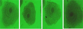
Follicles from pregnant and non-pregnant camels were aspirated separately with disposable 18-gauge needles connected to a 10 ml plastic syringe. The aspirated follicular fluids (FF) were left in a vertical position for 30 min at 4ºC. The lower phase of the FF containing immature cumulus–oocytes complex (COC) was transferred to a small Petri dish and examined under a stereomicroscope (SZ10, Olympus, Japan). In total, 1703 COCs (1160 from non-pregnant and 543 from pregnant) were aspirated. Only COCs with at least three layers of CCs and homogenous granular ooplasm were selected (Fig. 3). The total number of the selected COCs was 1220 (810 from non-pregnant and 410 from pregnant). Immature oocytes were mechanically (by gentle pipetting) and chemically (by hyaluronidase at a concentration of 1 mg/ml) separated from surrounding CCs and washed several times with collection medium. Denuded oocytes only were kept at −80ºC until gene expression profiling was conducted. Afterward, the upper phase of the FF, which contained GCs were transferred to 2 ml Eppendorf tubes and centrifuged at 750 g for 10 min at 4ºC to allow the GC pellet to settle out. GCs and FF were stored at −80°C until gene expression profiling and steroid hormones, E2 and P4, were detected, respectively.
Excellent oocyte: compact multilayered cumulus investment (five or more layers of compact cumulus cells); homogeneous ooplasm; total COCs light and transparent.

Estradiol and progesterone assay in the follicular fluid
The FF concentrations for oestradiol (E2) and progesterone (P4) were determined using commercial ELISA kits (Chemux BioScience, South San Francisco, USA) according to the assay procedures described by the manufacturers. The inter-assay and intra-assay precision were 8.2% and 8.8% for E2 and 8.6% and 10.6% for P4.
The relative expression of candidate genes in oocytes and GCs
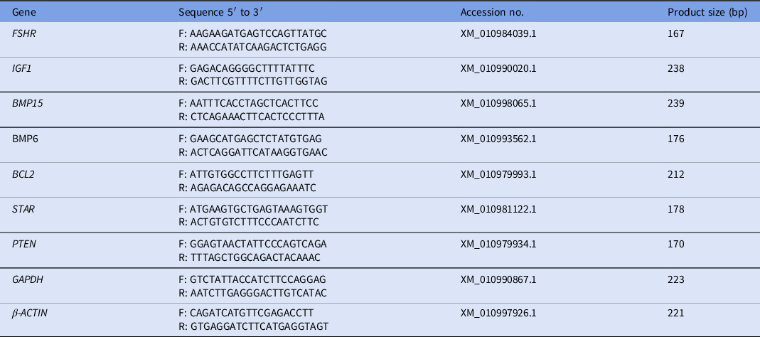
Total RNA was extracted separately from 150 immature oocytes and their related GCs in each group (pregnant and non-pregnant) using the miRNeasy Minikit (Qiagen) according to the manufacturer’s instructions and as previously described (El-Magd et al., Reference El-Magd, Ghoniem, Helmy, Abdelfattah-Hassan, Saleh, Abd Allah, Essawi and Kahilo2019). Within each group, samples were subdivided into five subgroups (class I to class V) based on follicle diameter and each contained 30 immature oocytes and GCs. The integrity of isolated RNA was tested by 2% agarose gel electrophoresis. The concentration and purity of the extracted total RNA was determined using a NanoDrop 2000c spectrophotometer (Thermo Fisher Scientific, Wilmington, USA). The cDNA synthesized using HiSenScript cDNA synthesis kits (iNtRON Biotechnology, South Korea) according to the manufacturer’s recommendations. In brief, a reaction mixture containing 5 µl RNA, 10 µl reverse transcription reaction solution, 1 µl enzyme, and DNase-/RNase-free water up to 20 µl was run on a programmable thermocycler (Bio-Rad, USA) at 42ºC for 60 min., then 85ºC for 10 min. The relative expression levels of BMP6, BMP15, IGF1, STAR, FSHR, PTEN, and BCL2 genes were detected in the oocytes and GCs of pregnant and non-pregnant camels using primers designed based on published camel sequences (Table 1), and QuantiTect SYBR Green qPCR Master Mix. Thermal cycling conditions included 95ºC for 15 s, 55ºC (annealing temperature) for 30 s and 72ºC for 30 s. The specificity of each amplicon was evaluated by analysis of the melting curve. The NormFinder program was used to determine housekeeping genes (Andersen et al., Reference Andersen, Jensen and Ørntoft2004). The results indicated that GAPDH and β-ACTIN genes were the most stable endogenous controls for both oocytes and GCs. The relative expression of each gene was calculated using the 2–∆∆CT method (Livak and Schmittgen, Reference Livak and Schmittgen2001).
List of primers used for qRT-PCR of selected genes

Statistical analysis
All data were expressed as mean ± standard error of mean (SEM). One-way analysis of variance (ANOVA) was performed to identify the effect of reproductive status on all follicle categories and between them. Duncan’s multiple range test was used to separate between significant means. The difference between means within and between the same follicle classes was detected using Student’s t-test. The correlation between gene expression and P4 levels in FF was calculated using Pearson’s correlation coefficient (r). GraphPad Prism 8.0 software was used for data analysis (GraphPad software. Inc., San Diego, CA, USA). Data were considered statistically significant at P < 0.05.
Results
Effect of reproductive status on follicle number
The number of follicles (3–8 mm) per ovarian pairs was significantly (P < 0.05) lower in pregnant (9.14 ± 2.71) compared with non-pregnant (13.67 ± 2.98) camels (Table 2). Additionally, the numbers of class I (3–3.9 mm) and class II (4–4.9 mm) follicles were significantly (P < 0.05) lower in pregnant, compared with non-pregnant, camels. However, the number of follicles in other classes was non-significantly different between pregnant and non-pregnant camels (Table 2).
Effect of reproductive status on follicular populations in dromedary camels

Data presented in form of mean ± SEM.
a,bValues within the same column and carrying different superscript letters are significantly different at P < 0.05.
The numbers within parenthesis represent the number of follicles in each class.
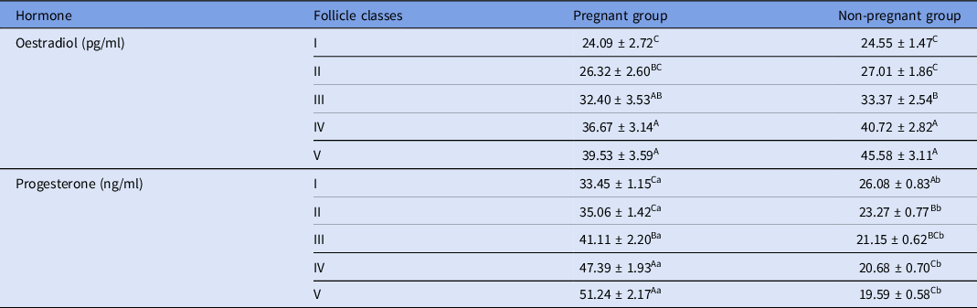
Effect of reproductive status on oestradiol and progesterone concentrations in FF
The FF concentration of P4 was significantly (P < 0.05) lower in all five classes in pregnant, compared with the non-pregnant, camels (Table 3). However, no significant variation was noticed in the FF concentration of E2 in all five classes between non-pregnant and pregnant camels. In the pregnant camels, P4 levels were higher (P < 0.05) in large (classes IV and V) compared with small (classes I–III) follicles. In non-pregnant camels, E2 levels in FF were significantly (P < 0.05) higher in large (classes IV and V) compared with small (classes I–III) follicles. However, P4 concentrations were significantly (P < 0.05) lower in large (classes IV and V) compared with small (classes I–III) follicles in non-pregnant camels (Table 3).
Effect of reproductive status on concentrations of estradiol and progesterone in the follicular fluid

Data presented in form of mean ± SEM.
Values within the same row and carrying different lowercase letters (a and b) are significantly different at P < 0.05.
Values within the same column for each hormone and carrying different uppercase letters (A–D) are significantly different at P < 0.05.
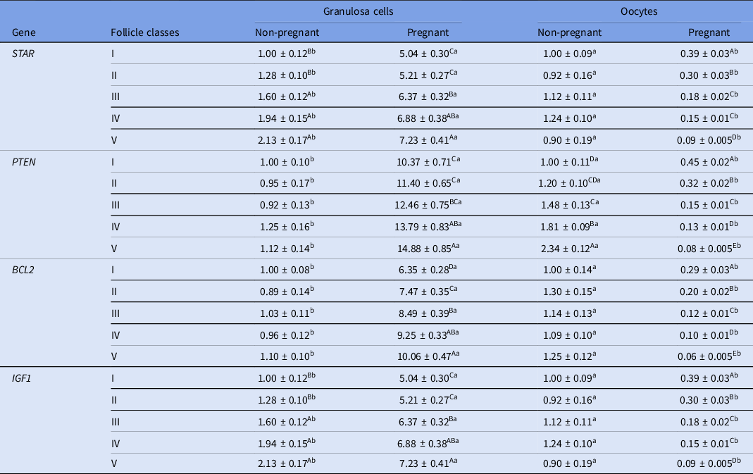
Effect of reproductive status on STAR, PTEN, IGF1 and BCL2 genes in oocytes and granulosa cells
The obtained real-time PCR results revealed that STAR, PTEN, IGF1, and BCL2 mRNA levels were significantly higher in GCs and significantly lower in oocytes of pregnant, compared with non-pregnant, camels at all five follicle classes (Table 4). In pregnant camels, these four genes were significantly upregulated in GCs and significantly downregulated in oocytes obtained from large follicles (classes IV and V) compared with small follicles (classes I–III). In non-pregnant camels, STAR and IGF1 genes were significantly upregulated in GCs and PTEN was significantly upregulated in oocytes obtained from large follicles (classes IV and V) compared with small follicles (classes I–III).
Differential expression of STAR, PTEN, BCL2, and IGF1 genes in granulosa cells and oocytes of non-pregnant and pregnant camels as detected by qRT-PCR

Data presented in form of mean ± SEM. Values within the same row and carrying different lowercase letters (a and b) are significantly different at P < 0.05.
Values within the same column for each gene and carrying different uppercase letters (A–E) are significantly different at P < 0.05.
The expression level of each target gene in class I non-pregnant animals was considered the baseline.
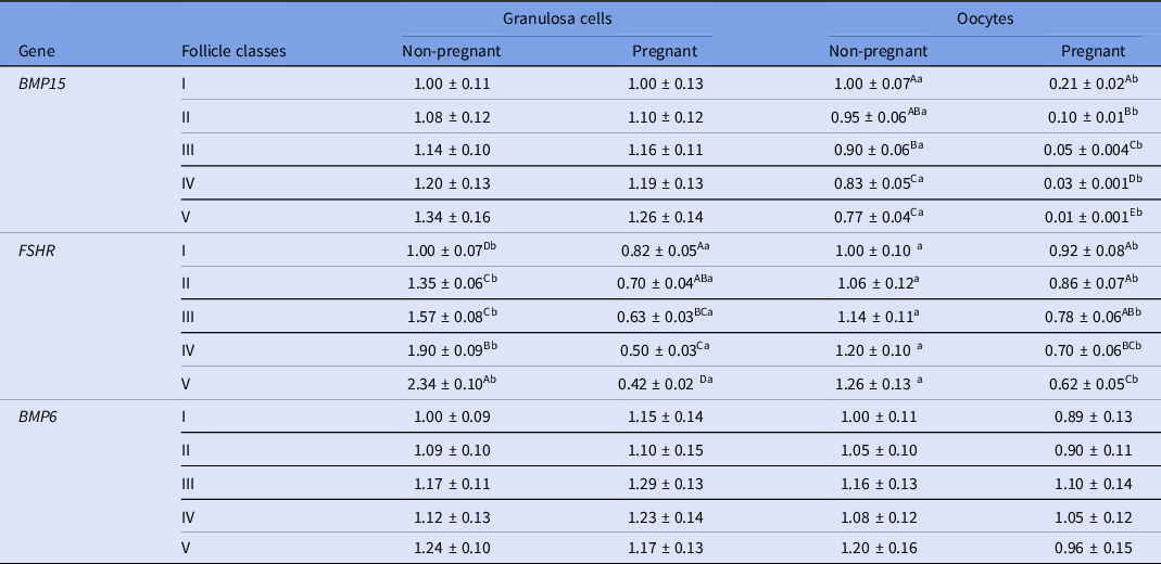
Effect of reproductive status on BMP15, FSHR and BMP6 genes in oocytes and granulosa cells
FSHR mRNA levels were significantly lower in GCs and oocytes, and the BMP15 mRNA level was significantly lower in oocytes of pregnant, compared with non-pregnant, camels (Table 5). However, BMP6 mRNA levels in the oocytes or GCs did not significantly differ between pregnant and non-pregnant camels. In the pregnant camels, P4 levels were higher (P < 0.05) in large (classes IV and V) compared with small (classes I–III) follicles. In non-pregnant camels, E2 levels in FF were significantly (P < 0.05) higher in large (classes IV and V) compared with small (classes I–III) follicles. In pregnant camels, the FSHR gene was significantly downregulated in GCs and oocytes obtained from large follicles (classes IV and V) compared with small follicles (classes I–III). In these animals, BMP15 was also significantly downregulated in oocytes obtained from large follicles (classes IV and V) compared with small follicles (classes I–III). In non-pregnant camels, the FSHR gene was significantly upregulated in GCs obtained from large follicles (classes IV and V) compared with small follicles (classes I–III).
Differential expression of BMP15, FSHR and BMP6 genes in granulosa cells and oocytes of non-pregnant and pregnant camels as detected by qRT-PCR

Data presented in form of mean ± SEM.
Values within the same row and carrying different lowercase letters (a and b) are significantly different at P < 0.05.
Values within the same column for each gene and carrying different uppercase letters (A–E) are significantly different at P < 0.05.
The expression level of each target gene in class I non-pregnant animals was considered the baseline.
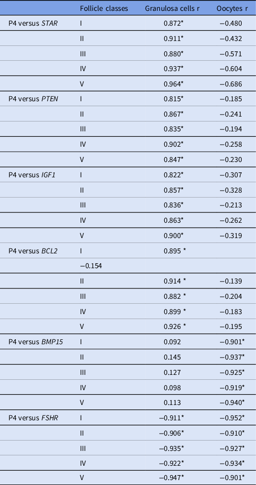
Correlation between gene expression and P4 concentration in FF in pregnant camels
Among the measured steroid hormones, only the P4 level was significantly increased in pregnant camels. Therefore, we checked whether this change was correlated with the expression of the candidate genes. The obtained results showed that the P4 level in FF obtained from the five follicle classes was positively correlated with STAR, PTEN, IGF1, and BCL2 mRNA levels in GCs and negatively correlated with the BMP15 mRNA level in oocytes and the FSHR mRNA level in GCs and oocytes of pregnant camels (Table 6).
Correlation between levels of P4 in follicular fluid and mRNA levels in granulosa cells and oocytes of pregnant camels

r: Pearson’s correlation coefficient.
*: P < 0.05.
Discussion
The reduced number of follicles (3–8mm) in pregnant versus non-pregnant camels agreed with the results of previous studies in camel (Abdoon, Reference Abdoon2001; Torner et al., Reference Torner, Heleil, Alm, Ghoneim, Srsen, Kanitz, Tuchscherer and Fattouh2003) and goat (Quan et al., Reference Quan, Zheng, Ling, Fang, Chu, Zhang, Liu and Li2019). This reduction could be attributed to the downregulation of FSHR expression in pregnant versus non-pregnant camels (this study) and goats (Quan et al., Reference Quan, Zheng, Ling, Fang, Chu, Zhang, Liu and Li2019). FSHR and its ligand, follicle-stimulating hormone (FSH), are required for the emergence of the follicle cohort (4 mm) at the onset of the follicular wave (Caixeta et al., Reference Caixeta, Ripamonte, Franco, Junior and Dode2009). Moreover, the higher IGF1 mRNA levels in the oocytes of non-pregnant, compared with pregnant, camels may explain the increased number of ovarian follicles in the non-pregnant camel as IGF1 is known to stimulate the recruitment of more follicles within the cohort of developing follicles (Silva et al., Reference Silva, Figueiredo and Van den Hurk2009; Grimard et al., Reference Grimard, Marquant-Leguienne, Remy, Richard, Nuttinck, Humblot and Ponter2013).
Expression levels of BCL2 and PTEN genes were significantly lower in the oocytes and significantly higher in GCs in pregnant, compared with non-pregnant, camels. Therefore, lower expression levels of these two genes in the oocytes may be compensated for by higher levels in their associated GCs during pregnancy. This finding indicates the potential effect of BCL2 and PTEN on follicle survival, especially in non-pregnant camels. The survival effect of PTEN acts in two ways. The first way is that the oocyte PTEN-P13K pathway governs follicle activation through controlling oocyte growth (Reddy et al., Reference Reddy, Liu, Adhikari, Jagarlamudi, Rajareddy, Shen, Du, Tang, Hämäläinen and Peng2008). The second way is that GCs-PTEN stimulates GC proliferation and survival by increasing the levels of cell cycle activators cyclin D2 and E2F1, and reducing the levels of the cell inhibitor, P27KIP, therefore inhibiting apoptosis in GCs (Fan et al., Reference Fan, Ren and Hou2018). Froment et al. (Reference Froment, Bontoux, Pisselet, Monget and Dupont2005) reported that the level of PTEN increased in ovine GCs during follicular growth and that PTEN regulated the expression of both E2F1 and P27KIP. BCL2 is a member of the BCL2 family that acts as an anti-apoptotic factor. The ratio of anti-apoptotic BCL2/pro-apoptotic BAX genes is critical for cell fate, as raised BCL2 levels favoured the survival of cells, whereas raised BAX levels accelerated the death of cells (Yang and Rajamahendran, Reference Yang and Rajamahendran2002; Huang et al., Reference Huang, Liu, Xu, Zhang, Yin, Liu, He and He2018). Yang and Rajamahendran (Reference Yang and Rajamahendran2002) confirmed that the propensity to apoptosis is continuously counterbalanced in cells by genes stimulating cell survival and proliferation. Similar compensation was also recorded for IGF1 and STAR, which were upregulated in GCs and downregulated in oocytes of pregnant camels. Consistent with this notion, Emori and Sugiura (Reference Emori and Sugiura2014) reported that BMP signals produced from GCs might sufficiently compensate for oocyte-derived BMP signals when deficient as in BMP6/15 mutant mice.
Steroidogenesis in the mammal ovary is controlled by gonadotropins that act through their receptors, LHR and FSHR, on both theca and GCs, respectively (Richards, Reference Richards1994). The steroidogenic activity of GCs is modulated by locally produced growth factors such as STAR (Carpintero et al., Reference Carpintero, Suárez, Mangas, Varea and Rioja2014), IGF1, BMP6, and BMP15 (Mani et al., Reference Mani, Fenwick, Cheng, Sharma, Singh and Wathes2010). These growth factors act either alone or in combination through endocrine, paracrine, and autocrine mechanisms. The higher expression levels of STAR and IGF1 genes in the GCs of pregnant versus non-pregnant camels and oocyte from non-pregnant versus pregnant camels showed the need for proteins encoded by these two genes for synthesis of E2 and P4 in pregnant and non-pregnant camels. Expression of the STAR gene is necessary for movement of cholesterol (precursor of steroid hormones) from the outer to the inner mitochondrial membrane; this is considered the rate-limiting step for steroidogenesis (LaVoie and King, Reference LaVoie and King2009). The higher expression levels of IGF1 in the oocyte of non-pregnant versus pregnant and in the GCs of pregnant versus non-pregnant may explain the non-significant variation in E2 concentration in FF between pregnant and non-pregnant camels. IGF1 plays a pivotal role in regulating follicular development by GCs proliferation, differentiation, and steroid production by increasing the expression levels of FSHR that modulates the stimulatory effect of FSH (Ipsa et al., Reference Ipsa, Cruzat, Kagize, Yovich and Keane2019). IGF1 can induce steroidogenic regulatory pathways through the activation of IGFR1 and FSHR (Mani et al., Reference Mani, Fenwick, Cheng, Sharma, Singh and Wathes2010).
In this study, we found a negative correlation between P4 levels in FF and BMP15 mRNA levels in the oocytes. Therefore, it is likely that the lower P4 concentrations in FF of non-pregnant camels may be attributed to upregulation of BMP15 in the oocytes of non-pregnant compared with pregnant camels. BMP15 is known to block P4 synthesis as a result of its inhibitory effect on luteinization (Shimasaki et al., Reference Shimasaki, Moore, Otsuka and Erickson2004). Previous studies have reported that BMP15 can inhibit FSHR expression in the oocytes in non-pregnant animals (Otsuka et al., Reference Otsuka, Yamamoto, Erickson and Shimasaki2001; Shimasaki et al., Reference Shimasaki, Moore, Otsuka and Erickson2004). However, we found higher BMP15 and FSHR mRNA levels in oocytes of non-pregnant camels. This contradiction may be as a result of the high expression levels of IGF1 that could enhance the expression of FSHR in camel oocytes. Furthermore, the follicular wave pattern characteristic of the cyclic activity in camels that is characterized by the absence of the luteal phase may explain the simultaneous presence of higher expression levels of both FSHR and BMP15. In support, it was reported that the strong expression of follistatin by the dominant follicle might modulate BMP15 action, such that it is sufficient to express FSHR in GCs to permit the development of follicles (Shimasaki et al., Reference Shimasaki, Moore, Otsuka and Erickson2004). The upregulation of FSHR in the non-pregnant camels favours follicle emergence, growth, and differentiation throughout the follicular wave, although the upregulation of the BMP15 that was demonstrated to have an anti-luteinization effect favoured normal follicular growth until either ovulation or regression (Shimasaki et al., Reference Shimasaki, Moore, Otsuka and Erickson2004).
BMP6, a member of the TGFβ superfamily regulates growth, differentiation, and apoptosis of oocytes and GCs (Grimard et al., Reference Grimard, Marquant-Leguienne, Remy, Richard, Nuttinck, Humblot and Ponter2013; Emori and Sugiura, Reference Emori and Sugiura2014). The similar expression levels of BMP6 in both GCs and oocytes in pregnant and non-pregnant camels suggested a similar regulatory effect regarding the biological reproductive process in pregnant and non-pregnant camels.
In conclusion, pregnancy-induced variations in the expression profiles of STAR, PTEN, IGF1, BCL2, FSHR, and BMP15 genes were associated with a decrease in the number of follicles and an increase in the P4 concentrations in FF of pregnant, compared with non-pregnant, camels. These molecular and hormonal changes could be attributed to the presence of corpus lutea on ovaries of pregnant females. Further investigations are required to determine the physiological significance of these changes and their effect on oocyte maturation and subsequent development in dromedary camels.
Financial support
None.
Conflict of interest
The authors declare that there is no conflict of interest.
Ethical standards
The authors declare that all procedures were performed according to national and institutional guidelines on the care and use of animals.