Implications
Studies on the metabolism and clearance of skatole, a major component of boar taint, have been hampered by the lack of specific tools to measure the activity of the cytochrome P450 (CYP450) enzymes involved. Here we have used a defined cell-based system to individually express six different isoforms of porcine CYP450 and cytochrome b5A, and measure their role in the metabolism of skatole. Our results show that CYP2A19 is the primary isoform responsible for the formation of 6-hydroxyskatole, although its importance to in vivo clearance will also be affected by its level of expression in vivo.
Introduction
Skatole (3-methylindole, 3MI) is a metabolite of tryptophan produced by the bacterial microflora in the gastrointestinal tract of animals, including humans (reviewed in Jensen and Jensen, Reference Jensen and Jensen1998). Skatole is a pneumotoxin in some species, causing acute pulmonary oedema and interstitial emphysema culminating in death of the animal (Thornton-Manning et al., Reference Thornton-Manning, Appleton, Gonzalez and Yost1996). Although pigs do not display pneumotoxicity to skatole, it is one of the main compounds contributing to boar taint, an unfavourable taste and odour that is liberated when pork fat from some entire male pigs is heated (Bonneau, Reference Bonneau1982). Castration reduces boar taint, but this also eliminates the natural anabolic androgens, which increase the conversion of feed into lean tissue in entire males. Raising entire males can thus have a dramatic impact on pork industry profits (de Lange and Squires, Reference de Lange and Squires1995) and welfare of the animals (Prunier et al., Reference Prunier, Bonneau, von Borell, Cinotti, Gunn, Fredriksen, Giersing, Morton, Tuyttens and Velarde2006), but this requires methods other than castration to control boar taint.
Skatole is extensively metabolized in the liver (Agergaard and Laue, Reference Agergaard and Laue1992), and at least seven different metabolites have been identified, including indole-3-carbinol (I3C), 2-aminoacetophenone (2AAP), 3-hydroxy-3-methyloxindole (HMOI), 3-methyloxyindole (3MOI), 3-hydroxy-3-methylindolenine (3-OH-MI), 5-hydroxy-3-methylindole (5-OH-3MI) and 6-hydroxy-3-methylindole (6-OH-3MI) (Friis, Reference Friis1993; Diaz et al., Reference Diaz, Skordos, Yost and Squires1999). Diaz and Squires (Reference Diaz and Squires2000a) found that 3-OH-MI and 3MOI accounted for 45.1% and 27.9%, respectively, of the total metabolites that were produced from skatole metabolism with in vitro microsomal systems. The major metabolite found in the plasma of pigs that efficiently metabolize and clear skatole was shown to be the sulphoconjugate of 6-OH-3MI, whereas pigs that were poor metabolizers of skatole had HMOI as the major metabolite in plasma (Friis, Reference Friis1995; Bæk et al., Reference Bæk, Hansen-Møller, Friis, Cornett and Hansen1997). These latter studies generated the hypothesis that the formation of 6-OH-3MI sulphate was an important excretory pathway of skatole, and that deficiencies in the metabolism and clearance of skatole contribute to boar taint.
Many studies to elucidate the physiological basis of variation in skatole content in porcine fat tissue have been conducted using microsomes and hepatocyte systems to identify the porcine cytochrome P450s (CYP450s) that are important in skatole metabolism. These studies utilized indicator substrates, inhibitors and antibodies that were characterized for human CYP450s to identify CYP2E1 and CYP2A as the major porcine CYP450 isoforms in skatole metabolism (Friis, Reference Friis1995; Diaz et al., Reference Diaz, Skordos, Yost and Squires1999; Diaz and Squires, Reference Diaz and Squires2000a; Terner et al., Reference Terner, Gilmore, Lou and Squires2006). These studies assume that the specificity of substrates, inhibitors and antibodies in one species will be the same in another species; however, this is often not the case. It is also difficult to isolate the effect of an enzyme of interest when studying the metabolism of skatole in hepatocytes or microsomes, as multiple isoforms of CYP450 are present, and two or more isoforms may be responsible for catalysing the same reaction. Thornton-Manning et al. (Reference Thornton-Manning, Ruangyuttikarn, Gonzalez and Yost1991 and Reference Thornton-Manning, Appleton, Gonzalez and Yost1996) investigated the metabolic potential of several vaccinia-expressed human CYP450s towards skatole, and found that CYP1A2, CYP2A6, CYP2B6, CYP2C8, CYP3A4, CYP2E1 and CYP2D6 were capable of metabolizing skatole. Matal et al. (Reference Matal, Matuskova, Tunkova, Anzenbacherová and Anzenbacher2009) reported that purified porcine CYP2A19, CYP2E1 and CYP1A2 could metabolize skatole, but they only measured the formation of the metabolites 3MOI and I3C.
Pigs are often explored as a potential model for humans in pharmaceutical studies because of their wide availability and physiological, dietary and anatomical similarity to humans (Balk, Reference Balk1987; Monshouwer et al., Reference Monshouwer, van't Klooster, Nijmeijer, Witkamp and van Miert1998; Tsiaoussis et al., Reference Tsiaoussis, Newsome, Nelson, Hayes and Plevris2001). Drug development is a complex process requiring multiple steps before reaching clinical trial testing. Individual porcine CYP450 enzymes have been studied to determine how metabolic capabilities correlate with those found in humans and for studies in veterinary pharmacology (Juskevich, Reference Juskevich1987; Anzenbacherová et al., Reference Anzenbacherová, Baranová, Zuber, Pěchová, Anzenbacher, Souček and Martínková2005). In addition, the microsomal haemoprotein cytochrome b5A (CYB5A) has been shown to augment the activity of human CYP2E1 (hCYP2E1) towards chlorzoxazone (CLZ) hydroxylation by apparently acting as an electron donor (Yamazaki et al., Reference Yamazaki, Nakano, Gillam, Bell, Guengerich and Shimada1996; Yamazaki et al., Reference Yamazaki, Nakamura, Komatsu, Ohyama, Hatanaka, Asahi, Shimada, Guengerich, Shimada, Nakajima and Yokoi2002; Wiercinska and Squires, Reference Wiercinska and Squires2010). CYB5 also interacts with a number of other CYP450 isoforms including CYP17A1 (Billen and Squires, Reference Billen and Squires2009); a CYB5 knockout mouse model has a dramatically altered expression of drug metabolizing enzymes and low levels of testicular androgens (McLaughlin et al., Reference McLaughlin, Ronseaux, Finn, Henderson and Wolf2010).
Therefore, to definitively characterize the porcine CYP450 isoforms that contribute to skatole metabolism, we have cloned and individually expressed various porcine CYP450s in a cell line system, thereby eliminating the need for inhibitors and antibodies to identify which CYP450 isoforms are present. We expressed the major porcine CYP450s, including CYP2E1, CYP1A1, CYP2A19, CYP2C33v4, CYP2C49 and CYP3A, as well as hCYP2E1 in human embryonic kidney HEK-293FT cells along with porcine P450-oxidoreductase (POR) and cytochrome b5 reductase (CYB5R3). We also investigated the effect of co-transfection with an expression vector for CYB5A on the formation of the different skatole metabolites, as little is known about the effects of CYB5A on the activity of individual porcine CYP450s. In these studies, we measured the formation of six different metabolites of skatole by each CYP450 isoform using HPLC calibrated with known standards. To further illustrate the utility of this system, we also report on the specificity of substrates for porcine CYP2A19 and CYP3A, and assessed the specificity of commercially available antibodies against human CYP2A6 and rat CYP2E1 for porcine CYP450 isoforms by Western blot analysis.
Material and methods
Cloning
The expression vectors for porcine P450-oxidoreductase/CYP450 NADPH reductase (POR), cytochrome b5 reductase (CYB5R3) and CYB5A were constructed previously (Billen and Squires, Reference Billen and Squires2009). The entire coding sequence of the different porcine CYP450s (CYP1A1, CYP2A19, CYP2E1, CYP2C33v4, CYP2C49 and CYP3A) were amplified from porcine liver cDNA by PCR and cloned into pcDNA3.1/V5-His TOPO (Invitrogen, Burlington, ON, Canada), as described by Wiercinska and Squires (Reference Wiercinska and Squires2010). A plasmid containing hCYP2E1 was obtained from Origene (Rockville, MD, USA) and used to amplify the coding region of hCYP2E1 that was cloned into pcDNA3.1/V5-His TOPO (Invitrogen), as described by Wiercinska and Squires (Reference Wiercinska and Squires2010).
Cell culture
Human embryonic kidney cells HEK-293FT were cultured under conditions described previously (Billen and Squires, Reference Billen and Squires2009). Cells were plated at 106 cells per well in 6-well plates (BD Biosciences, Mississauga, ON, Canada) and were transfected using Lipofectamine 2000 (Invitrogen, Burlington, ON, Canada) 24 h after plating. Each transfection contained 2.35 μg of individual P450 expression plasmid (CYP1A1, CYP2A19, CYP2C33v4, CYP2C49, CYP2E1, CYP3A or hCYP2E1) along with 0.40 μg of expression plasmid for POR, 0.25 μg of expression plasmid for CYB5R3, with or without 1.0 μg of expression plasmid for CYB5A. These plasmid amounts gave similar band intensities for each protein on Western blots (Wiercinska and Squires, Reference Wiercinska and Squires2010). When plasmid amounts were varied, empty pcDNA3.1/V5-His TOPO vector was used to bring the total DNA amount up to 4.0 μg for each transfection. Substrates (skatole, coumarin or testosterone) were dissolved in dimethylsulphoxide and added to the cells 24 h after transfection. The amount of organic solvent did not exceed 0.1% in culture.
For the assay of skatole metabolism, the substrate skatole (Sigma, St. Louis, MO, USA) was added to a final concentration of 400 μM in medium. After 6, 14, 24 and 48 h of incubation, a 0.5 ml aliquot of medium containing skatole and metabolites was collected and added to an equal volume of acetonitrile to precipitate proteins. The samples were cleared by centrifugation at 13 000 × g for 15 min and the supernatant was analysed by HPLC, as described in section ‘HPLC assays’. Cells were scraped into 300 μl of radioimmuno precipitation assay (RIPA) buffer containing 0.5% sodium deoxycholate, 1% SDS and 1% Nonidet P-40 (Sigma, St. Louis, MO, USA) with a Complete protease inhibitor tablet (Roche, Mississauga, ON, Canada), sonicated for 1 min and then centrifuged to pellet cell debris. The protein concentration was measured using the Bio-Rad protein assay (Bio-Rad Labs, Richmond, CA, USA).
The substrates testosterone and coumarin (Sigma, St. Louis, MO, USA) were incubated with the transfected cells at various concentrations. A 1.0 ml aliquot of media was collected after 24 h incubation, and 0.1 ml of 40.0% trichloroacetic acid (v/v) was added to samples to precipitate proteins. The samples were then cleared by centrifugation at 13 000 × g for 15 min and the supernatant was analysed by HPLC, as described below.
HPLC assays
The production of the skatole metabolites I3C, 5-OH-3MI and 6-OH-3MI was monitored with a fluorescent detector with an excitation of 285 nm and emission of 350 nm. The metabolites 2AAP, HMOI and 3MOI were monitored by a UV detector at 250 nm. A volume of 100 μl of sample was separated on a C18 reverse-phase column (150 × 4.6 mm, 3 μm) with a solvent system composed of buffer A: 90% of 5 mM potassium phosphate, pH 3.9 and 10% of acetonitrile; buffer B: 100% acetonitrile; and buffer C: water. Separation was achieved using the following gradient: 0 min, 90% A and 10% B; 5 min, 80% A and 20% B; 10 min, 70% A and 30% B; 20 min, 30% A and 70% B; 20.1 min, 90% B and 10% C; and 25.1 min, 90% A and 10% B with a flow rate of 0.5 ml/min. The retention times for I3C, 5-OH-3MI and 6-OH-3MI were 13.3, 15.0 and 17.5 min, respectively. The retention time for HMOI, 3MOI, 2AAP and skatole were 6.0, 17.0, 20.0 and 26.5 min, respectively.
The coumarin metabolite 7-hydroxycoumarin (7-OH-CM) was analysed by HPLC on a C18 reverse-phase column (250 × 4.6 mm, 5 μm). The solvent system consisted of buffer A: 5% acetonitrile, 95% water and 0.2% acetic acid and buffer B: 100% acetonitrile with a flow rate of 1 ml/min. The gradient used was as follows: 0 min, 80% A; 10 min, 10% A; 15 min, 10% A; 15.1 min, 80% A; and 20 min, 80% A. Coumarin and 7-OH-CM were monitored by absorbance at 320 nm; the retention times for 7-OH-CM and coumarin were 7.7 and 11.1 min, respectively.
The production of testosterone metabolites was quantified by HPLC, using a C18 reverse-phase column (250 × 4.6 mm, 5 μm) with a solvent system composed of buffer A: 25% acetonitrile, 75% water and 0.1% trifluoroacetic acid and buffer B: 100% acetonitrile. Separation was achieved by using the following gradient: 0 min, 100% A; 15 min, 80% A; 15.1 min, 0% A; 20.0 min, 0% A; 20.1 min, 100% A; 25 min, 100% A with a flow rate of 1 ml/min. The retention times for 6β-OH-T, 11β-OH-T and testosterone were 11.6, 15.9 and 20.3 min, respectively. The metabolites were monitored using a UV/visible detector set at 254 nm. The production of all metabolites was quantified using a standard curve produced using authentic standards.
Western blotting
Western blot analysis was carried out as described previously (Billen and Squires, Reference Billen and Squires2009; Wiercinska and Squires, Reference Wiercinska and Squires2010), with the following modifications. Cells were transfected with 2.35 μg of expression plasmids for the individual CYP450s, and cell extracts were prepared in RIPA buffer as described above. Proteins (20 μg total) were separated on SDS-12.0% polyacrylamide gels and incubated with a 1 : 5000 dilution of primary mouse-anti-V5 antibody (Invitrogen, Burlington, ON, Canada) and a 1 : 8000 dilution of secondary goat-anti-mouse-horseradish peroxidase (HRP) antibody (Sigma, St. Louis, MO, USA). To test the cross-reactivity of anti-human 2A6 antibody, a 1 : 2000 dilution of mouse-anti-human 2A6 antibody (BD Gentest, Mississauga, ON, Canada) and 1 : 2000 dilution of goat-anti-human-HRP (BD Gentest) were used. To test the cross-reactivity of anti-rat 2E1 antibody, a 1 : 20 000 goat-anti-rat primary antibody (Sigma, St. Louis, MO, USA) and 1 : 8000 dilution of rabbit-anti-goat (Sigma, St. Louis, MO, USA) were used.
Data analysis
Statistical analysis was performed using SAS/STAT (version 9.1; SAS Institute, Cary, NC). The MIXED procedure with linear and quadratic regression analysis and contrasts was used to analyse the production of metabolites over time with and without co-transfection with CYB5A. The significant difference of each metabolite production with and without CYB5A at different time points was tested using the Student t test. The kinetic parameters for coumarin and testosterone metabolism were determined using the SigmaPlot 8.0 with Kinetic Module software. Examination of the Eadie–Hofstee plot was used to determine the most appropriate model. Where a ‘hook-shaped’, opposed to a linear, Eadie–Hofstee plot was observed, the data were fitted using the Hill equation as it is characteristic of a sigmoidal data (Madan et al., Reference Madan, Usuki, Burton, Ogilvie and Parkinson2002).
Results
An intact cell system was used to assess the activity of the individual porcine CYP450 isoforms CYP1A1, CYP2A19, CYP2E1, CYP2C33v4, CYP2C49, CYP3A and hCYP2E1 on skatole metabolism, and to determine the relative amounts of different metabolites that are produced by these isoforms. Control incubations of cells without CYP450 expression plasmid but transfected with expression plasmids for POR, CYB5R3 and CYB5A and treated with skatole did not produce any detectable metabolites (data not shown). Western blotting confirmed that the different CYP450 isoforms were expressed at the same level (Wiercinska and Squires, Reference Wiercinska and Squires2010).
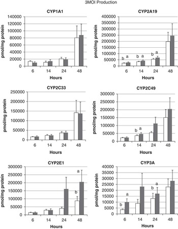
The production of the major skatole metabolite 3MOI by porcine CYP450s over time is shown in Figure 1. There was a significant linear increase in the production of 3MOI over time by all CYP450 isoforms. Co-transfection with CYB5A increased the production of 3MOI by CYP2A19, CYP2C49, CYP2E1 and CYP3A at some of the time points, whereas CYP1A1 and CYP2C33 were not affected by CYB5A. The largest amounts of 3MOI were produced by CYP2A19, CYP2C49 and CYP2E1, with the lowest amounts produced by CYP3A.
Formation of skatole metabolite 3-methyloxyindole (3MOI) by porcine cytochrome P450s (CYP450s). Human embryonic kidney HEK 293 cells were transfected with expression plasmids for porcine CYP450s along with expression plasmids for P450-oxidoreductase and cytochrome b5 reductase, with or without expression plasmid for cytochrome b5A (CYB5A). Twenty-four hours after transfection, cells were incubated with 400 μM skatole and the concentration of skatole metabolites in the media was measured at 6, 14, 24 and 48 h later by HPLC. Data are presented as the mean ± s.d. with n = 8. Open bars are experiments without CYB5A and filled bars are co-transfection experiments with CYB5A. Values with different lower case letters indicate a significant (P < .05) effect of co-transfection with expression plasmid for CYB5A at the indicated time point.

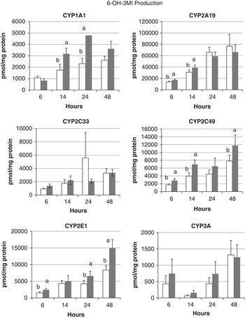
The production of the physiologically important metabolite 6-OH-3MI by porcine CYP450s over time is shown in Figure 2. The production of this metabolite increased linearly over time for CYP1A1, CYP2A19, CYP2C49 and CYP2E1, and this production was increased by co-transfection with CYB5A at some of the time points. CYP2A19 produced the greatest amount of 6-OH-3MI, which was 5- to 15-fold higher than that produced by CYP2E1 and CYP2C49, depending on the time point. Only trace amounts of this metabolite were produced by CYP2C33 and CYP3A.
Formation of skatole metabolite 6-hydroxy-3-methylindole (6-OH-3MI) by porcine cytochrome P450s. Experiments were conducted and data are presented as described in the legend to Figure 1.

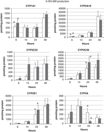
The production of the related metabolite 5-OH-3MI over time by porcine CYP450s is shown in Figure 3. Production of this metabolite increased linearly over time for CYP2A19, CYP2C33, CYP2C49 and CYP2E1, with some increase in production at 24 h by co-transfection of CYB5A with CYP2C49 and CYP2E1. Production of 5-OH-3MI was highest by CYP2A19, which was 3- to 10-fold higher than that produced by CYP2E1 and CYP2C49, depending on the time point.
Formation of skatole metabolite 5-hydroxy-3-methylindole (5-OH-3MI) by porcine cytochrome P450s. Experiments were conducted and data are presented as described in the legend to Figure 1.

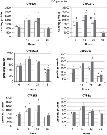
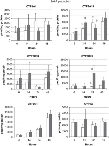
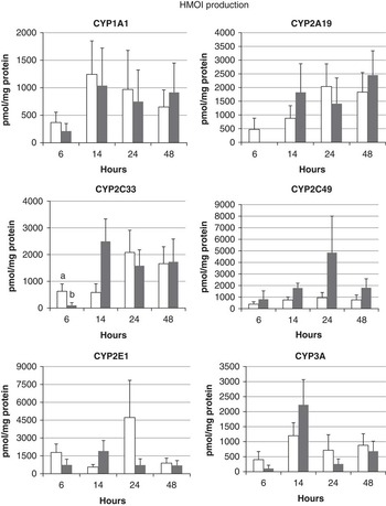
The production of I3C by all the porcine CYP450s occurred in a quadratic manner over time, with an inital increase in production followed by a decrease at 48 h (Figure 4), suggesting that I3C is further metabolized to other products. The highest amount of this metabolite was produced by CYP2A19, with increased production at some time points after co-transfection of CYP2A19, CYP2C49 or CYP2E1 with CYB5A. The production of 2AAP increased linearly over time for only porcine CYP2A19 and CYP2E1; co-transfection of CYB5A with CYP2A19 increased production of 2AAP at 14 and 24 h (Figure 5). The largest amounts of 2AAP were produced by CYP2E1. The production of the minor metabolite HMOI by porcine CYP450s did not increase significantly over time (Figure 6).
Formation of skatole metabolite indole-3-carbinol (I3C) by porcine cytochrome P450s. Experiments were conducted and data are presented as described in the legend to Figure 1.

Formation of skatole metabolite 2-aminoacetophenone (2AAP) by porcine cytochrome P450s. Experiments were conducted and data are presented as described in the legend to Figure 1.

Formation of skatole metabolite 3-hydroxy-3-methyloxindole (HMOI) by porcine cytochrome P450s. Experiments were conducted and data are presented as described in the legend to Figure 1.

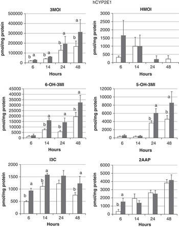
We next examined the production of skatole metabolites by human CYP2E1 (Figure 7). The production of 3MOI, 6-OH-3MI and 5-OH-3MI increased linearly over time, with significantly increased metabolite production after co-transfection with CYB5A at some time points. Production of 6-OH-3MI by hCYP2E1 was less than that for porcine CYP2E1, but hCYP2E1 produced more 3MOI and 5-OH-3MI than porcine CYP2E1. 2AAP production by hCYP2E1 increased linearly over time, whereas there was a significant quadratic effect of time on I3C production; levels of 2AAP and I3C were higher at some time points after co-transfection with CYB5A. Production of HMOI by hCYP2E1 did not increase over time.
Formation of skatole metabolites by human CYP2E1. Human embryonic kidney HEK 293 cells were transfected with human CYP2E1 (hCYP2E1) expression plasmid along with expression plasmids for P450-oxidoreductase and cytochrome b5 reductase, with or without expression plasmid for cytochrome b5A. Experiments were conducted and data are presented as described in the legend to Figure 1.

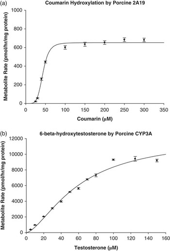
We next showed the utility of this system for determining the specificity of substrates and commercially available antibodies for porcine CYP450s, so that they could be used to identify specific isoforms of porcine CYP450s in tissues. We first investigated the specificity of the porcine CYP450s for coumarin hydroxylase and testosterone 6β-hydroxylase activity. Coumarin was found to be a specific substrate for CYP2A19 (Figure 8a), and formation of 6β-hydroxy-testosterone was specific for CYP3A (Figure 8b). CYP2A19 could also metabolize testosterone, but three unidentified metabolites were produced that did not correspond to 6β-hydroxy-testosterone. Both CYP3A and CYP2A19 displayed sigmoidal shaped curves, which were modelled by the Hill equation. The apparent kinetic parameters for CYP3A were K m = 60.6 μM and V max = 12 210 pmol/h per mg of protein. Apparent parameters for CYP2A19 were K m = 43.5 μM and V max = 652.3 pmol/h per mg of protein.
(a) Coumarin hydroxylation by CYP2A19 modelled by the Hill equation. Each point represents a mean of four trials conducted in triplicate (n = 12) ± s.d. incubated for 24 h with varying amounts of coumarin as substrate. (b) Regio-specific hydroxylation of testosterone at the 6β position by cytochrome 3A (CYP3A) modelled by the Hill equation. Each point represents a mean of four trials in triplicate (n = 12) ± s.d. incubated for 24 h with varying amounts of testosterone as substrate.

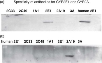
We next used this expression system to investigate the specificity of commercially available antibodies against rat CYP2E1 and human CYP2A6 for the porcine CYP450 isoforms. Western blot analysis was conducted on cell extracts containing individually expressed porcine CYP450s to test the specificity of anti-rat CYP2E1 antibody and anti-human CYP2A6 antibody (Figure 9). The anti-rat CYP2E1 antibody showed good affinity for hCYP2E1, but weaker affinity for porcine CYP2E1; a faint band can also be seen with CYP2A19. The anti-human CYP2A6 antibody shows good affinity for porcine CYP2A19 and does not appear to cross-react with the other porcine CYP450s tested.
Western blot of individually expressed cytochrome P450s (CYP450s) with (a) anti-rat cytochrome P450 2E1 (CYP2E1) antibody and (b) anti-human CYP2A6 antibody. Human embryonic kidney HEK 293FT cells were transfected with expression plasmids for the individual CYP450s, cell extracts were prepared and subjected to Western blot analysis. The anti-rat CYP2E1 antibody cross-reacts with human CYP2E1 and to a lesser extent pig CYP2E1 and slight band can be detected with pig CYP2A19. The anti-human CYP2A6 antibody cross-reacts only with pig CYP2A19, with no detectable cross-reactivity with other CYP450s.

Discussion
The characterization of CYP450s responsible for the production of specific skatole metabolites is of much interest, as it has been proposed that the production of certain metabolites, especially 6-OH-3MI, affects the rate of skatole clearance. Here we show that pig CYP1A1, CYP2A19, CYP2C33v4, CYP2C49, CYP2E1 and CYP3A and hCYP2E1 are all capable of producing the major skatole metabolite 3MOI, as well as I3C, 5-OH-3MI, 6-OH-3MI, 2AAP and HMOI at varying amounts. Diaz et al. (Reference Diaz, Skordos, Yost and Squires1999) reported that the metabolite produced in the highest amount by porcine liver microsomes was 3-OH-MI; however, 3-OH-MI is converted to 3MOI by aldehyde oxidase in hepatocytes (Diaz and Squires, Reference Diaz and Squires2000b). As 3-OH-MI was not produced in our incubations, it can be assumed that sufficient aldehyde oxidase exists within the human embryonic kidney HEK 293FT cell line to convert it to the major metabolite 3MOI. CYP2A19 produced the highest amount of 6-OH-3MI, which was 5- to 15-fold higher than for porcine CYP2E1 and CYP2C49. In addition, CYP2A19 also yields high levels of 5-OH-3MI and I3C. hCYP2E1 differs from porcine CYP2E1, as it produces more 3MOI and 6-OH-3MI and less 2AAP than porcine CYP2E1. This suggests that human and porcine CYP2E1 may differ in their capacity for metabolism of drugs and endogenous substrates, which is of relevance for toxicological and pharmaceutical studies that use the pig as a model for humans.
The involvement of CYP450 isoforms in the production of skatole metabolites by porcine liver microsomes or hepatocytes has been based on studies using inhibitors, probe substrates and antibodies that were specific for isoforms of human CYP450. However, the specificity of these tools in the pig has not been adequately tested. Friis (Reference Friis1995) suggested that the decrease in clearance of skatole in some male pigs is due to decreased activity of CYP2E1, based on their decreased ability to metabolize CLZ, a substrate used as a probe for CYP2E1. Skaanlid and Friis (Reference Skaanlid and Friis2007) have since questioned the specificity of CLZ as a probe for CYP2E1 and we have shown that porcine CYP2A19, CYP1A1 and CYP2C33 also contribute to CLZ hydroxylation (Wiercinska and Squires, Reference Wiercinska and Squires2010). Further, Squires and Lundström (Reference Squires and Lundström1997) showed that when CYP2E1 expression was high in liver, skatole levels were low in fat. The rate of sulphation of 6-OH-3MI has also been shown to be negatively correlated with skatole levels in fat and is decreased in pigs with high levels of skatole (Babol et al., Reference Babol, Squires and Lundström1998). In these two latter studies, the expression of CYP2E1 was monitored via western blot using anti-rat CYP2E1 antibody. Here we show that the anti-rat 2E1 antibody can detect porcine CYP2E1, but also cross-reacts to some degree with porcine CYP2A19. This may have given an overestimate of CYP2E1 levels in liver preparations that contain a high level of CYP2A19 protein. Thus, the suggestion that CYP2E1 is the only isoform involved in skatole clearance may not be the case, and we have showed here that other porcine CYP450 isoforms may also be involved.
Further characterization of porcine CYP450s by Diaz and Squires (Reference Diaz and Squires2000a) revealed that only treatment with inhibitors for human CYP2A6 and CYP2E1 (4-methylpyrazole, diethyldithiocarbamate, 8-methoxypsoralen and menthofuran) affected the production of skatole metabolites including 5/6-OH-3MI. Furthermore, analysis of porcine CYP2A content and activity, measured by coumarin hydroxylation, were found to be negatively correlated with skatole fat content. This implicated a new porcine isoform, CYP2A, in skatole metabolism. Terner et al. (Reference Terner, Gilmore, Lou and Squires2006) further extended this work and suggested that CYP2A19 was important in the metabolism of skatole in primary cultured porcine hepatocytes. Our study indicates that coumarin hydroxylation is indeed specific to porcine CYP2A19 and this isoform produces 6-OH-3MI in greatest amounts compared with all other isoforms tested.
The system employed here of using individually expressed CYP450 enzymes in a cell line can be used to investigate the specificity of inhibitors, substrates and antibodies of CYP450s from human and other species of pig. This eliminates the contribution from other CYP450s, allowing for the characteristics to be studied individually. However, both the tissue content of various CYP450 isoforms and their activities determine their contribution to the metabolism of a substrate in vivo (Bertz and Granneman, Reference Bertz and Granneman1997; Rendic and Carlo, Reference Rendic and Carlo1997). Currently, information about the abundance of various porcine CYP450 isoforms is limited. Kojima and Morozumi (Reference Kojima and Morozumi2004) used reverese transcriptase-PCR to show that hepatic expression of CYP2C33v4 and CYP2E1 mRNA was the greatest, followed by CYP2C49 > CYP1A1 and CYP2A19 > CYP2B22. Therefore, it is possible that CYP2E1 may play a more important role than CYP2A19 in skatole metabolism in vivo; definitive conclusions about the contribution of individual porcine CYP450s to skatole metabolism in vivo await conclusive measures on the abundance of the individual CYP450s. Moe et al. (Reference Moe, Lien, Bendixen, Hedegaard, Hornshøj, Berget, Meuwissen and Grindflek2008) used microarray analysis to determine the gene expression profiles from pigs with high and low levels of boar taint. They found that CYP2C49 and CYP2C33 were significantly upregulated in boars with high boar taint, whereas CYP2E1 and CYP2A19 were downregulated. This change in gene expression profiles may explain the low production of 6-OH-3MI in tainted boars.
The effect of CYB5A on the catalytic properties of porcine CYP450 enzymes has not been investigated extensively. The ability of CYB5A to modify the activity of CYP17A1 in the pig has been reported (Billen and Squires, Reference Billen and Squires2009), and a c.-8G>T polymorphism in the CYB5A gene was associated with low androstenone levels in fat and low levels of both CYB5A mRNA and CYB5A protein (Peacock et al., Reference Peacock, Lou, Lundström and Squires2008). The contribution of CYB5A to CYP450-mediated catalysis is complex and is dependent on the substrate and CYP450 isoform (Schenkman and Jansson, Reference Schenkman and Jansson2003). In some cases, CYB5A can modify the reaction by having an inhibitory or stimulatory effect, and in other cases it has been shown to be obligatory. Our results show that the effects of CYB5A on the production of individual skatole metabolites are specific to different CYP450 isoforms. Regression analysis indicated that CYB5A increased the production of 3MOI by CYP2A19, CYP2E1, hCYP2E1 and CYP3A, increased production of 6-OH-3MI by CYP1A1, CYP2A19, CYP2C49, CYP2E1 and hCYP2E1, increased production of I3C by CYP2A19, CYP2C49, CYP2E1 and hCYP2E1, increased production of 5-OH-3MI by CYP2C49, CYP2E1 and hCYP2E1 and increased production of 2AAP by CYP2A19 and CYP2C49; however, these effects were not significantly different at all time points. CYB5A thus plays an important role both in the biosynthesis of androstenone and in the metabolism of skatole.
In summary, we present evidence here that supports CYP2A19, CYP2E1 and CYP2C49 as potential key regulators of skatole metabolism and clearance through the production of 6-OH-3MI. As the actual contribution of the individual CYP450 isoforms in vivo is also determined by the expression levels of these isoforms, a better understanding of porcine CYP450 isoform abundance is required before conclusions can be drawn about the key CYP450s that regulate skatole clearance in vivo.
Acknowledgements
This study was supported by NSERC Discovery Grant no. 400066 and funding from the Ontario Ministry of Agriculture and Food (Grant no. 026398) to E.J.S.