INTRODUCTION
The Decapoda stomach is a complex structure with great interspecific variation (Icely & Nott, Reference Icely and Nott1992). There, after manipulation of the mouthparts, the secondary processing and the real trituration of the food occurs by the action of the gastric mill (Ceccaldi, Reference Ceccaldi2006). This structure varies between species reaching its highest development (greater number of ossicles and calcification) in Brachiuran crabs (Icely & Nott, Reference Icely and Nott1992), in which the gastric mill was described as a ‘three-branched claw through which the food must pass in order to reach the pyloric region’ (see Ceccaldi, Reference Ceccaldi2006). This ‘claw’ is made up of a pair of lateral teeth protruding from the side walls of the heart chamber, a medium tooth extending from the dorsal wall of the chamber (Allardyce & Linton, Reference Allardyce and Linton2010) and a number of supportive ossicles. On the other hand, in other groups of Decapoda the stomach can be very simple with just a few calcified ossicles or total absence of them (Felgenhauer & Abele, Reference Felgenhauer and Abele1985; Icely & Nott, Reference Icely and Nott1992).
Several studies suggest that the anatomy of the gastric mill varies according to the diet of species in different groups of Decapoda (e.g. Kunze & Anderson, Reference Kunze and Anderson1979; Icely & Nott, Reference Icely and Nott1992; Allardyce & Linton, Reference Allardyce and Linton2010). According to this hypothesis the gastric mill of macrophagous species is expected to be calcified and dentate with few setae, while those that feed on micro material should present small blunt teeth and high setae density (Caine, Reference Caine1975; Kunze & Anderson, Reference Kunze and Anderson1979). Furthermore, the teeth of carnivorous species is expected to present molar processes for grinding soft animal tissue, while herbivores should present high transverse ridges in order to cut fibrous material (Skilleter & Anderson, Reference Skilleter and Anderson1986). Omnivorous species are characterized by ossicles with cusps but also smooth surfaces for grinding (Salindeho & Johnston, Reference Salindeho and Johnston2003). An alternative view suggests the gastric mill's structure is related to phylogenetic aspects rather than to the diet of a species (Felgenhauer & Abele, Reference Felgenhauer and Abele1985), therefore, ossicles and teeth characteristics of the cardiac stomach have been used as phylogenetic markers (e.g. Felgenhauer & Abele, Reference Felgenhauer and Abele1985; Sakai, Reference Sakai2004; Brösing et al., Reference Brösing, Richte and Scholtz2007). According to this hypothesis phylogenetically close species should have similar gastric mills despite having very different feeding habits. Finally, Brösing & Türkay (Reference Brösing and Türkay2011) propose a synthesis stating that teeth of the brachyuran cardiac stomach have a relatively stable structure, nonetheless this does not exclude the existence of adaptations to food preferences or to different resources use.
Ovalipes trimaculatus (De Haan, 1833) is a portunid crab distributed from southern Brazil to southern Argentina in the Atlantic Ocean (Vinuesa, Reference Vinuesa2005; Melo, Reference Melo2010), and from southern Peru to south of Chile in the Pacific Ocean (Retamal, Reference Retamal1981). In San Matias Gulf, Argentina, it has been exploited since 2007 by artisanal fishermen who have historically developed a diving shellfish fishery (Narvarte et al., Reference Narvarte, González and Filippo2007). The relevance of the activity has been growing ever since because of the products’ low cost–benefit ratio, but also because during red tide outbreaks O. trimaculatus is the only resource the diving fishermen can extract. This new extractive activity involves the need to understand, evaluate and assess biological and ecological aspects of the populations of O. trimaculatus. This species has been described as carnivorous and/or scavenger feeding mainly on small benthic invertebrates and fish (Fenucci & Boschi, Reference Fenucci and Boschi1975). Dietary analysis on the San Matías Gulf population has revealed that the more frequent occurrences in stomach contents are small fishes, crustaceans and bivalves, with occasional occurrence of echinoderms (Ophiuroidea and Echinoidea) and chitons (de la Barra et al., Reference de la Barra, Romero, González and Narvarte2014). We believe it is necessary to deepen the study of trophic aspects of this population to draw potentiality and consumption patterns.
The aim of this paper is to describe morphofunctionally the gastric mill of O. trimaculatus. In turn, the morphology of the gastric mill is evaluated in light of the species diet and compared with homologous structures in other portunid crabs and other decapods in order to establish the influence of phylogeny or diet on morphology.
MATERIALS AND METHODS
Ovalipes trimaculatus specimens were obtained at the north of San Matias Gulf, Argentina (40°58′S 65°06′W). Crabs were collected manually by scuba diving at a depth of 10–20 m and preserved at a temperature of −20 °C. Thirty individuals were dissected and their stomachs were preserved in 70% alcohol in order to be put through different treatments afterwards. The stomachs were cleaned and the contents were removed under stereo-microscope. Muscle remains that were attached to the external parts were eliminated. In order to optimize the observation of the structures of interest, 10 stomachs were digested in boiling KOH (10%) for 1 h and then stained with Alzarin-red; another 10 stomachs were stained without prior digestion, and the remaining 10 were neither digested nor stained. The different treatments allowed the observation of different structures with more detail and clarity. The stomachs were observed and photographed in a stereo-microscope Leica S8 APO with built-in camera (Canon EOS Rebel t2). Three more stomachs were removed cleaned and preserved as indicated previously and then placed in a desiccator with silica-gel with a moisture indicator for at least 24 h. After conditioning, these samples were mounted on a metal stub using double-sided tape and/or silver paint, samples were gold coated for 120 s using Combined Deposition System metal/carbon, SPI Supplies, AX-12157, operated under argon atmosphere (18 mA) for 120 s. Then, observations were made using a JSM-35C scanning electron microscope (JEOL, China), equipped with a system of digital image acquisition Sem Afore, at an accelerating voltage of 20 kV.
Descriptions of the structures of the cardiac chamber that could match food processing and outline trophic possibilities were made from the photographs and micrographs using the nomenclature proposed by Ngoc-Ho (Reference Ngoc-Ho1984) and Kunze & Anderson (Reference Kunze and Anderson1979).
RESULTS
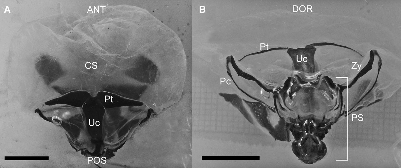
The oesophagus connects ventrally with the stomach which consists of two regions: the cardiac and the pyloric stomach. The cardiac stomach is a chitinous, thin-walled translucid chamber, of greater volume than the pyloric stomach. It has a series of well calcified pieces on the walls symmetrically disposed at the posterior end (Figure 1).
Stereo-microscopy images of the cardiac stomach of Ovalipes trimaculatus. (A) Dorsal view showing urocardiac and pterocardiac ossicles; (B) posterior view showing gastric mill ossicles and pyloric stomach. ANT, anterior direction; CS, cardiac stomach; DOR, dorsal direction; Pc, pectineal ossicles; POS, posterior direction; PS, pyloric stomach; Pt, pterocardiac ossicles; Uc, urocardiac ossicle; Zy, zygocardiac ossicles. Scale bars: 1 mm.

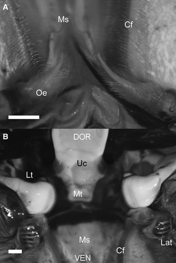
On the internal laterals of the cardiac stomach's entrance there is a pair of oesophageal valves regulating the opening of the stomach. These valves are elongated, cirrus-like structures, fully covered with setae (Figure 2A). On the ventral wall of the stomach, posterior to the esophageal valves, the medial strip extends reaching the posterior end of the cardiac stomach. On either side of the medial strip there are longitudinal folds of the stomach's floor forming a pair of channels covered by setae called ‘cardiac filter’ (Figure 2A).
Stereo-microscopy images of Ovalipes trimaculatus. (A) Inside ventral view of the cardiac stomach showing oesophageal valves followed by medial strip; (B) frontal view of the gastric mill. Cf, cardiac filter; DOR, dorsal direction; Lat, lateral accessory teeth; Lt, lateral teeth; Ms, medial strip; Mt, medial tooth; Oe, oesophageal valves; VEN, ventral direction. Scale bars: 1 mm.

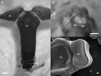
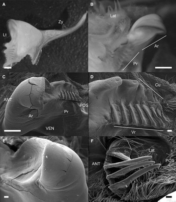
The gastric mill, the most conspicuous structure of the cardiac stomach, is found posteriorly (Figure 2B). It consists of a medial tooth supported by the urocardiac ossicle (Figure 3), two lateral teeth, supported by the zygocardiac ossicles (Figure 4), a pair of less calcified lateral accessory teeth (Figure 4B, F) and the cardio-pyloric valve (Figure 5).
(A) Urocardiac ossicle carrying the medial tooth on its posterior region; (B) view of the medial tooth showing setose pads around it; (C) medial tooth detail, showing its smooth concave surface. ANT, anterior direction; Mt, medial tooth; POS, posterior direction; Sp, Setose pads; Uc, Urocardiac ossicle; A–B Stereo-microscopy images; C SEM image. Scale bars: A–B, 1 mm; C, 0.1 mm.

(A) Zygocardiac ossicle with lateral tooth; (B) posterior view of the lateral tooth showing relative position to lateral accessory tooth, anterior and posterior region; (C) anterior and posterior region of lateral tooth; (D) posterior region of the lateral tooth showing ventral cusps and dorsal vertical ridges; (E) spoon shaped anterior region of the tooth; (F) lateral accessory tooth surrounded by setae. ANT, anterior direction; Ar, Anterior region of the lateral tooth; Cu, cusps; Lat, lateral accessory tooth; Lt, lateral tooth; Pr, posterior region of the lateral tooth; VEN, ventral direction; Vr, vertical ridges; Zy, zygocardiac ossicle. A–B are stereo-microscopy images; C–F are SEM images. Scale bars: A–C, 1 mm; D–F 0.1 mm.

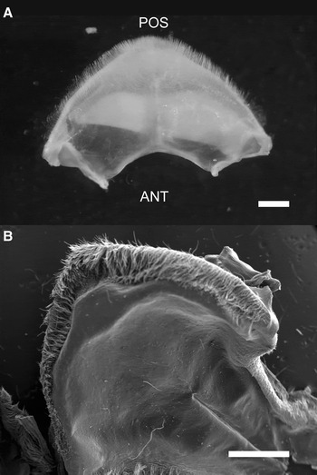
(A) Stereo-microscopy of the ventral view of the cardio-pyloric valve; (B) scanning electron microscopy of the dorsal view of the cardio-pyloric valve. ANT, anterior direction POS, posterior direction. Scale bars: 1 mm.

The urocardiac ossicle extends from the centre of the dorsal wall of the cardiac stomach to posterior. It is dorsoventrally flattened and elongated in anteroposterior direction (Figure 3A). Posterior and internally to the chamber this ossicle bears the medial tooth of the gastric mill, a calcified structure pointing toward the anteroventral region from the dorsal wall of the stomach (Figure 3). This tooth has a quadrangular shape and a concave, smooth surface; at each corner of the square there is a projection with round edges (Figure 3C). A setose pad surrounds this structure (Figure 3B).
The zygocardiac ossicles are paired, calcified structures on the posterior laterals of the cardiac chamber (Figure 1B, 4A). At their posterior end, the zygocardiac ossicles bear the lateral teeth, which are heavily calcified structures that protrude inside the chamber (Figure 4). The lateral teeth are antero-posteriorly elongated. An anterior and a posterior region can be clearly distinguished in the teeth (Figure 4B). The anterior region is wider, with a concave, smooth surface; it has smooth, sharp edges and an anterior concave cusp at the anterior end (Figure 4E). The posterior region of the lateral teeth narrows down from anterior to posterior. It has 6 to 9 sharp vertical ridges on the dorsal side; 2 to 5 rounded cusps of decreasing size can be found ventrally (Figure 4D). The anterior region of the lateral teeth is more robust and somewhat wider than the posterior region, and protrudes inwards, pointing postero-ventrally.
Pectineal ossicles are located on the lateral walls of the cardiac stomach (Figure 1B). From a lateral view, they are found below each zygocardiac ossicle. These paired structures have the shape of a curved rod and articulate anteriorly with zygocardiac and pterocardiac ossicles. On their posterior end, slightly ventroanteriorly to the lateral teeth the lateral accessory teeth can be observed (Figure 4B, F). These are chitinous structures, paired and un-calcified, constituted by 4–6 sharp denticles pointing inward and backward of the cardiac chamber. These structures are much smaller and softer than the previously described teeth.
On the posterior end of the cardiac stomach the medial strip curves downward forming the cardio-pyloric valve, a very thin calcareous structure with a semicircular shape, an apical edge covered by setae and a smooth protuberance on the tip (Figure 5).
DISCUSSION
The gastric mill of O. trimaculatus is similar to the gastric mills of other Portunidae (Brösing, Reference Brösing2010). It is a complex structure with robust, well calcified teeth. These characteristics would be functional when breaking down large pieces of food, in accordance with a macrophagous diet (Caine, Reference Caine1975; Kunze & Anderson, Reference Kunze and Anderson1979).
When food enters the cardiac stomach the oesophageal valves and the lateral accessory teeth direct the larger pieces into the mill (Kunze & Anderson, Reference Kunze and Anderson1979). The function of the lateral accessory teeth is probably to support and/or to direct the food, rather than that of grinding or processing for they are significantly softer than the other teeth and not calcified. The function of transferring food to the grinder area is consistent with the characteristics described for carnivorous species of Decapoda (Caine, Reference Caine1975; Kunze & Anderson, Reference Kunze and Anderson1979; Salindeho & Johnston, Reference Salindeho and Johnston2003). Ovalipes trimaculatus lateral accessory teeth are very similar to those described for other carnivorous species of Decapoda that are not closely related to Portunidae, like pagurids Dardanus setifer (Kunze & Anderson, Reference Kunze and Anderson1979) and Petrochirus diogenes (Caine, Reference Caine1975), or like the grapsids Geograpsus grayi and Geograpsus crinipes (Allardyce & Linton, Reference Allardyce and Linton2010), and the calappid, Calappa granulata (Cartes et al., Reference Cartes, Abelló, Lloris, Carbonell, Torres, Maynou and Sola2002; Brösing, Reference Brösing2010). Lateral accessory teeth of these species consist of a row of sharp denticles which are in some cases accompanied by groups of setae. Skilleter & Anderson (Reference Skilleter and Anderson1986) propose that denticle size of the lateral accessory teeth could be related to the size of the pieces that reach the stomach, for in detritivore pagurids setae seem more relevant than denticles in the structure of lateral accessory teeth, as opposed to what they observed in carnivore pagurids, in which denticles are larger. Nonetheless there are predatory species such as Pseudocarcinus gigas that do not even present lateral accessory teeth (Heeren & Mitchell, Reference Heeren and Mitchell1997). Whitin portunids, Nectocarcinus tuberculosus (Salindeho & Johnston, Reference Salindeho and Johnston2003) and Callinectes sapidus (Cochran, Reference Cochran1935) both have lateral accessory teeth forming a row of 5–7 sharp denticles, no special setae arrangement is mentioned in their description. In this sense, the structures found in O.trimaculatus fit a carnivorous diet, but also resemble the morphology of its close relatives.
Medial and lateral teeth of the gastric mill grind the larger pieces by performing simultaneous movements: the medial tooth moves anteroventrally and lateral teeth swing dorsomedially (McGaw & Curtis, Reference McGaw and Curtis2013). As these elements are the ones forming the chewing apparatus of the gastric mill, their structure is essential in terms of digestion capacity (Jaszkowiak et al., Reference Jaszkowiak, Keiler, Wirkner and Richter2015). In this sense, the aspects in which we focused during this work are directly related to the digestive function of the gastric mill. The presence of smooth concave structures in O. trimaculatus gastric teeth suggests a mortar-like action on the food: this feature is usually associated with the action of grinding or softening animal fibres (e.g. Creswell & Marsden, Reference Creswell and Marsden1990; Heeren & Mitchell, Reference Heeren and Mitchell1997; Salindeho & Johnston, Reference Salindeho and Johnston2003; Allardyce & Linton, Reference Allardyce and Linton2010). Conversely, in species with higher vegetal content in their diets, the gastric mill is more complex and presents more hooks and sharp peaks to disassemble hard vegetable fibres (e.g. Giddins, Reference Giddins1986; Cannicci et al., Reference Cannicci, Gomei, Boddi and Vannini2002).
The medial tooth is not as robust as observed in other species and presents a concave shield where other portunid crabs present ridges (Salindeho & Johnston, Reference Salindeho and Johnston2003). A setose pad is observed at each lateral and dorsally to the medial tooth. Some authors suggest their function could be sweeping the ground particles from the teeth into the light of the stomach (Skilleter & Anderson, Reference Skilleter and Anderson1986; Salindeho & Johnston, Reference Salindeho and Johnston2003). In the anterior region of the lateral teeth there is a concave zone similar to the ones observed in carnivores from other families such as C. granulata (Calappidae, Cartes et al., Reference Cartes, Abelló, Lloris, Carbonell, Torres, Maynou and Sola2002; Brösing, Reference Brösing2010), G. grayi and G. crinipes (Grapsidae, Allardyce & Linton, Reference Allardyce and Linton2010); and also in portunids as C. sapidus (Cochran, Reference Cochran1935). Other species, such as the omnivore grapsid Pachygrapsus marmoratus, present ridges or peaks in the anterior region of the lateral teeth (Cannicci et al., Reference Cannicci, Gomei, Boddi and Vannini2002).
The presence of concavities in the medial and lateral teeth could have the function of retaining the food in order to maximize the mechanical action of the mill and the exposure to digestive enzymes. The bulged dimensions of the cardiac chamber along with these concavities could indicate the food ingested requires a considerable amount of time to be processed mechanically and physiologically. It could also be indicative of a trophic strategy, for example the scavenger crustaceans of the deep benthic habitats can ingest large amounts of food in short time (Eastman & Thiel, Reference Eastman, Thiel, Thiel and Watling2015). Furthermore, in species that spend a considerable amount of time sheltered, such as Thenus orientalis, the folds that increase the storage capacity are a possible adaptation to optimize the reduced time spent in capturing prey (Johnston & Alexander, Reference Johnston and Alexander1999). In species with this kind of behaviour the calcification of the mill is considerably low although they present a macrophagous diet and a predatory behaviour. Ovalipes trimaculatus is generally buried in sandy bottoms and presents rapid ingestion after quick external processing of food, therefore the dimensions of the stomach and the concavities of the teeth probably respond to its feeding behaviour.
At the posterior region of the lateral teeth of O. trimaculatus there are vertical ridges, very typical structures of the lateral teeth also found in many other species of Brachyura (e.g. Cochran, Reference Cochran1935; Cannicci et al., Reference Cannicci, Gomei, Boddi and Vannini2002). Similar structures but with hooked ends can be found in herbivorous species (Giddins, Reference Giddins1986). In micromaterial feeders these teeth present comb-like processes (e.g. Brösing & Türkay, Reference Brösing and Türkay2011) or even a saetiform structure (Kropp, Reference Kropp1986). In O. trimaculatus the blunt ends of these structures can be indicative of adaptation to animal tissue processing, which does not require a strong mechanical treatment opposed to vegetal fibres. Also, similar ridges are present in the lateral teeth of other Portunidae and related families (Brösing, Reference Brösing2010).
In some decapods the cardiopyloric valve contributes to the grinding action of the mill by forcing the accumulation of the material and providing a structure on which the food can be disintegrated (Icely & Nott, Reference Icely and Nott1992). The latter seems unlikely in the case of O. trimaculatus because of the low calcification of its cardiopyloric valve and the absence of annexed structures that could collaborate with the mechanical destruction of the material. Despite being a carnivorous species, the cardiopyloric valve of O. trimaculatus lacks masticatory structures as seen in other carnivores such as the anomuran Petrochirus diogenes (Caine, Reference Caine1975).
To our knowledge, the masticatory structures of the gastric mill of three other portunid crabs have been described to date: Callinectes sapidus (Cochran, Reference Cochran1935; Maynard & Dando, Reference Maynard and Dando1974), Ovalipes guadulpensis (Caine, Reference Caine1974) and Nectocarcinus tuberculosus (Salindeho & Johnston, Reference Salindeho and Johnston2003). Brösing (Reference Brösing2010) also described the gastric mill of other portunids but information on the characters of interest to this study is lacking. When comparing the gastric mill of O. trimaculatus with those of C. sapidus, O. guadulpensis and N. tuberculosus similar structures can be easily found: lateral teeth bearing a front cusp and two lines of posterior denticles, medial tooth relatively quadrangular pointing in anterior direction, semicircular cardiopyloric valve with setous edge (except in O. guadulpensis, which lacks description). However certain differences can be observed when comparing the gastric mill of N. tuberculosus gastric mill with those of O. trimaculatus, O. guadulpensis and C. sapidus. Nectocarcinus tuberculosus has a medial tooth that presents ridges and cusps (Salindeho & Johnston, Reference Salindeho and Johnston2003) while in the other three portunids the medial tooth has a smooth surface. When comparing lateral teeth within portunids, N. tuberculosus presents more pronounced cusps and more evident grooves than the other species; also, it presents an anterior cusp with a convex surface while the other portunids present an anterior cusp forming a concave region. Although Portunidae are generally predators and scavengers, species have been observed that integrate vegetal material in their diet (Williams, Reference Williams1981). This is the case of N. tuberculosus, which has an omnivorous diet in which sea grasses are a predominant component (Salindeho & Johnston, Reference Salindeho and Johnston2003). Conversely, C. sapidus and O. guadulpensis have a diet mainly composed of small benthic invertebrates and fishes, with a low presence of algal material (Caine, Reference Caine1974; Laughlin, Reference Laughlin1982; 4% for C. sapidus). The described diet for O. trimaculatus is very similar to the ones of C. sapidus and O. guadulpensis: benthic invertebrates, small fishes and a very low content of algal material (Fenucci & Boschi, Reference Fenucci and Boschi1975; de la Barra et al., Reference de la Barra, Romero, González and Narvarte2014). The gastric mill of N. tuberculosus, the portunid with more relevant vegetal component in the diet has greater presence of uneven surfaces and structures with more cusps. However, all three species conserve the typical structure of portunidae gastric teeth (Brösing, Reference Brösing2010). These structures can also be found in closely related families such as Cancridae (e.g. Schubart & Reuschel, Reference Schubart, Reuschel, Martin, Crandall and Felder2009; Spiridonov et al., Reference Spiridonov, Neretina and Schepetov2014). In species of this family quadrangular, smooth medial teeth are found along with lateral teeth bearing an anterior cusp, posterior ventral ridges and posterior dorsal cusps (Cancer pagurus Brösing, Reference Brösing2010; Cancer novaezelandiae Creswell & Marsden, Reference Creswell and Marsden1990). They also present a semicircular cardiopiloric valve crowned by setae. The lateral accessory teeth, on the other hand are much more robust in Cancridae (Brösing, Reference Brösing2010).
Gastric mill ossicles have been used in various Decapoda groups to discern their phylogeny (e.g. Brösing et al., Reference Brösing, Richte and Scholtz2007; Reimann et al., Reference Reimann, Richter and Scholtz2011). Although, mainly, supportive ossicles traits are used to discern between brachyuran families (see Brösing et al., Reference Brösing, Richte and Scholtz2007), medial tooth morphology has proven useful to distinguish even closely related species (e.g. Naderloo & Schubart, Reference Naderloo and Schubart2010; Naderloo et al., Reference Naderloo, Türkay and Chen2010). Also, numerous examples exist of closely related species with different traits in their gastric teeth associated with their diets (e.g. Icely & Jones, Reference Icely and Jones1978; Allardyce & Linton, Reference Allardyce and Linton2010; Brösing & Türkay, Reference Brösing and Türkay2011). The strength of the relationship between structures and phylogeny or diet most likely depends on adaptive pressure and physical constraints. What is clear is that within brachyurans these structures are very plastic and can take saetiform shape as well as robust hard teeth, although conserving a similar ground pattern (Brösing, Reference Brösing2010). Other feeding structures (i.e. chela and mouth appendages) have also been tested for phylogenetic diagnosis (e.g. Naderloo et al., Reference Naderloo, Türkay and Chen2010; Sahlmann et al., Reference Sahlmann, Chan and Chan2011) and trophic habits (e.g. Sahlmann et al., Reference Sahlmann, Chan and Chan2011).
The phylogenetic position of the genus Ovalipes has been discussed in the past years because molecular phylogenetic reconstructions do not place Ovalipes within the Portunidae family (Schubart & Reuschel, Reference Schubart, Reuschel, Martin, Crandall and Felder2009; Spiridonov et al., Reference Spiridonov, Neretina and Schepetov2014). Schubart & Reuschel (Reference Schubart, Reuschel, Martin, Crandall and Felder2009) placed Ovalipes provisionally in family Geryonidae along with genus Benthochascon, and later Spiridonov et al. (Reference Spiridonov, Neretina and Schepetov2014), proposed the new family Ovalipidae with Ovalipes as the type genus. In that sense, although in the present study O. trimaculatus was considered a portunid crab (Ng et al., Reference Ng, Guinot and Davie2008), it is possible that this classification is not accurate. To our knowledge there is no description of any Geryonidae's foregut traits, and within Ovalipes the only description available is the study by Caine (Reference Caine1974).
The constraint observed between phylogeny and trophic habit is also observed in other feeding structures that have shaped the habits of consumption of portunids as well. Some cheliped traits conserved throughout Portunoidea, for example, are considered the morpho-functional basis of the predatory and scavenger habit typical of this group (Spiridonov et al., Reference Spiridonov, Neretina and Schepetov2014). Moody & Steneck (Reference Moody and Steneck1993) on the other hand, have seen that patterns of behaviour in handling and consuming molluscs may vary between phylogenetically related species more than between ecologically similar species. In summary, the results attained in the present work and their comparison with the existing literature on other portunid species and other Decapoda families show that while the gastric mill of O. trimaculatus' shares its main traits with other portunids, it also shows the typical characters expected for a carnivorous diet. In this sense, our results agree with the views expressed by Brösing & Türkay (Reference Brösing and Türkay2011): there is a stable ground pattern given by shape and elements forming the gastric mill: O. trimaculatus' gastric mill is probably more similar to C. sapidus' or N. tuberculosus' than that of Pseudocarcinus gigas (Heeren & Mitchell, Reference Heeren and Mitchell1997); upon that pattern, trophic habits can adaptively shape certain characteristics (e.g. smooth medial teeth, concave lateral teeth). In future work, to thoroughly analyse the morphology modelling trophic habits we should analyse the morphology of the mouthparts and chelipeds of O. trimaculatus. In addition, information of digestive enzymes and feeding behaviour will further improve the knowledge we have on trophic habits of this species.
ACKNOWLEDGEMENTS
This paper is part of the Doctoral thesis of P. de la Barra at University of Buenos Aires (UBA). We would like to thank Captain G. Aguayo for providing the biological samples. Support provided by the scientific staff of Instituto Nacional de Limnología and Instituto de Biología Marina y Pesquera A. Storni is gratefully acknowledged.
FINANCIAL SUPPORT
The work described herein was funded by grants PICT CONAE CONICET No. 04/2010, PICT Préstamo BID 2159 (Agencia Nacional de Promoción Científica y Tecnológica) and PIP CONICET 052.