Introduction
Mandel and Métais (Ref. Reference Mandel and Metais1) reported in 1948 the presence of nucleic acids in plasma of both healthy individuals and patients with various diseases. Following this discovery, the interest about cell-free DNA (cfDNA) reawakened in the 1960s when the detection of circulating cfDNA was reported in the serum of patients with systemic lupus erythematous (Ref. Reference Tan2). Interestingly, Leon et al. (Ref. Reference Leon3) demonstrated in 1977 that cancer patients had higher levels of serum cfDNA than healthy individuals and that significantly higher cfDNA levels were present in patients with metastatic disease when compared with those without metastases. Furthermore, these investigators found that the levels of cfDNA decreased in up to 90% of the patients after radiation therapy (especially in those who responded well to treatment), whereas patients with persistently high or increased cfDNA levels had a poor prognosis. The observation that cancer patients had higher levels of cfDNA than healthy individuals was later extended by a study showing that patients with malignant disease of the gastrointestinal tract had significantly higher levels of cfDNA when compared with patients with benign disease (Ref. Reference Shapiro4). A decade later, Stroun et al. (Ref. Reference Stroun5) extracted and purified DNA from plasma of cancer patients which was shown to consist of double-stranded DNA fragments of human origin ranging up to 21 kb in length. Soon after it was shown that the DNA extracted from plasma of cancer patients presented decreased strand stability and enhanced DNA synthesis, features that are typical of DNA from malignant cells and that were not observed in plasma DNA from healthy individuals (Ref. Reference Stroun6). These findings clearly showed that part of plasma DNA in cancer patients had its origin from tumour cells, being referred to as cell-free tumour DNA (ctDNA).
The issue of cfDNA attracted little attention during these early days, but its importance was recognised in 1994 with the detection of mutated KRAS gene in the plasma of patients with pancreatic carcinoma, which harboured the same mutation (Ref. Reference Sorenson7). Soon after, several studies reported the presence of microsatellite alterations (Refs Reference Chen8, Reference Nawroz9), TP53 mutations (Ref. Reference Silva10), and aberrant promoter hypermethylation of tumour suppressor genes (Refs Reference Esteller11, Reference Wong12) in cfDNA from cancer patients. Over the last two decades, many other tumour-related genetic and epigenetic alterations have been detected in plasma and serum from cancer patients, such as point mutations, rearranged genomic sequences, copy number variations (CNVs), microsatellite instability, loss of heterozygosity and DNA methylation (Ref. Reference Marzese13).
There are several features that make analysis of cfDNA a promising strategy in cancer patients: first, ctDNA provides a better representation of the tumour genome since it comprises DNA released by tumour cells from both primary tumour and metastases, minimising the problem of inter- and intra-tumour heterogeneity (Ref. Reference Burrell14); second, it is possible to quantify cfDNA and its concentration seems to reflect the tumour burden, making cfDNA measurements potentially useful to monitor tumour dynamics (Ref. Reference Diehl15) and third, blood samples can be drawn by minimally invasive methods, presenting lower risk than tissue biopsy, allowing repeated sampling and ‘real-time’ monitoring during the entire course of the disease.
The origin and biological characteristics of cfDNA
Recent studies have confirmed that, although cfDNA levels vary between individuals, higher levels are found in cancer patients. Indeed, small amounts are detected in the plasma from healthy individuals, with a plasma concentration range of 1.8–118 ng/ml (Refs Reference Shapiro4, Reference Gautschi16), while cancer patients present a range of 59–819 ng/ml. Furthermore, it has been shown that the proportion of plasma ctDNA varies from 10 to 90% of the total cfDNA (Ref. Reference Jahr17) and can be derived from the primary tumour, metastases and circulating tumour cells (CTCs) in cancer patients, thereby representing the entire tumour burden (Ref. Reference Alix-Panabieres and Pantel18).
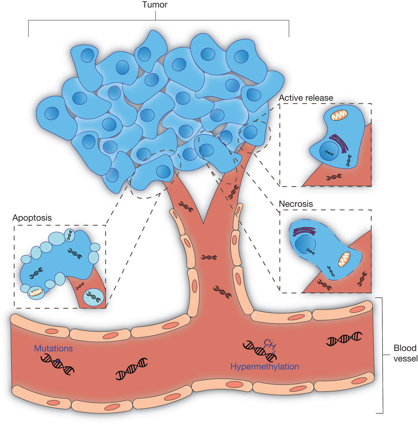
Some aspects concerning the biology of cfDNA remain poorly understood, such as the mechanism behind DNA release by cells, the process of clearance from the circulation and the biological impact of cfDNA. The assumption that cfDNA has its origin from dying cells that release their content into the bloodstream has become the most highlighted hypothesis. In healthy individuals, cfDNA is thought to derive from apoptotic cells, since normal plasma samples mainly contain DNA fragments of 180 bp and multiples of this (characteristic of apoptotic cells) and fragments larger than 2000 bp are not detected (Ref. Reference Suzuki19). In cancer patients, apoptosis has also been proposed as the origin of cfDNA based on several observations showing that their plasma and serum DNA presents a ladder pattern after electrophoresis, which is identical to the pattern displayed by apoptotic cells (Ref. Reference Jahr17). However, this hypothesis is questionable because apoptosis is a mechanism supposedly lost by cancer cells and this characteristic ladder pattern was also observed for actively released DNA (Ref. Reference Stroun20). Necrosis has also been pointed out as a mechanism of tumour DNA release into the bloodstream, since the presence of plasma DNA fragments larger than 10 000 bp, typical of cells dying via necrosis, have been observed (Ref. Reference Jahr17). However, the decrease of plasma DNA levels reported following radiation therapy (Ref. Reference Leon3) confronts this hypothesis, as an initial increase of cfDNA following radiation therapy would be expected if necrosis was the main mechanism of DNA release by tumour cells. Another hypothesis commonly advanced is the lysis of CTCs, although this is clearly not the predominant pathway for DNA release because CTCs are considered extremely rare (Ref. Reference Plaks21) and unlikely concentrations of CTCs would be necessary to explain the ctDNA concentrations found in plasma and serum of cancer patients (Refs Reference Alix-Panabieres and Pantel22, Reference Sorenson23). A fourth possible and plausible possibility is that proliferating tumour cells actively release DNA into the bloodstream (Ref. Reference Stroun20) through the same mechanism as observed in in vitro cultivated lymphocytes (Ref. Reference Anker24). Be that as it may, the main mechanism of DNA release has still not been determined and it is very likely that more than one mechanism is involved (Fig. 1).
The origins of cell-free tumour DNA. Tumour cells can release DNA into the blood stream by three different mechanisms, such as apoptosis, necrosis and spontaneous active release, making tumour-related genetic and epigenetic alterations detectable in blood.

The stability and clearance mechanism of cfDNA from plasma is also not completely understood and a few possibilities have been proposed. Although there are no studies in cancer patients, it is likely that ctDNA may be rapidly removed from bloodstream analogously to the rapid clearance of foetal DNA from maternal blood (Ref. Reference Lo25). Several studies have shown that cfDNA has a relatively short half-life (Refs Reference Lo25, Reference Fleischhacker and Schmidt26) through a process that involves plasma nucleases (Ref. Reference Lo25), but several studies in experimental animals indicate that liver, spleen, and kidneys are also involved in the removal of cfDNA from plasma (Refs Reference Tsumita and Iwanaga27, Reference Chused28, Reference Emlen and Mannik29).
Regardless of the mechanism involved in the origin of cfDNA, the fact is that elevated levels are present in the plasma of cancer patients and several questions have been raised about its potential biological impact. Experimental studies with tumour-bearing rats showed that their plasma DNA could be incorporated into the genome of cultured cells and transform them (Ref. Reference Garcia-Olmo and Garcia-Olmo30). Based on these experiments and the reported presence of oncogenes in plasma of cancer patients, the ‘hypothesis of genometastasis’ was proposed which postulates the transformation of normal cells through the uptake of ctDNA (Ref. Reference Garcia-Olmo and Garcia-Olmo30). This theory could eventually explain the development of metastases in distant target organs due to transfection of dominant oncogenes released from the primary tumour and present in the plasma (Ref. Reference Garcia-Olmo and Garcia-Olmo30).
Clinical applications of ‘liquid biopsies’ in ovarian cancer
Ovarian cancer is the seventh most common cancer and the eighth most common cause of cancer-related death in women. In 2010, 239 000 new cases and 152 000 deaths were registered worldwide (Ref. Reference Ferlay31). The lack of specific symptoms and effective screening methods explains the high mortality rate, as most ovarian carcinomas are diagnosed at an advanced stage in which surgery and chemotherapy are not curative. The 5-year survival of early detected ovarian cancers (stage I) is about 90%, but decreases to 30–50% and 13% for stage III and stage IV disease, respectively (Ref. Reference Guppy32), so a new strategy for early detection of this disease is an unmet need.
Epithelial ovarian cancer (EOC) is the most common type of ovarian cancer, accounting for over 95% of the cases, and is divided into five subtypes: high-grade serous (HGSC), endometrioid, clear cell, mucinous and low-grade serous (LGSC) carcinomas. These subtypes are considered as five distinct diseases because they present different morphologies, patterns of genetic alterations, precursor lesions, responses to therapy and prognosis (Refs Reference Prat33, Reference Seidman34). The mutational spectrum of the various histological subtypes is the following: HGSC is genetically characterised by frequent TP53 mutations and less frequent but recurrent mutations in BRCA1, BRCA2, NF1, RB1 and CDK12; LGSC presents frequently BRAF and KRAS mutations; mucinous tumours have recurrent KRAS mutations; endometrioid carcinomas have high rates of PTEN, ARID1A, CTNNB1 and PIK3CA; and clear cell carcinomas have prevalent mutations of HNF1, ARID1A and PIK3CA (Refs Reference Prat33, Reference Kanchi35, Reference Meinhold-Heerlein and Hauptmann36, 37). Ovarian tumours are also characterised by epigenetic changes, and among other genes, BRCA1, RASSF1, MLH1, CDKN2A and ARH1 are found frequently silenced trough promoter hypermethylation (Refs Reference Bast38, Reference Barton39). Moreover, DNA copy number aberrations and chromosomal abnormalities are frequently identified in ovarian carcinomas (Refs 37, Reference Bai40). The well-established genetic characterisation of ovarian carcinomas makes possible the use of ctDNA as a blood based liquid biopsy.
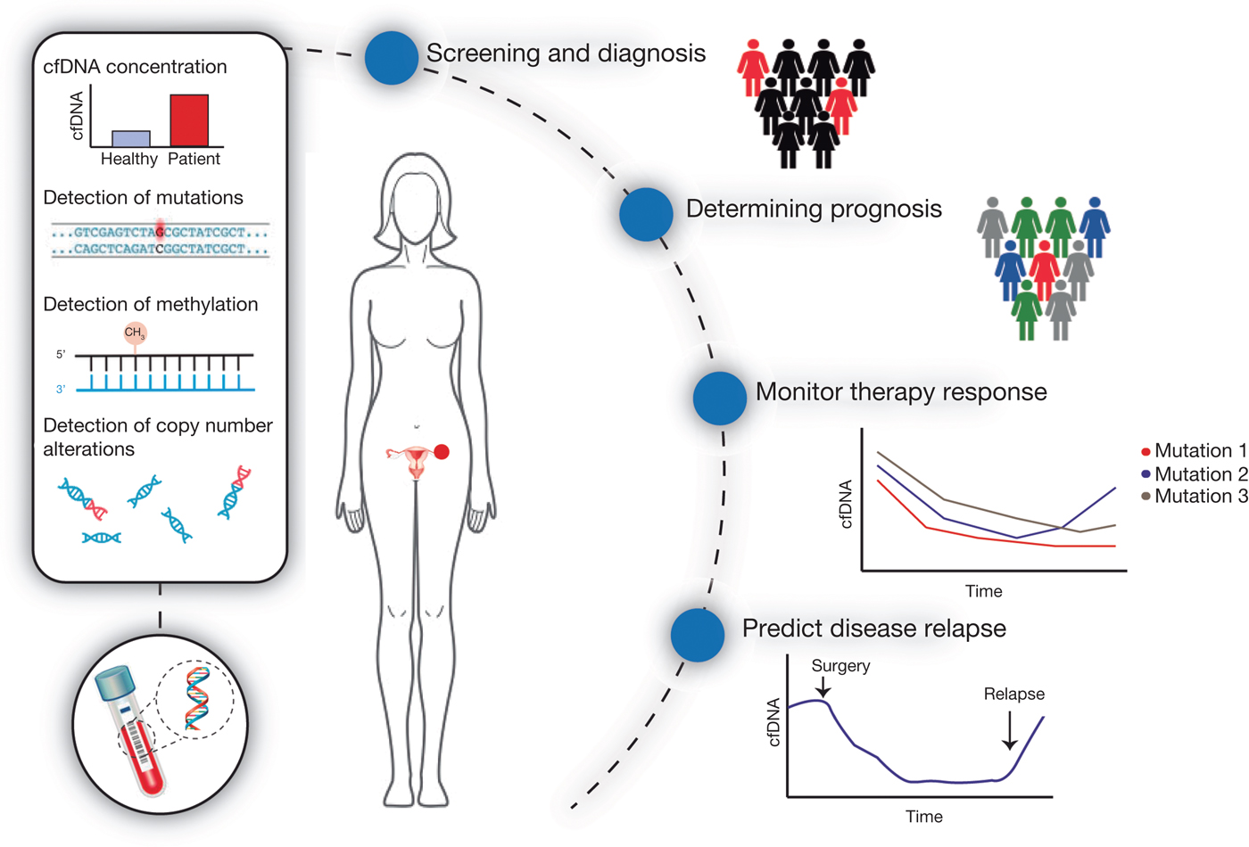
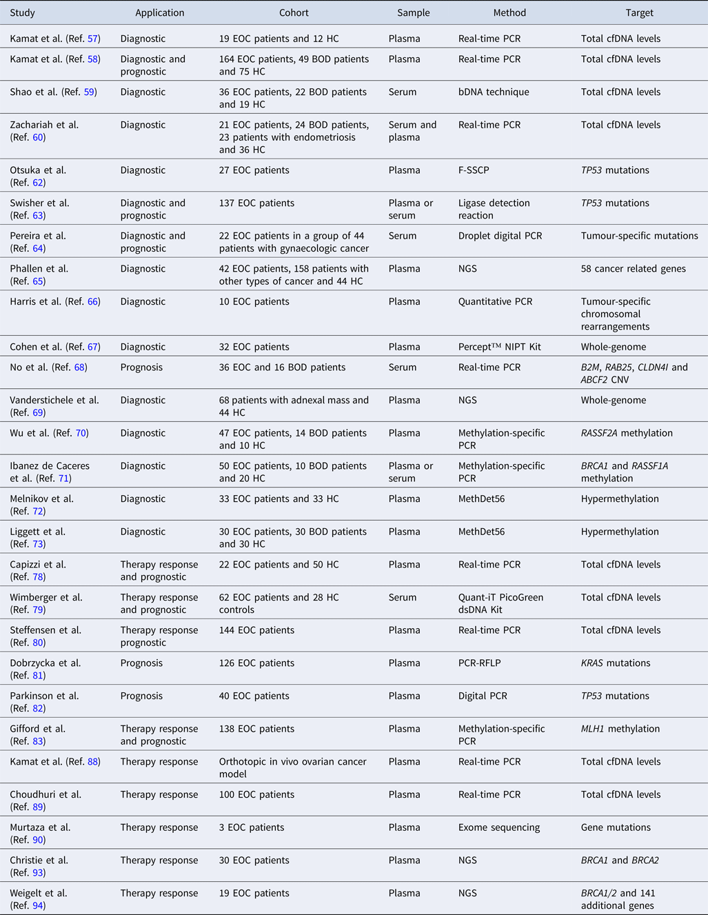
The potential of ‘liquid biopsy’ in ovarian cancer management has gained increasing attention and has several possible applications, including diagnosis, prognosis, evaluation of therapy response, monitoring the emergence of resistance during the course of treatment and disease relapse prediction (Fig. 2). Several of these applications have also been evaluated and demonstrated to be promising in other types of cancer (Refs Reference Sozzi41, Reference Mori42, Reference Gautschi43, Reference Kuang44, Reference Misale45, Reference Dawson46, Reference Hamakawa47, Reference Garcia-Murillas48, Reference Chen49, Reference Reinert50). Herein, we will present an overview of the relevant discoveries using cfDNA and its potential applications in ovarian cancer. The literature review was performed using the PubMed database and the search terms included ‘ovarian cancer’, ‘circulating cell-free DNA’, ‘circulating tumor DNA’, ‘circulating nucleic acid’, ‘plasma DNA’ and ‘liquid biopsy’. All the relevant articles published until the end of 2017 were included in the review (Table 1).
Clinical applications of liquid biopsies in ovarian cancer patients. cell-free tumour DNA (cfDNA) can be obtained from peripheral blood of ovarian cancer patients. Analyses of cfDNA can involve quantification of total cfDNA levels or detection and measurement of genetic and epigenetic alterations such as point mutations, hypermethylation and copy number alterations. This strategy may allow screening and diagnosis of disease and can be used to estimate patient prognosis. The periodic monitoring of cfDNA might give information about therapy response, allowing detection of drug resistance mutations and prediction of disease relapse.

Studies evaluating the use of cfDNA in ovarian cancer patients

BOD, benign ovarian disease; bDNA, branched DNA; CNV, copy number variation; EOC, epithelial ovarian cancer; F-SSCP, fluorescence-based single-strand conformation polymorphism; HC, healthy controls; NGS, next-generation sequencing; NIPT, non-invasive prenatal testing; PCR-RFLP, PCR-restriction fragment length polymorphism.
Screening and diagnosis of ovarian cancer
Nowadays, screening for ovarian cancer in symptomatic and high-risk women is done through ultrasound examination and measurement of the biomarker CA125 (Refs Reference Visintin51, Reference Ricci and Shih52) but none of these methods are sensitive and specific enough for this type of cancer. The use of CA125 in early detection is questionable, because increased levels are found in only 50–60% of patients with early stage ovarian cancer and can also appear in other types of cancer and benign diseases (Ref. Reference Duffy53), being therefore a non-specific marker for ovarian cancer. Like CA125, ultrasound also detects benign ovarian lesions (Ref. Reference DePriest and DeSimone54). In the presence of elevated levels of CA125 and ovarian masses detected through ultrasound examination, surgery is usually necessary for diagnosis and disease staging, as well as for the initial treatment (Ref. Reference Schwartz55). This screening strategy of ovarian cancer based on the combination of biomarker levels and imaging has therefore limited positive predictive value and leads to excessive and unnecessary surgeries (Ref. Reference Reade56). For these reasons, there is a clear need to develop new biomarkers for screening and diagnosis of ovarian cancer.
There has been an increasing interest in the use of cfDNA and several studies have evaluated this approach for disease diagnosis. Some studies focused on the quantification of total levels of cfDNA and all of them found significantly increased levels in ovarian cancer patients when compared with healthy individuals and with patients with benign ovarian diseases (Refs Reference Kamat57, Reference Kamat58, Reference Shao59, Reference Zachariah60). When comparing healthy controls with women with benign ovarian diseases, none of the studies found statistically significant differences in total cfDNA levels. The sensitivity and specificity ranges of total cfDNA levels for ovarian cancer diagnosis were 87–91.5% and 85–89.5%, respectively (Refs Reference Kamat58, Reference Shao59). Zhou et al. (Ref. Reference Zhou61) conducted a meta-analysis using data from nine studies, including 462 ovarian cancer patients and 407 controls, to evaluate the potential of cfDNA as a biomarker for the diagnosis of ovarian cancer. The polled sensitivity and specificity of cfDNA were 70 and 90%, respectively, suggesting that the diagnostic accuracy of cfDNA has unsatisfactory sensitivity but acceptable specificity. Shao et al. (Ref. Reference Shao59) reported that cfDNA presents a sensitivity and specificity of 88.9 and 89.5%, which are higher than those of other ovarian cancer biomarkers such as CA125 (75 and 52.6%) and HE-4 (80.6 and 68.4%). When they combined cfDNA levels with CA125 and HE-4, they observed an improvement of the sensitivity up to 91.67%. The study developed by Zachariah et al. (Ref. Reference Zachariah60) included an additional group of women with ovarian endometriosis and the cfDNA levels in ovarian cancer patients were not significantly increased when compared with this group. However, total cfDNA levels presented a sensitivity and specificity of 74 and 69%, respectively, to discriminate between ovarian cancer patients and healthy controls and 74 and 67%, respectively, to separate ovarian cancer patients from patients with benign ovarian diseases (Ref. Reference Zachariah60). Interestingly, Kamat et al. (Ref. Reference Kamat58) found that preoperative cfDNA levels in patients with early stage disease were significantly higher when compared with controls and patients with benign ovarian diseases. Regarding the correlation between cfDNA levels and clinicopathological characteristics, patients with stage I–II tumours presented lower cfDNA levels compared with patients with stage III–IV, indicating that cfDNA levels reflect the burden of disease (Ref. Reference Shao59).
Other studies were more specific and attempted to quantify the presence of tumour-specific genetic alterations in cfDNA to evaluate its potential for screening and diagnosis of ovarian cancer. One of these studies, developed by Otsuka et al. (Ref. Reference Otsuka62), was the first to demonstrate that tumour-specific somatic mutations can be detected in plasma DNA of ovarian cancer patients. They found that 44% of the patients involved in the study had detectable TP53 mutations in the tumour tissue and 16.7% of those had the same mutations detectable in the plasma DNA. Regarding patients with advanced disease, the detection rate of TP53 mutations was 28.6%. This study also showed that TP53 mutations were undetectable in plasma after surgery, but in one patient the TP53 mutation became again detectable 16 months after surgery and the patient died 2 months later. Regarding the detection rate, similar results were obtained in another study also based on tumour-specific TP53 mutations in cfDNA, with a concordance rate between tumour and plasma DNA of 30% (Ref. Reference Swisher63). On the other hand, Pereira et al. (Ref. Reference Pereira64) quantified tumour-specific mutations in serum of 44 patients with a range of gynaecological cancers, including 22 with ovarian cancer, to explore the use of cfDNA to predict tumour presence, detected through computed tomography (CT) imaging and findings at the time of surgery. Interestingly, they found six cases with detectable ctDNA levels and negative CT imaging in which tumours were later detected in surgery. When compared with the presence of tumour at surgery, ctDNA presented a sensitivity and specificity of 81 and 99%, respectively, to detect the presence of ovarian cancers and, on average, ctDNA predicted the disease recurrence approximately 7 months earlier than CT scans (Ref. Reference Pereira64). In a more recent study reporting the development of an ultrasensitive approach to detect alterations in frequently altered cancer genes in cfDNA, the authors analysed plasma samples from 42 ovarian cancer patients and detected alterations in 71% of them. Furthermore, 68% of these patients presented stage I–II disease suggesting the potential of cfDNA for early detection of disease (Ref. Reference Phallen65).
Chromosomal alterations might also contribute to increase sensitivity in cfDNA analysis. Aberrant chromosomal rearrangements previously identified in ovarian carcinomas were subsequently detected in pre-surgery plasma DNA samples of eight of 10 patients (Ref. Reference Harris66). Undetectable chromosomal junctions in post-surgery plasma DNA samples were consistent with the lack of detectable disease, while the continued presence of genomic rearrangements in post-surgery DNA plasma samples was consistent with the observed presence of the disease (Ref. Reference Harris66). Cohen et al. (Ref. Reference Cohen67) used a commercially non-invasive prenatal testing that identifies foetal aneuploidy through sequencing of DNA in the maternal plasma to evaluate its potential for ovarian cancer diagnosis. The algorithm chosen to identify genomic gains or losses presented a sensitivity of 40% for ovarian cancer detection, with approximately the same frequency for early and late stage disease, and a specificity of 94%. Another study was able to demonstrate a difference in copy number changes of a few genes between patients with ovarian cancer and those with benign ovarian tumours (Ref. Reference No68). Furthermore, Vanderstichele et al. (Ref. Reference Vanderstichele69) have recently reported a promising method for presurgical prediction of disease in patients with adnexal masses. This method for ovarian cancer detection is based on chromosomal instability detected by whole-genome sequencing on cfDNA obtained from plasma of women with adnexal masses. They observed that chromosomal instability was significantly higher in ovarian cancer patients compared with healthy individuals and patients with benign disease. CNAs detected in tumour biopsies were also detectable in cfDNA, with a median Spearman correlation coefficient of 0.51. This method yielded a greater sensitivity and specificity (74 and 91%) to detect malignancy when compared with CA125 (86 and 64%).
Some investigators tried to use epigenetic alterations to evaluate the use of cfDNA in screening and diagnosis of ovarian cancer (Refs Reference Wu70, Reference Ibanez de Caceres71, Reference Melnikov72, Reference Liggett73). In most studies, the samples collected from patients with benign ovarian diseases and healthy controls showed no promoter hypermethylation, suggesting tumour-specificity. When looking at methylation status of the RASSF2A gene, Wu et al. (Ref. Reference Wu70) found that the gene promoter was methylated in 51.1% of tumour samples and in 36.2% of the corresponding plasma samples. In a similar study, Ibanez de Caceres et al. (Ref. Reference Ibanez de Caceres71) tested a panel of six genes and observed a concordance rate of 82% in gene methylation pattern between tumour DNA and serum DNA, with a specificity to detect ovarian cancer of 100%. Two other studies used a panel of 56 genes and presented sensitivity and specificity ranges of 85–90% and 61–86.7%, respectively, for the detection of ovarian cancer (Refs Reference Melnikov72, Reference Liggett73). For the discrimination between malignant and benign ovarian diseases the sensitivity and the specificity were 73.3 and 80%, respectively. Furthermore, for the first time, the possibility to differentiate benign ovarian diseases from healthy controls was demonstrated, with a sensitivity of 90% and a specificity of 76.7% (Ref. Reference Liggett73).
The screening of asymptomatic individuals is the most ambitious application of liquid biopsies. Efforts are being conducted by several companies to develop a liquid biopsy test for early detection of cancer in the general population (Ref. Reference Aravanis74). In early 2018, Cohen et al. (Ref. Reference Cohen75) reported the results of a liquid biopsy test, called CancerSEEK, which can detect and localise eight types of cancer (ovarian, breast, liver, stomach, pancreatic, oesophageal, lung and colorectal) through the assessment of protein biomarkers levels and cfDNA mutations. The specificity of this test was greater than 99% and the median sensitivity among the eight types was 70%, which is low for the purpose of cancer screening. In addition, CancerSEEK was able to localise the cancer in a median of 83% of the patients. Despite the promising results, the authors recognise several limitations of the study and the need of prospective studies in a larger population.
Determining prognosis
Prognostic factors are used to estimate the likelihood of recurrence and survival. The prognostic factors established for ovarian cancer include: age of patient, tumour histology, tumour stage, tumour grade, residual disease after initial debulking surgery, volume of ascites and disease status at second-look and CA125 levels (Refs Reference Holschneider and Berek76, Reference Agarwal and Kaye77). Several studies have been conducted to evaluate the potential of cfDNA as a prognostic factor for ovarian cancer.
Based on preoperative total levels of cfDNA, ovarian cancer patients with higher levels had decreased median survival compared with patients with low preoperative cfDNA levels, presenting a 2.38-fold increased risk of death from disease (Ref. Reference Kamat58). Concerning patients submitted to treatment, higher cfDNA levels before chemotherapy were associated with high risk of disease relapse and presence of distant metastasis, whereas after chemotherapy higher levels were associated with higher risk of disease progression (Ref. Reference Capizzi78) and poor overall survival (Ref. Reference Wimberger79). In multiresistant ovarian cancer patients treated with Bevacizumab, higher total levels of cfDNA were correlated with worse progression free and overall survival (Ref. Reference Steffensen80).
Some other studies were based on the identification of genetic or epigenetic alterations in certain genes instead of relying on quantification of total cfDNA levels. No et al. (Ref. Reference No68) found that lower copy number of the RAB25 gene before surgery was associated with an increased risk of death from ovarian cancer, being suggested as an independent prognostic factor for poor disease-free and overall survival. The detection of tumour-specific TP53 or KRAS mutations in plasma from ovarian cancer patients was associated with decreased overall survival, with a mean of 21–28 months for ovarian cancer patients with detectable mutations and a mean of 54–58 months for patients with no detected mutations in plasma (Refs Reference Swisher63, Reference Dobrzycka81). Recently, Parkinson et al. reported that patients in whom a decrease of up to 60% of TP53 mutant allele frequency was observed after one cycle of chemotherapy had a significantly longer time to progression than patients whose TP53 mutant allele frequency after decreased by 60% or less (Ref. Reference Parkinson82). On the other hand, Pereira et al. (Ref. Reference Pereira64) found that undetectable levels of tumour-specific mutations in serum samples of ovarian cancer patients obtained after chemotherapy were associated with improved progression free and overall survival. Finally, the detection of acquired MLH1 promoter methylation in cfDNA of ovarian cancer patients at disease relapse after chemotherapy was found significantly associated with poor overall survival after disease progression (Ref. Reference Gifford83).
Monitoring the response to therapy
Ovarian cancer is treated with one or a combination of strategies. Surgery, apart from its role for diagnosis and disease staging, is usually the first step in the treatment of ovarian cancer and adjuvant chemotherapy is administrated after surgery to patients with a significant risk of recurrence (Ref. Reference Aletti84). Although ovarian cancer is sensitive to chemotherapeutic drugs and most patients respond to initial treatment, most patients will develop disease recurrence. Routinely, measurement of CA125 levels should be done during the course of treatment and increasing levels can be indicative of treatment failure (Ref. Reference Jelovac and Armstrong85). The main factor of disease recurrence is the emergence of drug resistance by tumour cells and several molecular mechanisms have been shown to be involved (Refs Reference Johnson86, Reference Liu87). Evaluation during the treatment course is important to anticipate the emergence of drug resistance, so that other strategies can be used promptly.
cfDNA may play a relevant role in disease monitoring and several studies have been performed for this purpose. Using an orthotopic in vivo ovarian cancer model, Kamat et al. (Ref. Reference Kamat88) evaluated the kinetics of cfDNA levels in response to therapy and observed a variation over time. More specifically, they observed an initial rise correlated with increased apoptosis rate of tumour cells (an increase of 63% at post-treatment day 1), followed by a rapid decline (20% lower at day 3 and 83% at post-treatment day 10). The key finding of this study is that cfDNA levels closely reflect tumour burden. Studies in which total levels of cfDNA were evaluated in ovarian cancer patients presented similar results, showing higher levels of cfDNA before chemotherapy and a significant decrease in total cfDNA levels after chemotherapy (Refs Reference Capizzi78, Reference Wimberger79, Reference Choudhuri89).
Murtaza et al. (Ref. Reference Murtaza90) evaluated the genomic evolution in response to chemotherapy using exome sequencing of serial cfDNA samples from advanced cancer patients, three of which were affected with ovarian cancer. In one of the ovarian cancer cases, they observed an increasing allelic frequency of a truncating mutation in the RB1 gene, previously described as being associated with therapy resistance (Ref. Reference Knudsen and Knudsen91), thereby showing that exome sequencing analysis of cfDNA represents a novel non-invasive strategy to evaluate the evolution of therapy response and to identify the emergence of drug resistance.
On the other hand, Martignetti et al. (Ref. Reference Martignetti92) reported a case of a patient with ovarian cancer treated with surgery and chemotherapy who experienced multiple tumour recurrences for a period of four years. At the time of diagnosis, the patient presented a CA125 level of 40 U/ml and during the subsequent four years the CA125 levels were above the normal limit only three times, whereas a series of biopsies proved the presence of tumour. When looking for genomics-based biomarkers, they found a fibroblast growth factor receptor 2 fusion transcript in the tumour tissue that was also detectable in plasma or serum DNA from all 28 blood samples collected since the initial surgery, indicating the continued presence of tumour cells and explaining the multiple recurrences of the disease.
Finally, there are several studies regarding the detection of reversion germline mutations of BRCA1/2, which are associated with acquired resistance to chemotherapy. Christie et al. (Ref. Reference Christie93) evaluated the possibility to detect reversion germline mutations of BRCA1/2 in cfDNA from patients with HGSOC. Reversion mutations were only detected in tumours from patients with recurrence disease and in 60% of these patients the reversion mutations were detected in cfDNA. In another study conducted by Weigelt et al. (Ref. Reference Weigelt94), BRCA1 or BRCA2 reversion mutations were identified in cfDNA from 21% of platinum resistant ovarian cancer patients, indicating that cfDNA sequencing may aid the selection of patients for PARP inhibition therapy (Refs Reference Christie93, Reference Weigelt94). In fact, with the emergence of targeted therapies in oncology, one of the main applications of liquid biopsies is the detection of gene mutations to steer therapy decisions. Furthermore, this approach can help to detect mutations which are known to be associated with acquired resistance. For example, nearly all EGFR mutated non-small cell lung cancer patients develop resistance under tyrosine kinase inhibitors (TKIs) therapy, being the T790M mutation the most common mechanism (Ref. Reference Kuang44). According to ESMO and NCCN guidelines, liquid biopsy is a valid alternative to tissue re-biopsy in the identification of resistance mechanism to EGFR TKI therapy (Ref. Reference Planchard95). Furthermore, according to Committee for Medicinal Products for Human Use (CHMP) of the European Medicines Agency, liquid biopsy can be used to predict response to EGFR TKI therapy when a tumour sample is not available (Ref. 96). It is well possible that similar strategies to detect and monitor the molecular mechanisms of targeted therapy may enter clinical practice for other cancer types, namely in serous ovarian cancer in which treatment with PARP inhibitors may be used in BRCA1/BRCA2 mutation carriers (Ref. Reference Ledermann97).
Conclusions
The lack of an effective screening method for early detection, combined with the acquired resistance to commonly used chemotherapeutics, makes this gynaecological malignancy a worldwide health burden. Thus, there is a growing need for better tools to improve the poor outcome underlying ovarian cancer. The findings summarised here emphasise the potential use of cfDNA as a liquid biopsy for early diagnosis and molecular characterisation of ovarian cancer, as well as for monitoring response to therapy and identifying the emergence of drug resistance mechanisms.
Among the studies reviewed, better results were obtained in those using genetic or epigenetic alterations in ctDNA, which present both higher sensitivity and specificity when compared with those which used total cfDNA levels. Genetic and epigenetic alterations found in ctDNA are representative of the tumour and are absent in normal cfDNA, whereas the total levels of cfDNA can be increased as a result of benign conditions or other malignancies. Furthermore, regarding the correlation between findings in tumour and plasma, better results were observed when more sensitive techniques were used, such as next-generation sequencing (NGS) and droplet digital polymerase chain reaction (ddPCR). However, patient's recruitment was enriched for individuals diagnosed with ovarian cancer in the majority of the studies, so the reported sensitivities and specificities of cfDNA for ovarian cancer detection do not necessarily apply to the general population.
The molecular characterisation of tumours through cfDNA is particularly interesting with the emergence of targeted therapies for ovarian cancer, mainly in situations in which a tumour sample is not available and biopsy is not viable. Due to the distinct patterns of genetic alterations of each histological subtype, as well as the molecular heterogeneity in the pathogenesis and therapy resistance mechanisms in ovarian cancer, NGS of cfDNA with a customised gene panel, may be a good strategy to improve the diagnosis and clinical management of this disease. Using bioinformatic tools for the detection of CNVs using NGS data may improve the accuracy of cfDNA to detect the different histologic subtypes of ovarian carcinomas.
Although there are several advantages in the use of ctDNA as a screening tool, some limitations also exist. Major limitations are the low concentrations of ctDNA in plasma and the need of high sensitive techniques such as digital PCR (dPCR) and NGS for the detection of tumour specific alterations. NGS has the advantage of simultaneously analyse alterations in several genes and recent advances, such as the introduction of molecular barcodes, will enable the detection of low frequency variants.
Although the report of the detection of ctDNA in cancer patients preceded the detection of foetal DNA in blood of pregnant women, a reliable diagnostic test based on cfDNA was first established as a non-invasive pre-natal test (NIPT). NIPT rapidly evolved to a frequent test used for screening Down syndrome and other aneuploidies and is already implemented in public prenatal care in several countries worldwide (Ref. Reference van Schendel98). In comparison, the development of a liquid biopsy approach to be applied in oncology has been more difficult and is taking longer due to several factors. Cancer is a pathologic and complex process, a dynamic and heterogeneous disease with multiple variables, while pregnancy is a physiological process with minimal variables. Furthermore, the percentage of foetal cfDNA in the mother's circulation is relatively high from 9 weeks of pregnancy, while the fraction of ctDNA in cancer patients is frequently low and variable even among patients with the same disease stage. Consequently, it is more difficult to standardise liquid biopsy assays based on ctDNA analysis for the management of cancer patients (Ref. Reference Heitzer99). Liquid biopsies are being evaluated in large studies in order to validate their use in the management of cancer patients, with 322 clinical trials registered on the National Institute of Health database on early October 2018. Standardisation of the methodology from blood collection to cfDNA analysis is essential and there is an ongoing clinical trial for the development and comparison of different cfDNA detection techniques (NTC01350980). Furthermore, there are two ongoing clinical trials for the development of a blood test for ovarian cancer diagnosis based on cfDNA (NCT02441140 and NCT03485651). In addition, the International Ovarian Tumor Analysis Group (IOTA) is exploring the potential of cfDNA to be included in their disease prediction models (NCT01698632). It is expected that results from large-scale and multicentre clinical trials will support the use of liquid biopsies in clinical practice also for ovarian cancer in a not too distant future.
Author ORCIDs
Manuel R. Teixeira 0000-0002-4896-5982.
Financial support
Ana Barbosa is a research fellow supported by the project NORTE-01-0145-FEDER-000027, supported by Norte Portugal Regional Operational Programme (NORTE 2020), under the PORTUGAL 2020 Partnership Agreement, through the European Regional Development Fund (ERDF). Pedro Pinto and Manuela Pinheiro are research fellows of the Núcleo Regional do Norte da Liga Portuguesa Contra o Cancro and FCT (SFRH/BPD/113014/2015), respectively.
Conflict of interest
None.
Ethical standards
This review work did not involve human and/or animal experimentation.