Introduction
External auditory exostosis (EAE), known as surfer’s ear, is a condition of progressive benign, hyperostotic overgrowths of the external auditory canal (EAC). They typically occur in sportspeople with repetitive cold-water exposure, such as swimmers and surfers, with a prevalence of up to 73 per cent.Reference Attlmayr and Smith1, Reference Wong, Cervantes, Doyle, Karamzadeh, Boys and Brauel2 It is hypothesized that vasodilation occurs secondary to cold exposure, causing increased vascular tension and subsequent osteoblastic activity with lamellar bony growth and ossification, possibly as an evolutionary mechanism to protect the vulnerable tympanic membrane (TM) from irritation.Reference Harrison3
EAE is often asymptomatic and does not require treatment. Once symptoms present, these range from water or cerumen entrapment to recurrent otitis externa and conductive hearing loss and typically correlate with the degree of stenosis. Canaloplasty remains the only surgical intervention for EAE following failure of conservative management, with consensus amongst most otolaryngologists to intervene in symptomatic stenoses of greater than 80 per cent.Reference Vallée4
Several techniques have been described for the removal of EAE, typically via osteotome or microdrill. Approaches include trans-canal or post-aural and may be microscopic or endoscopic. Main complications associated with each technique include sensorineural hearing loss, post-operative stenosis and TM perforation, with tinnitus, infection and temporomandibular joint (TMJ) dislocation also reported.Reference Shohet, Justeson and Wilkins5
We describe the Swansea technique; an alternative method for canaloplasty in EAE; endoscopic underwater excision of exostoses using a piezoelectric saw and continuous irrigation. The piezoelectric saw is widely used in oral and maxillofacial surgery, as it preferentially induces cavitation and fracture of mineralised tissue, facilitating reduced soft tissue damage.Reference Lyutenski, James and Bloching6 Whilst not yet widely used in otology, the precise angled tip and reduction in acoustic trauma are postulated to reduce the risk of sensorineural hearing loss.Reference Marchioni, Ronzani, Gazzini, Gianfranco and Rubini7
The SUPER reporting guideline has been utilised to describe this approach.Reference Zhang, Ma, Wu, Shi, Barchi and Scarci8
Method
Pre-operative preparation
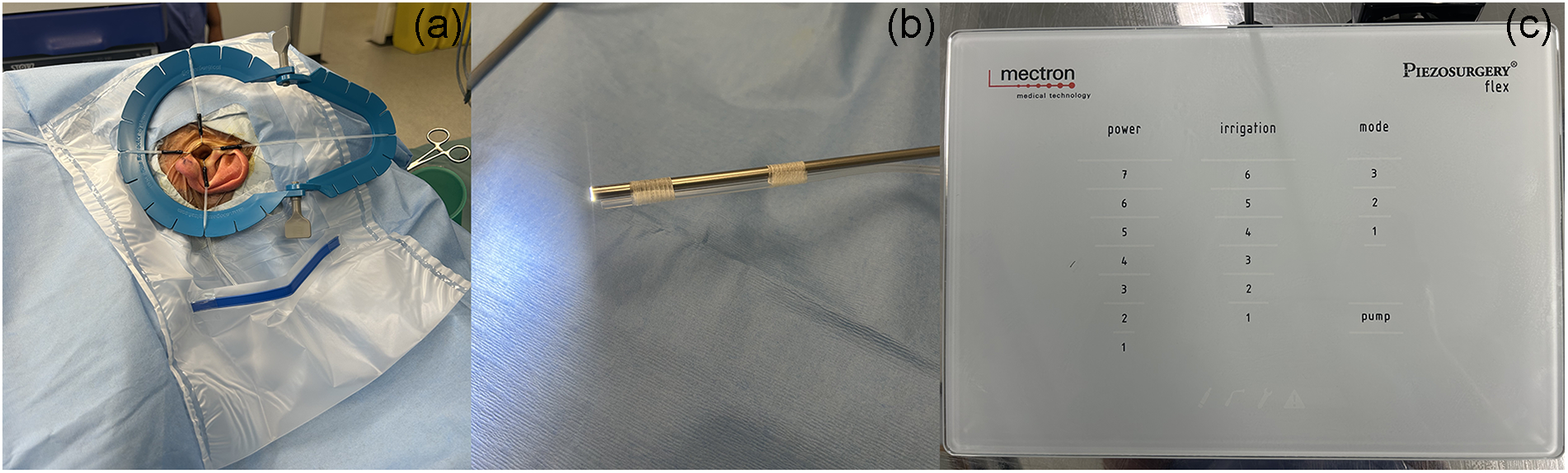
Following general anaesthesia and facial nerve monitoring, the patient is positioned in the reverse Trendelenburg position, with the head tilted away from the operated ear. The EAC and pinna are prepped with aqueous iodinated povidone and draped to ensure sterility. EAC access is optimised by placement of a Lone Star Retractor System and four 5-mm blunt hook elastic stays, in 12, 3, 6 and 9 o’clock positions. A plastic drape is placed over the pinna to collect irrigation fluid (Figure 1A).
Pre-operative preparation. (a) Lonestar retractor system with plastic drape for irrigation. (b) Irrigation tubing connected to zero-degree rigid endoscope using steristrips. (c) Control panel settings for Piezoelectric saw.

The irrigation tubing is removed from the piezoelectric saw (Piezosurgery flex, Mectron) and attached to a 4-mm 0-degree Karl Storz rigid endoscope using steristrips wrapped around the endoscope and tubing at two points (Figure 1B). The tubing end is secured immediately behind the tip of the endoscope to prevent obstruction of the surgical field. The irrigation system is connected to a 1000-ml bag of 0.9 per cent NaCl infiltrated with 1ml 1:1000 adrenaline. The angled 45-degree MP3a30 attachment is connected to the Piezoelectric handpiece with the following settings: power 6, irrigation 6, mode 2 (Figure 1C) and the system primed. This operative system is designed for a single surgeon; the modified endoscope is held in the left hand, with the piezoelectric saw in the right in the right-handed surgeon.
Intra-operative technique
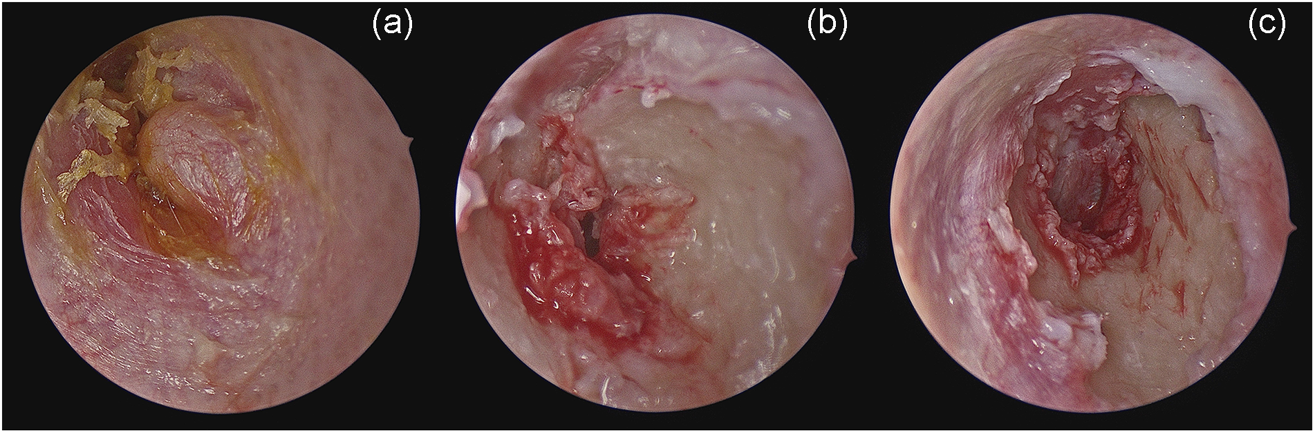
The 2-ml of 2 per cent lidocaine with 1:80,000 adrenaline is injected immediately lateral to the centre of each exostosis (Figure 2A). A circumferential meatal flap is raised with a Rosen elevator over the exostoses in a bloodless field to expose underlying bone (Figure 2B).
Intra-operative endoscopic appearances of Swansea technique. (a) Exostoses prior to intervention. (b) Elevation of meatal flap to expose underlying bone. (c) Exostoses lowered to level of anatomical canal wall, leaving a smooth, uniform tubular canal.

The piezoelectric saw is activated outside the EAC, with the endoscope in situ, to flood the surgical field. The saw is then introduced, with ongoing continuous irrigation throughout the procedure, and placed between the meatal flap and exostoses, with bone excised from medial to lateral. The side of the rounded tip is used for most effective dissection. Once exostoses have been reduced to the level of the anatomical EAC wall, bone is levelled to a smooth, tubular canal (Figure 2C). Excess irrigation is suctioned, and the meatal flap replaced. The EAC is then packed with Bismuth Iodoform Paraffin paste coated ribbon gauze (BIPP) or Gelfoam with Otocomb Otic ointment. Average operative time is 90 minutes.
Several key points should be noted. Irrigation is continuous to prevent formation of air bubbles and expel excised bony fragments, preventing obstruction of the surgical field. This also allows cooling of the tip of the piezoelectric saw, preventing thermal damage to surrounding structures. The surgeon must always keep anatomical reference points in view, to minimise the risk of damage to structures such as the facial nerve and TMJ. We recommend achieving a view of the TM as early as possible, to delineate salient anatomical landmarks.
Post-operative care
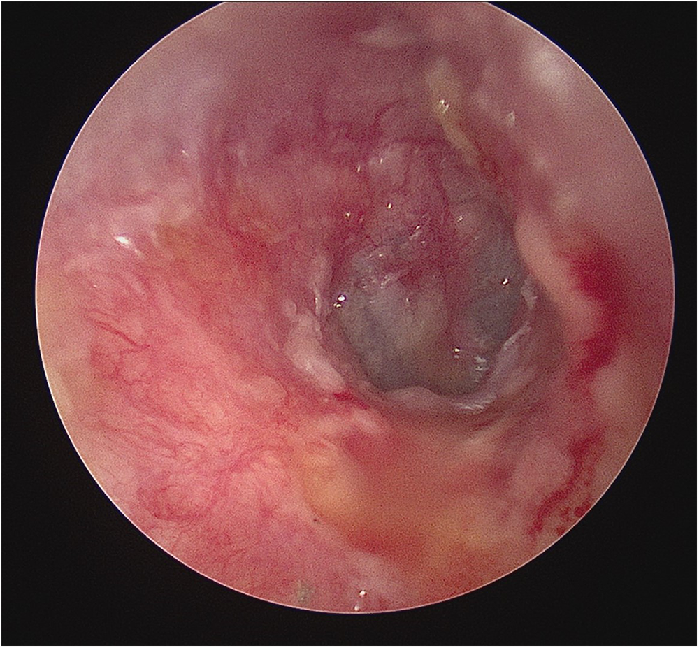
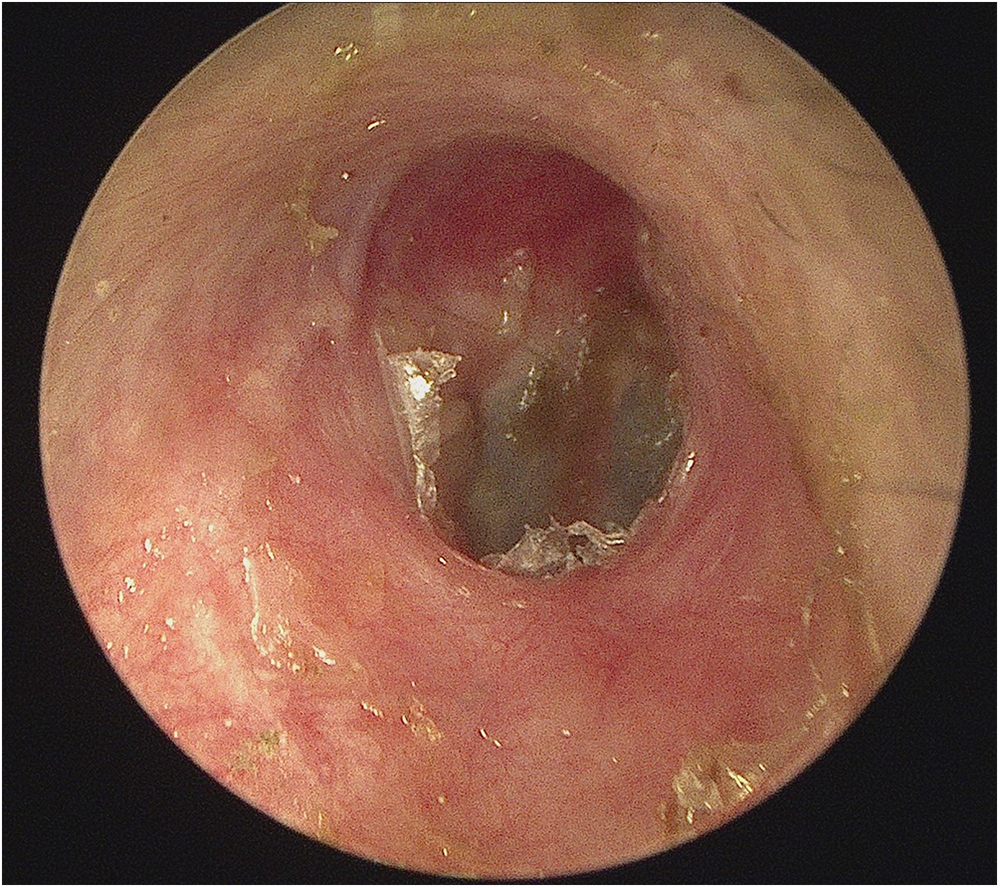
Post-operatively, the patients are monitored for 4 hours before same-day discharge. They are instructed to keep the operated ear dry until follow-up at three weeks, when the ear is examined microscopically, packing removed and Otocomb Otic ointment applied (Figure 3). A second follow-up occurs four weeks later with pure tone audiogram, following which (in the absence of complications) the patient is discharged (Figure 4).
Post-operative appearance at 3 weeks.

Post-operative appearance at 7 weeks.

Results
To date, a total of 34 procedures have been performed on 32 patients in this single surgeon series. All patients were male, with an average age of 54 years (range, 37–69 years). Mean estimated degree of exostosis was 90 per cent (range, 60–100 per cent). Two patients (8.3 per cent) had pinhole pars tensa perforations at the time of surgery (healed completely by initial follow up). There was no reduction in hearing thresholds with pure tone audiogram post-operatively; two patients achieved improved post-operative hearing thresholds; one patient had significant pre-operative conductive hearing loss secondary to exostoses, and one patient had concurrent myringoplasty for pre-existing perforation. There were no post-operative complaints of new onset tinnitus or TMJ pain or dysfunction.
Discussion
Our innovative use of continuous irrigation with adrenaline and detailed description of the use of the piezoelectric saw in the management of EAE are unique.
To fully appreciate the benefits of the piezoelectric saw, one must understand the inverse Piezoelectric effect that powers it. Electric current directed from the generator over the piezo-cermic rings in the handpiece causes their deformation, with subsequent ultrasonic linear vibration of the tool tip at frequencies of 24-30kHz. Bone excision thus is not just due to the mechanical function of the instrument, but also due to cavitation; microbubbles form within the saline solution used, which implode on contact with and lead to fracture of mineralised tissue only (as opposed to soft tissue).Reference Lyutenski, James and Bloching6
This reduces the likelihood of injury to the meatal flap and, therefore, the risk of poor wound healing and stenosis. Additionally, it lowers risk of sensorineural hearing loss; there is less acoustic trauma (compared to use of the drill and/or regular suctioning). At sound levels above 115dB, sensorineural hearing loss can occur after 15 minutes, with correlation between threshold shift and duration of exposure.Reference Parkin, Wood, Wood and McCandless9 It is pertinent therefore to note that the average operating time for the microdrill is 104 minutes.
A risk of sensorineural hearing loss secondary to instrument contact with the lateral process of the malleus still exists. However, as the tip is flat and angled, it enables excision of bone from medial to lateral, allowing more efficient and safe excision of exostoses once the level of the TM has been established. The ability to perform precise cuts also lowers the risk of TMJ damage, seen more commonly with the osteotome.
Continuous irrigation allows consistent clearance of excised bony fragments from the EAC, providing an unobstructed view throughout the procedure. The addition of adrenaline to the irrigation fluid (1:1,000,000 dilution) assists haemostasis.
Limitations of this technique are those inherent to all endoscopic ear surgery; the use of a single hand for dissection and the requirement for proficiency in use of the endoscope as not all surgeons have access to or are trained in this approach.
Comparing our early outcomes to a recent meta-analysis of microdrill and osteotome EAE excision, our patient population is comparable; predominantly male, although slightly older at time of surgery (54 vs. 46 years).Reference Swisher, Singh, Debbaneh and Rivero10 We demonstrated superiority in the rate of sensorineural hearing loss (0.0 per cent vs. 0.69 per cent [osteotome] and 4.3 per cent [drill]) and post-operative stenosis (0.0 per cent vs. 1.1 per cent [osteotome] and 4.1 per cent [drill]). Data describing post-operative tinnitus and TMJ prolapse were less well defined, with a range of 0.0–8.8 per cent and 0.0–25.9 per cent, respectively (combined osteotome and drill), compared to our rates of 0.0 per cent for both. We did however have a slightly higher rate of TM perforation (8.3 TM vs. 5.3 TM [osteotome] and 3.8 TM [drill]), although both were pinhole and could be considered inevitable, as these exostoses were in direct contact with the TM. One must note however that there is significant heterogeneity amongst studies reporting complication rates from the use of the osteotome or otologic drill.
From a financial perspective, the piezoelectric saw is comparable to the otologic drill, with Piezoelectric insert costing £172.70 + VAT (May 2025) and drill burrs (based on an average of 3 sizes) costing £280.
• External auditory exostosis (surfer’s ear) is a progressive condition of benign hyperostotic overgrowths of the external auditory canal, secondary to repetitive cold-water exposure, in which symptoms correlate to degree of exostosis.
• Surgical intervention is the only available treatment, most commonly using the osteotome or microdrill, but these carry a risk of sensorineural hearing loss, post-operative stenosis, temporomandibular dysfunction and tympanic membrane perforation.
• We describe the Swansea technique, an innovative method of canaloplasty with endoscopic underwater excision of exostoses using a piezoelectric saw and continuous irrigation with 1:1,000,000 adrenaline.
• The inherent properties of the piezoelectric saw reduce the overall risk of complications; bone excision occurs secondary to ultrasonic linear vibration at frequencies of 24-30kHz, resulting in less acoustic trauma and a lower risk of sensorineural hearing loss.
• Additionally, the inverse piezoelectric effect causes fracture of mineralised tissue only, leading to reduced injury to the soft tissue meatal flap.
• This novel technique is cost-equivocal and easily implemented in departments with endoscopic capabilities.
Conclusion
We describe a novel use of the piezoelectric saw with continuous underwater irrigation with 1:1,000,000 adrenaline for the removal of EAE. Whilst this technique is in its infancy, early results are promising, with complication rates comparable or superior to established techniques and equal financial outlay.