In the mammary gland, invaded pathogens induce an immune response by stimulating the innate immune system. Along with this process considerable changes in milk composition occur due to changes in secretion but also through changes of the permeability of the blood-milk barrier leading to an increase of blood components in milk (Burton & Erskine, Reference Burton and Erskine2003; Schmitz et al. Reference Schmitz, Pfaffl, Meyer and Bruckmaier2004; Wellnitz & Bruckmaier, Reference Wellnitz and Bruckmaier2012). These changes influence the nutritional value and taste, and the further processing of the milk. In addition, the change of milk composition plays an important role in the detection of mastitis through measurement of changes in concentration of mastitis indicators in milk. For this purpose a detailed knowledge of chronological changes and the mechanisms of the transfer of various factors into milk during early stages of mastitis is essential.
Changes of specific components in milk that have immune function can influence the mammary immune defence. The infiltration of polymorphonuclear neutrophils (PMN), the main effectors of the mammary immune defence, into the mammary gland during mastitis is measured as an increase of the milk somatic cell count (SCC) (Burton & Erskine, Reference Burton and Erskine2003; Wellnitz & Bruckmaier, Reference Wellnitz and Bruckmaier2012). A low SCC is, therefore, used as a marker of good hygienic milk quality (Harmon, Reference Harmon1994).
Lipopolysaccharide (LPS) is an endotoxin that is released from gram-negative bacteria. Intramammary administration of LPS from Escherichia coli, a common mastitis pathogen, is a well-established method to experimentally induce mastitis under defined conditions for studying the immune response of the mammary gland (Bruckmaier et al. Reference Bruckmaier, Schallibaum and Blum1993; Schmitz et al. Reference Schmitz, Pfaffl, Meyer and Bruckmaier2004; Baumert et al. Reference Baumert, Bruckmaier and Wellnitz2009a; Wellnitz et al. Reference Wellnitz, Arnold and Bruckmaier2011).
Serum albumin is a blood protein whose appearance in milk can be used as an indicator of blood-milk barrier permeability (Stelwagen et al. Reference Stelwagen, Politis, White, Zavizion, Prosser, Davis and Farr1994) and as an indicator of mastitis (Verhoeff & Smith, Reference Verhoeff and Smith1981). Immunoglobulin (Ig) G is the major immunoglobulin in milk of ruminants (Butler, Reference Butler1983). The predominant antibody type in milk from healthy udders, although at a very low concentration, is IgG1 due to its active and selective transport across the blood-milk barrier (Baker et al. Reference Baker, Quiao, Kuo, Kobayashi, Yoshida, Lencer and Blumberg2009). During mastitis IgG1 and IgG2 increase in milk and IgG2 is found in higher concentrations than IgG1 in mastitis milk (Lehmann et al. Reference Lehmann, Wellnitz and Bruckmaier2013). For the mammary immune defence IgG2 seems to play a more important role than IgG1 because bovine neutrophils upregulate Fc receptors for IgG2 when they migrate into infected tissue but not for IgG1 or IgA (Paape et al. Reference Paape, Shafer-Weaver, Capuco, Van Oostveldt and Burvenich2000; Burton & Erskine, Reference Burton and Erskine2003). In addition, IgM is not a major antibody subclass in normal or mastitic milk (Rainard & Caffin, Reference Rainard and Caffin1983).
Milk L-lactate and lactate dehydrogenase (LDH) increase in milk during intramammary infection and they have been considered as indicators of mastitis (Davis et al. Reference Davis, Farr, Prosser, Nicholas, Turner, Lee and Hart2004; Chagunda et al. Reference Chagunda, Larsen, Bjerring and Ingvartsen2006). However, there is no obvious contribution of these factors to the immune defence.
For all these components a correlation to SCC increase is accepted (Harmon, Reference Harmon1994; Davis et al. Reference Davis, Farr, Prosser, Nicholas, Turner, Lee and Hart2004; Chagunda et al. Reference Chagunda, Larsen, Bjerring and Ingvartsen2006). However, a detailed chronological pattern of increase has, to our knowledge, not yet been described in the first 5 h of mastitis. To test the hypothesis that there are different component-specific patterns for the transfer of blood components into milk, this study aimed to investigate the sequence and the pattern of increase of SCC, the blood components IgG1, IgG2, serum albumin, L-lactate, and lactate dehydrogenase in milk. To record also a potential transfer in the opposite direction, i.e. from milk into blood, the milk protein α-lactalbumin was measured in blood during the first hours of an endotoxin- induced mastitis.
Material and methods
The experimental procedures followed the Swiss Federal Law on Animal Protection and were approved by the Committee of Animal Experiments of the Canton Fribourg, Switzerland.
Animals and management
Holstein (n = 6) and Swiss Fleckvieh (n = 2) cows in their 1st to 5th lactation (2·5 ± 0·6) and in month two to nine (5·5 ± 0·8) of lactation were used for this experiment. Cows were housed in tie stall barns during experiments and had constant low SCC below 130 000 cells/ml for at least 5 d before the experiment in all four quarters (morning foremilk samples). None of the udders showed clinical signs of mastitis. An intravenous catheter was inserted into one jugular vein on the day before the experiment to take blood samples.
Experimental design
After morning milking one quarter of each cow was challenged with 200 µg Escherichia coli LPS (serotype O26:B6; Sigma-Aldrich, Buchs, Switzerland), diluted in 10 ml of saline solution after a careful disinfection of teat openings with 70% ethanol. Another quarter within cow was used as control and treated with 10 ml of saline solution. The treated quarters were randomly selected (front or rear, left or right) and equally distributed. Foremilk samples (~10 ml) were taken hourly by hand from LPS challenged and control quarter at 0 h (prior to injection) until 5 h after challenge. In addition, between 2 and 5 h after challenge, samples were taken every 15 min. Both milk samples were taken within 50 s before milk ejection started. Immediately after collection SCC was measured with a DeLaval cell counter (DCC; DeLaval, Tumba, Sweden) and then samples were frozen at −20 °C for later analyses.
Blood samples were taken coinciding with milk sampling through the intravenous catheter. Blood was collected into tubes containing EDTA as anticoagulant. Blood samples were stored briefly on ice and then centrifuged for 20 min at 3000 g. Plasma was collected and frozen at −20 °C until further analyses.
Concentrations of serum albumin in milk and plasma were measured using a commercial ELISA kit (Bethyl Laboratories, Montgomery, TX, USA) according to the manufacturer's instructions, except for using a higher dilution for milk samples (1 : 12 000) than described in the manual (1 : 5000). Because of high divergent values in treated and control samples, in plate dilutions were performed using a multichannel pipette (one 1 : 2 dilution), to meet the standard curve of the assay.
The immunoglobulin G1 and G2 concentrations in whole milk and plasma were measured using a bovine IgG1 and IgG2 ELISA quantitation kit (Bethyl Laboratories Inc., Montgomery, TX). The procedure was performed as described in the manufacturer's protocol, except for slight modifications: After coating the plates with anti IgG1/IgG2 antibody (50 µl of capture antibody per 10 ml Coating buffer), the plates were washed with an automatic washing system (microplate washer Columbus Pro, Tecan Group Ltd., Männedorf, Switzerland). To reduce unspecific binding to anti IgG Antibodies, the blocking reagent was replaced with 150 µl/well of 5% fish skin gelatin Solution [5% of cold water fish skin gelatin (G7765; Sigma Aldrich, Steinheim, Germany] in distilled water). After washing, 100 µl of milk diluted in washing buffer (1 : 200) were added to each well. Because of the high sensitivity of the assay, in plate dilutions were performed (one serial dilution (1 : 2) using a multichannel pipette) to hit the standard range. Concentrations of IgG1/IgG2 were calculated by extrapolating from a standard curve generated by a microplate reader (Thermomax; Molecular Devices, Sunnyvale, CA).
Concentrations of α-lactalbumin in plasma were measured using a bovine α-lactalbumin ELISA quantitation set (Bethyl Laboratories Inc., Montgomery, TX) according to the manufacturer's instructions. Because of the high sensitivity of the assay, plasma samples had to be diluted 1 : 20. In addition, the samples were serial-diluted once by 1 : 3 on the plate using a multichannel pipette to meet within standard curve of the assay.
For all ELISAs one control sample was added to each plate and was used for the calculation of coefficients of variation. For IgG ELISAs an intra assay variation of 10% and an inter assay variation of 20% was reached, respectively. For all other ELISAs and intra assay variation of 8% and an inter assay variation of 12% was reached, respectively.
Lactate dehydrogenase activity and L-lactate concentrations in milk serum and blood plasma were measured by using the test kits LDH IFCC (Axon Lab AG, Baden, Switzerland) and Lactate PAP (bioMérieux, Marcy l`Etoile, France), respectively, with an automated analyser (COBAS MIRA, Roche Diagnostics, Switzerland) according to the manufacturer's instructions.
Statistical analysis
Data are presented as means ± sem. Data of SCC are presented and statistically evaluated at a logarithmic scale (log10) to achieve normal distribution. Differences between LPS and control within each time point and differences between time points within one group were tested for significance (P < 0·05) by ANOVA using PROC MIXED of SAS (SAS Institute Inc., Cary, NC; 2002–2008, Release 9.2). The model included time, treatment (LPS or control) and their interaction as fixed effects and the quarter as repeated subject.
Results
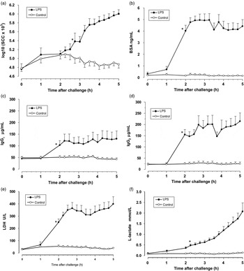
Concentrations of somatic cells, serum albumin, IgG1, IgG2, and L-lactate and LDH activity in milk before and until 5 h after LPS challenge in challenged and control quarters are shown in Fig. 1. The SCC in milk was increased in LPS challenged quarters after 2·75 h compared to the initial value before LPS challenge (P < 0·05) and increased further until the end of the experiment. No significant changes of SCC were detected in control quarters. Serum albumin, IgG1, IgG2, and LDH was low in milk of all quarters at the beginning of the experiment and increased within 2 h in LPS challenged quarters (P < 0·05). Concentrations then remained elevated until the end of experiment. In control quarters the concentrations of these factors did not change.
Somatic cell count (a), serum albumin (b), IgG1 (c), IgG2 (d), LDH (e), and L-lactate (f) in milk within 5 h after LPS challenge in LPS treated and control quarters. Data are presented as means ± sem. *: time point of first significant elevation compared to time point 0 until the end of experiment.

L-lactate was increased (P < 0·05) in milk of LPS challenged quarters at 2·25 h and increased further thereafter until the end of experiment. In control quarters L-lactate concentrations did not significantly change within the 5 h of experiment.
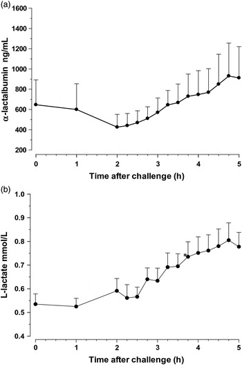
Except for L-lactate, no change of any parameter in blood was detectable in the 5 h of experiment. Blood concentration of serum albumin was 4·11 ± 0·11 ng/ml throughout the experiment. Concentrations of IgG1 and IgG2 in blood were 5·3 ± 0·2 and 19·7 ± 0·6 mg/l, respectively. The activity of LDH in blood was 923 ± 15 U/l throughout the entire experiment (mean of all values). Concentrations of α-lactalbumin in blood did not significantly change during the whole experiment, whereas between 2 h after the experiment and the end of the experiment an increase (P = 0·013) was detected (Fig. 2). The concentration of L-lactate in blood was increased at 3·75 h after challenge (P < 0·05) and further increased until the end of experiment (Fig. 2).
Alpha-lactalbumin (a) and L-lactate (b) in blood plasma within 5 h after LPS challenge. Data are presented as means ± sem. *: time point of first significant elevation compared to time point 0 until the end of experiment.

Discussion
The increase of somatic cell count and changes of milk composition during mastitis in dairy cows have been investigated over many decades. However, to our knowledge, the detailed description of the chronological patterns of the changes of blood components in milk within the first hours of mastitis has not been shown before. The hypothesis that different blood components are transferred into milk in different patterns was clearly confirmed by our study. Several blood components follow similar patterns of transfer into milk. This is of high importance for understanding the mechanisms of transfer of these components from blood into milk during mastitis. Furthermore the knowledge of the chronological appearance during the development of mastitis is important if factors are used for the early detection of mastitis and to figure out which is the most suitable parameter.
In LPS challenged quarters the increase of SCC within some hours was expected (Schmitz et al. Reference Schmitz, Pfaffl, Meyer and Bruckmaier2004; Lehmann et al. Reference Lehmann, Wellnitz and Bruckmaier2013). This increase is known to be depending on the dosage of LPS (Baumert et al. Reference Baumert, Bruckmaier and Wellnitz2009a) and the SCC before challenge (Baumert et al. Reference Baumert, Bruckmaier and Wellnitz2009b). In this study the SCC increase was significant within 2·75 h.
During a mammary immune response the permeability of tight junctions increases and, therefore, a breakdown of the milk-blood barrier occurs (Kehrli & Shuster, Reference Kehrli and Shuster1994; Burton & Erskine, Reference Burton and Erskine2003). This leads to a facilitated passage of several blood components into the milk. The increased appearance of serum albumin in milk is accepted as an indicator for leakage of blood constituents into the milk through the blood-milk barrier (Poutrel et al. Reference Poutrel, Caffin and Rainard1983; Shuster et al. Reference Shuster, Kehrli and Stevens1993; Stelwagen et al. Reference Stelwagen, Politis, White, Zavizion, Prosser, Davis and Farr1994) although there is conclusive evidence that the mammary gland also produces albumin and the expression is increased during mastitis (Shamay et al. Reference Shamay, Homans, Fuerman, Levin, Barash, Silanikove and Mabjeesh2005). The results of the present study agree with results from Shuster et al. (Reference Shuster, Kehrli and Stevens1993) who showed an increase of serum albumin in whey within the first 2 h after LPS infusion before SCC increased. It is possible that there is a retarded influx of cells due to slow diapedesis because the size of the cells necessitates a change in shape to fit through the gaps between endothelial and epithelial cells (Burton & Erskine, Reference Burton and Erskine2003). Chemoattractants have to be present and a number of events have to be initiated for an efficient influx of neutrophils into the mammary gland (Burton & Erskine, Reference Burton and Erskine2003). For other molecules like IgG, albumin, or sucrose a free transfer through openings within the cell layers is assumed (Nguyen & Neville, Reference Nguyen and Neville1998).
Interestingly the albumin concentrations in milk did not further increase after 3 h after challenge in the present study. Obviously, a maximal transfer from blood into milk was already attained at that time and shows a parallel transfer to osmotic regulated influx of water for milk production.
Immunoglobulin G1 and G2 concentrations increased in milk similar to albumin concentrations before a significant increase of SCC was detected. This phenomenon was described before (Guidry et al. Reference Guidry, Ost, Mather, Shainline and Weinland1983). The majority of IgG2 antibodies in milk are transferred from the blood (Leitner et al. Reference Leitner, Yadlin, Glickman, Chaffer and Saran2000). The early leakage of high levels of blood IgG2 was discussed to be crucial if pathogen-reactive blood IgG2 antibodies are present to determine the success of neutrophil defence system against acute clinical mastitis (Burton & Erskine, Reference Burton and Erskine2003). The results agree with previous findings of parallel increase of albumin and total IgG concentrations in milk (Harmon et al. Reference Harmon, Schanbacher, Ferguson and Smith1976) or albumin and IgG1 and IgG2 concentrations in milk (Rainard & Caffin, Reference Rainard and Caffin1983) indicating a similar facilitated influx of these factors from blood.
LDH was shown to increase after intramammary LPS challenge mainly due to the leakage from the blood (Lehmann et al. Reference Lehmann, Wellnitz and Bruckmaier2013). Although LDH is released from the cytoplasm of cells into the extracellular fluid during cell damage and cell death (Glick, Reference Glick1969) the rapid increase of LDH concomitantly to serum albumin and immunoglobulins in the present study confirms blood as the main source of milk LDH in the first hours of mammary inflammation. However, an increase of LDH in milk during mammary inflammation could play a role in later stages of the inflammatory process which was not investigated in the present study. The rapid increase of LDH in milk after LPS challenge before SCC increased shows again a rapid opening of the blood-milk barrier that enables a free influx of several blood components compared to slow diapedesis of cells.
For all the above mentioned factors that were increased in milk, no changes in blood could be observed. This is most likely due to the much bigger volume of blood compared to the produced milk during the 5 h of the experiment.
L-lactate concentrations are known to increase in milk during mastitis (Davis et al. Reference Davis, Farr, Prosser, Nicholas, Turner, Lee and Hart2004) and during LPS challenge (Lehmann et al. Reference Lehmann, Wellnitz and Bruckmaier2013). Due to the rapid increase in milk and the small size of the lactate molecule (90 Da) as compared to the other investigated constituents it is assumed that most of the L-lactate in milk in the beginning of mastitis is transferred from blood (Lehmann et al. Reference Lehmann, Wellnitz and Bruckmaier2013). However, it was shown before that L-lactate increases in blood in response to intravenous LPS administration (Giri et al. Reference Giri, Emau, Cullor, Stabenfeldt, Bruss, Bondurant and Osburn1990). In addition it increases in milk after intramammary LPS challenge to even higher concentrations compared to those in blood (Lehmann et al. Reference Lehmann, Wellnitz and Bruckmaier2013). This clearly shows that L-lactate is not only transferred from blood through an opened blood-milk barrier. Silanikove et al. (Reference Silanikove, Rauch-Cohen, Shapiro, Blum, Arieli and Leitner2011) found that L-lactate is produced by mammary epithelial cells as a result of a transient shift to anaerobic metabolism during mastitis which is responsible for an increase of L-lactate in milk during the further course of mastitis.
Alpha-lactalbumin in plasma did not increase significantly throughout the 5 h experiment compared to concentrations immediately before LPS challenge. The appearance of α-lactalbumin in blood was expected as this protein, which is produced exclusively in the mammary gland is increased in blood due to the impairment of the blood-milk barrier (McFadden et al. Reference McFadden, Akers and Capuco1988). It was measured to show the chronological sequence of the opening of the blood-milk barrier. However, already high concentrations of α-lactalbumin could be measured at the beginning of the experiment directly after milking with very large individual variations. Cows were in different stages of lactation. As the cows were milked just twice a day, it can be assumed that the udder in cows in earlier lactation has been relatively full immediately before milking and, therefore before the experiment. This opens the mammary tight junctions (Stelwagen et al. Reference Stelwagen, Farr, McFadden, Prosser and Davis1997). Therefore, it is likely that the α-lactalbumin leaked into the blood until the milking, and although the barrier was closed already at the start of the experiment (minutes after milking) the α-lactalbumin concentrations were still high, because a-lac needs 8 h to return to baseline levels (Stelwagen et al. Reference Stelwagen, Farr, McFadden, Prosser and Davis1997). On the contrary blood components in milk were removed with the milk during milking and were not transferred into milk in control quarters and in the first hour of the experiment in challenged quarters due to the sealed blood-milk barrier. Thus, serum albumin in milk for example, which is agreed to be a marker of a leaky blood-milk barrier was low in the beginning of the experiment but increased during the experiment which clearly shows the impairment of the barrier. Due to the high levels of alpha lactalbumin in the beginning of the experiment differences between 2 h after challenge and 5 h (end of experiment) were statistically investigated and detected. This shows that alpha-lactalbumin was transferred through the opening of the blood-milk barrier from milk into the blood in response to LPS challenge, but this effect was superposed in the first 2 h of the experiment by high concentrations in blood before the start of experiment.
Conclusion
In conclusion, through a high frequency of milk sampling during the early stage of induced mammary inflammation the rapid opening of the blood-milk barrier can be demonstrated. However, this early effect is not reflected by the influx of cells from blood into milk. Clearly before the increase of SCC in foremilk samples an increase of several other blood components like serum albumin, IgG1, IgG2, or LDH is observed. These components reach a plateau in concentration within 3 h after LPS challenge. On the contrary, the increase of SCC and L-lactate in milk after LPS challenge shows a consistent increase over the experimental period not reaching a plateau within 5 h. While a free influx for several molecules is widely accepted cells are known to move into the milk as a consequence of chemotactic diapedesis. The increase of L-lactate in mastitic milk however, includes other mechanisms than just a facilitated influx from the blood as shown for the other measured factors mechanisms including an additional production in blood and/or milk. These results are of most importance if the measurement of blood components in milk is used for early mastitis detection and show that milk composition can dramatically change before an increase of SCC can be measured.