INTRODUCTION
Trypanosoma cruzi, the causative agent of Chagas disease, exploits different strategies to invade mammalian cells. The invasion process is complex and seems to depend on the molecules engaged in the recognition process during T. cruzi-host cell interaction, triggering distinct signalling pathways (Fernandes et al. Reference Fernandes, Neira, Ferreira and Mortara2006; Ferreira et al. Reference Ferreira, Cortez, Atayde and Yoshida2006; Yoshida and Cortez Reference Yoshida and Cortez2008). Although trypomastigotes are classic infective forms, amastigotes are also able to invade mammalian cells (Ley et al. Reference Ley, Andrews, Robbins and Nussenzweig1988) and are crucial for the maintenance of tissue infection and inflammation (Scharfstein and Morrot, Reference Scharfstein and Morrot1999).
Molecules at the host cell surface, including carbohydrates (Barbosa and Meirelles, Reference Barbosa and de Meirelles1992; Araújo-Jorge and De Souza, Reference Araújo-Jorge and De Souza1998), extracellular matrix components (Calvet et al. Reference Calvet, Toma, De Souza, Meirelles and Pereira2003, Reference Calvet, Meuser, Almeida, Meirelles and Pereira2004), and also specialized microdomains at the plasma membrane (Barrias et al. Reference Barrias, Dutra, De Souza and Carvalho2007; Fernandes et al. Reference Fernandes, Cortez, Geraldo Yoneyama, Straus, Yoshida and Mortara2007), play key roles in the T. cruzi invasion process. Binding of amastigotes mainly at the surface microvilli of HeLa cells, while trypomastigotes invade preferentially by the cell edges, is suggestive of the participation of distinct membrane molecules in the recognition and invasion process of infective forms of T. cruzi (Mortara, Reference Mortara1991). Although efforts have been made to determine the molecules involved in the receptor-ligand recognition during trypomastigote-host cell interaction, little is known about the surface receptors and extracellular matrix molecules that orchestrate adhesion and invasion of amastigotes.
Evidence has demonstrated the involvement of the mannose receptor in the recognition of T. cruzi amastigotes by macrophages (Kahn et al. Reference Kahn, Wleklinski, Aruffo, Farr, Coder and Kahn1995). Also, the participation of caveolae and lipid rafts has been recently demonstrated in the internalization of amastigotes (Barrias et al. Reference Barrias, Dutra, De Souza and Carvalho2007; Fernandes et al. Reference Fernandes, Cortez, Geraldo Yoneyama, Straus, Yoshida and Mortara2007). The localization of cell surface heparan sulfate proteoglycans (HSPG) in signalling membrane microdomains, including lipid rafts (Chu et al. Reference Chu, Buczek-Thomas and Nugent2004) and caveolae (Stan, Reference Stan2002), which concentrates protein- and lipid-signalling molecules (Kawamura et al. Reference Kawamura, Miyamoto and Brown2003), lead us to postulate its involvement in amastigote invasion.
HSPG consist of heparan sulfate (HS) glycosaminoglycan (GAG), composed of alternating units of hexuronic acid [d-glucuronic acid (GlcA) or l-iduronic acid (IdoA)] and d-N-acetylglucosamine, covalently linked to specific core proteins, syndecans and glypicans (Bernfield et al. Reference Bernfield, Götte, Park, Reizes, Fitzgerald, Lincecum and Zako1999; Dietrich et al. Reference Dietrich, Tersariol, Toma, Moraes, Porcionatto, Oliveira and Nader1998). This molecule is ubiquitously distributed in the surface of mammalian cells and participates in many biological processes (Ori et al. Reference Ori, Wilkinson and Fernig2008; Yung and Chan, Reference Yung and Chan2007). A wide variety of bioactive proteins, including growth factors, chemokines and cytokines, are modulated by HSPG, which act as key regulators of mitogenic factors at the cell surface (Chen et al. Reference Chen, Sivakumar, Barley, Peters, Gomes, Farach-Carson and Dallas2007; Yung and Chan, Reference Yung and Chan2007). Furthermore, studies have demonstrated that HSPG mediate the invasion process of many intracellular pathogenic microorganisms (Jacquet et al. Reference Jacquet, Coulon, De Nève, Daminet, Haumont, Garcia, Bollen, Jurado and Biemans2001; Plotkowski et al. Reference Plotkowski, Costa, Morandi, Barbosa, Nader, de Bentzmann and Puchelle2001; Reddi and Lipton, Reference Reddi and Lipton2002; Chen et al. Reference Chen, Goette, Liu and Park2008), including Trypanosoma cruzi (Calvet et al. Reference Calvet, Toma, De Souza, Meirelles and Pereira2003; Oliveira-Jr et al. Reference Oliveira, Alves, Calvet, Toma, Bouças, Nader, Castro, Côrtes, Krieger, Meirelles and Pereira2008; Ortega-Barria and Pereira, Reference Ortega-Barria and Pereira1991). The presence of a heparin-binding protein has been demonstrated in both infective forms of T. cruzi (Oliveira-Jr et al. Reference Oliveira, Alves, Calvet, Toma, Bouças, Nader, Castro, Côrtes, Krieger, Meirelles and Pereira2008; Ortega-Barria and Pereira, Reference Ortega-Barria and Pereira1991), but its role in the amastigote invasion has not been demonstrated yet. In this study, we evaluated the participation of sulfated proteoglycans in the invasion process of amastigote forms of T. cruzi in cardiomyocytes.
MATERIALS AND METHODS
Cardiomyocyte cultures
Primary cultures of cardiomyocytes were obtained from 18-day-old mouse embryos (Meirelles et al. Reference Meirelles, de Araujo-Jorge, Miranda, de Souza and Barbosa1986). Briefly, ventricular cardiac cells were isolated by enzymatic dissociation using 0·025% trypsin (Sigma Chemical Co., Missouri, USA) and 0·01% collagenase (Worthington Co., New Jersey, USA). The isolated cells were plated at a density of 8×10⁴ cells/ml into 24-well culture dishes containing glass cover-slips coated with 0·01% gelatin (Sigma). The cells were cultivated in Dulbeccos’ modified Eagle medium (DMEM; Sigma) supplemented with 5% fetal bovine serum (FBS; Sigma), 2·5 mm CaCl2, 1 mml-glutamine (Sigma), 2% chicken embryo extract plus antibiotics and maintained at 37°C in a 5% CO2 atmosphere. The animal procedures were approved by the animal care and use committee (license LW-11/09, Oswaldo Cruz Foundation).
Amastigogenesis in vitro
Culture-derived trypomastigotes, clone Dm28c, were obtained from Vero cultures at day 4 post-infection. The in vitro amastigogenesis procedure was modified from Tomlinson and co-workers (Reference Tomlinson, Vandekerckhove, Frevert and Nussenzweig1995). Trypomastigotes (5×10⁶ parasites/ml) were incubated for 4 h at 37°C in RPMI 1640 medium, pH 5·0, plus 1% FBS. Afterwards, acid-induced parasites were centrifuged at 2100 g for 10 min and the sediment was resuspended in RPMI, pH 7·2, plus 1% FBS. The parasites were transferred to 25 cm2 flask culture (5×10⁷/10 ml) and maintained at 37°C in a 5% CO2 atmosphere. Parasite morphology was observed daily under a phase-contrast inverted microscope (Axiovert Zeiss).
Parasite-host cell interaction
Forty-eight hour cardiomyocyte cultures were infected with extracellular amastigote forms of T. cruzi, Dm28c, at a ratio of 30:1, 50:1 and 65:1 parasites/host cell. After 2 h and 4 h of interaction, the cultures were fixed with Bouin fixative and stained with Giemsa. The infection level was determined by quantifying a minimum of 200 cells in random selected microscopic fields. The invasion assay was performed in duplicate in 3 independent experiments. Amastigote differentiation indices lower than 95% were not employed in the parasite-host cell interaction assays.
Glycosaminoglycan competition assay
To investigate the participation of GAGs on parasite invasion, extracellular amastigotes (Dm28c) were pre-treated for 1 h on ice with 20 μg/ml of heparin from bovine intestine, heparan sulfate (HS) from bovine pancreas, chondroitin 4-sulfate (CS) from whale cartilage (Seikagaku America, USA), dermatan sulfate (DS) from bovine mucosa (Sigma) or keratan sulfate (KS) from bovine cornea (Sigma). Untreated amastigotes were used as control. After treatment, the amastigotes were incubated, at a ratio of 65:1 parasites/host cell, for 2 h at 37°C with the target cells followed by fixation with Bouin and Giemsa staining. The percentage of infection was determined as described above.
As a control of the endocytic process, we interacted Zymosan (Zy) particles (Sigma) with cardiomyocyte cultures. Similar to amastigotes, the Zy particles were pre-treated or not with 20 μg/ml of GAGs prior to interaction, at a ratio of 65:1 Zy particles/host cell, for 2 h at 37°C with cardiomyocytes, fixed and processed as described. The assay was performed 3 times in duplicate.
Chinese hamster ovary cells (CHO cells)
CHO (CHO-k1, wild-type) and glycosaminoglycan-deficient (CHO-745) cells were plated at 5×10⁴ cells/ml in Ham's F-12 medium (GIBCO, Grand Island, NY, USA) plus 7% FBS on 24-well culture dishes containing glass cover-slips and grown for 24 h at 37 °C in a 5% CO2 atmosphere. The cells were infected with extracellular amastigotes at a ratio of 65:1 parasites/host cell. After 2 h and 4 h of interaction, the cells were fixed with Bouin and stained with Giemsa. The percentage of infection was determined as described above. Also, an experimental assay was performed adding GAGs to CHO-745 cultures during amastigote infection.
Indirect immunofluorescence
Amastigotes were adhered to poly-l-lysine-coated glass cover-slips and fixed for 20 min at 4°C with 4% paraformaldehyde (PFA) in phosphate-buffered saline (PBS), pH 7·2. Following blockage, the parasites were incubated overnight at 4°C with anti-Ssp4 (1:100) monoclonal antibody, which was kindly supplied by Dr Norma Andrews. After rinsing, the cells were incubated for 1 h at 37°C with FITC-conjugated anti-mouse IgG (1:200; Sigma). The DNA was detected with 10 μg/ml 4,6-diamidino-2-phenylindole dye (DAPI; Sigma) and the cover-slips were mounted with 2·5% 1,4-diazabicyclo-(2,2,2)-octane (DABCO; Sigma) in PBS containing 50% glycerol, pH 7·2. Controls were performed by incubation with homologous serum or by omission of the primary antibody. Images were acquired using a Zeiss Axioplan epifluorescence microscope (Zeiss) equipped with a cooled CCD camera (Color View Soft Imaging System).
Flow cytometry study
To evaluate the effect of glycosaminoglycans on parasite viability, amastigotes were incubated for 1 h on ice with 20 μg/ml of heparin or HS followed by 2 h of treatment at 37°C. Afterwards, the parasites were stained with 30 μg/ml propidium iodide (PI) for 15 min as previously described (Menna-Barreto et al. Reference Menna-Barreto, Corrêa, Pinto, Soares and de Castro2007). As positive control, amastigotes were permeabilized with 0·1% saponin for 30 min and PI stained. Untreated amastigotes were used as negative control. Data acquisition and analysis were performed using a FACSCalibur flow cytometer (Becton-Dickinson, San Jose, CA, USA). In total 10 000 events were acquired in the regions previously established as those corresponding to each form of T. cruzi.
Transmission electron microscopy
Acid-induced amastigotes were treated with 20 μg/ml of HS or heparin for 48 h and then fixed for 1 h at 4 °C with 2·5% glutaraldehyde in 0·1m sodium cacodylate buffer, pH 7·2, containing 3·5% sucrose. Afterward, the parasites were post-fixed for 1 h at 4 °C with 1% osmium tetroxide (OsO4) in 0·1m sodium cacodylate buffer, pH 7·2, containing 3·5% sucrose. The samples were dehydrated in an acetone series and embedded in Epoxy resin. Ultrathin sections were stained with 5% uranyl acetate and 2% lead citrate and then observed in a JEOL transmission electron microscope (JEM/1011). Untreated amastigotes were used as control.
Statistical analysis
Student's t-test was used to determine the significance of differences between means in the invasion assays; a P value ⩽0·05 was considered significant.
RESULTS
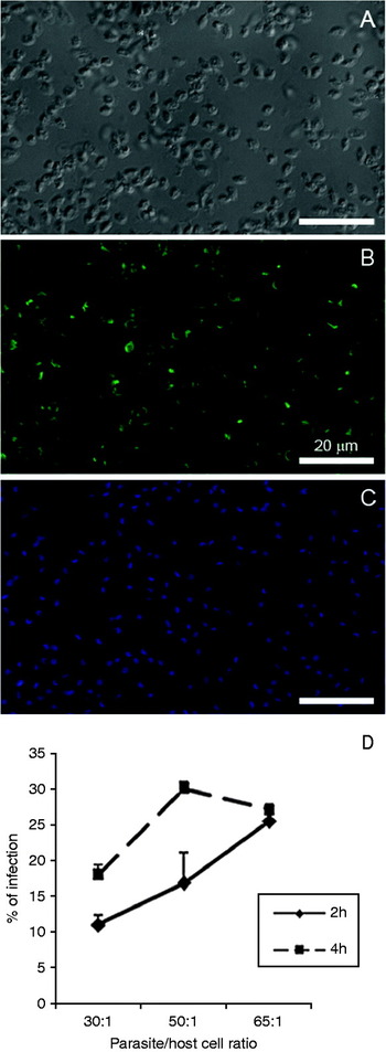
To analyse the participation of host cell sulfated proteoglycans in the invasion process by T. cruzi amastigotes, we performed a competition assay using soluble sulfated glycosaminoglycans (GAGs). First, we evaluated the level of infection of extracellular amastigotes, Ssp4 positive (Fig. 1), in cardiomyocyte cultures at early time-points (2 h and 4 h). The kinetic study with different host cell/parasite ratios (30:1, 50:1 and 65:1) revealed levels of infection varying between 10% and 30% after 2 h and 4 h of interaction at ratios of 30:1 and 50:1, respectively (Fig. 1). Thus, in order to avoid shedding of bound molecules from the parasite surface in the invasion assay, we performed the competition experiments at the shorter time of interaction (2 h) using a ratio of 65:1 parasite/host cell, which achieved approximately 25% infection.
Kinetic study of amastigote-host cell interaction. (A) Differential interferential contrast (DIC) image of extracellular amastigotes. (B) Amastigotes revealed an intense staining of Ssp4 (green), the stage-specific antigen of amastigotes, at the surface of the parasite. (C) DNA labelling with DAPI (blue)-stained nuclei and kinetoplast of parasites. (D) Cardiomyocytes were infected at the ratio of 30:1, 50:1 and 65:1 amastigotes/host cell for 2 h and 4 h of interaction. A high level of infection, 25%, was achieved after 2 h of interaction at the ratio of 65:1 amastigotes/ host cell. Scale bar=20 μm.

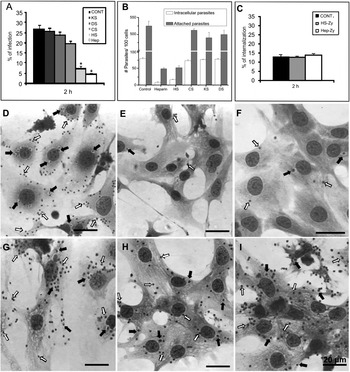
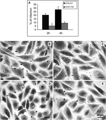
Next, we analysed the involvement of sulfated GAGs in the recognition and invasion process of amastigotes in cardiomyocytes. The amastigotes were pre-treated with 20 μg/ml of soluble sulfated GAGs, including heparan sulfate (HS), heparin, dermatan sulfate (DS), chondroitin sulfate (CS) and keratan sulfate (KS). Our results revealed that treatment of amastigotes with HS or heparin, GAGs consisting of repeated alfa-linked disaccharides, inhibit amastigote invasion in cardiomyocytes. A drastic reduction in the level of infection was noticed when the amastigotes were treated with heparin, decreasing from 25% in the control to 4·5% infection (P⩽0·0001), which represents 82% inhibition of parasite invasion (Fig. 2). A significant reduction was also observed after pre-treatment of the parasites with HS, achieving 8·7% (P⩽0·0004) infection that corresponds to 65% inhibition of invasion (Fig. 2). An approximately 10-fold decrease in the number of attached and intracellular parasites was observed when amastigotes were pre-treated with heparin and heparan sulfate (Fig. 2), indicating that blockage of heparin-binding protein impairs amastigote invasion. The effect of heparin and HS in T. cruzi invasion is clearly visualized by light microscopy, showing only a few cells with attached and interiorized parasites (Fig. 2). In contrast, the other sulfated GAGs examined herein, CS, DS and KS, which are GAGs consisting of beta-linked disaccharides, did not inhibit amastigote entry, reaching infection levels similar to the control (Fig. 2). However, to address the question of whether the GAGs are either involved in amastigote invasion or interfere with the endocytic process, we interacted an inert particle, zymosan (Zy), with the cardiomyocyte culture in the presence of HS or heparin. Neither treatment with HS nor heparin affected the cardiomyocyte endocytic process. The level of Zy particle internalization reached 13% after 2 h of interaction with cardiomyocyte cultures and remained unaltered in the presence of GAGs (Fig. 2). The involvement of GAGs in amastigote invasion was also evaluated using proteoglycan-deficient Chinese hamster ovary (CHO) cells. Wild-type CHO cells (CHO-K1) and GAG-deficient cells (CHO-745), a mutant defective in GAG biosynthesis due to the xylosyl transferase depletion, were infected for 2 h and 4 h with amastigotes at a ratio of 65:1 parasite-host cell. Our results demonstrate that amastigote invasion is impaired in CHO-745 mutant cells when compared to the wild-type CHO-K1 cells. The percentage of infection reduced significantly from 20·3% and 28% in CHO-K1 to 5·3% (P⩽0·0009) and 9% (P⩽0·013) in CHO-745 after 2 h and 4 h of infection, respectively (Fig. 3). Addition of soluble HS and heparin to CHO-745 cultures during amastigote infection did not revert the infection level (data not shown). The failure of soluble GAGs to restore the infection may be related to the fact that binding of GAGs to the core protein of proteoglycans is carried out by activity of specific enzymes (glycosyltransferases – EXT1 and EXT2) in the Golgi apparatus.
Sulfated glycosaminoglycans (GAGs) are involved in host cell invasion by Trypanosoma cruzi amastigotes. (A) Pre-treatment of amastigotes with 20 μg/ml of heparin and heparan sulfate (HS) prior to cardiomyocyte interaction resulted in a significant inhibition of 82% and 65% in the infection levels (2 h). No inhibition was detected with chondroitin sulfate (CS), keratan sulfate (KS) and dermatan sulfate (DS). (B) The relative number of attached and internalized parasites after treatment with GAGs. (C) Effect of GAGs on the endocytic process. Note that zymosan (Zy) particle internalization is not altered after GAGs treatment. (D) General aspect of amastigote-cardiomyocyte interaction (2 h of infection) and after treatment of amastigotes with 20 μg/ml of heparin (E), HS (F), CS (G), KS (H) and DS (I). Arrows indicate interiorized (➙) or attached (➾) parasites. (*) Student's t-test: statistically significant P⩽0·05. Scale bar=20 μm.

Amastigote invasion in Chinese hamster ovary (CHO) cells. CHO-K1 (wild-type) and CHO-745 (proteoglycan-deficient mutant) cultures were infected with amastigotes of Trypanosoma cruzi for 2 h and 4 h (A). Amastigote invasion was impaired in the CHO-745 mutant when compared to the wild-type CHO-K1, with a statistically significant inhibition in the infection level of 74% and 67% after 2 h and 4 h of interaction, respectively. (B–E) Aspect of Chinese hamster ovary (CHO) cell cultures infected with T. cruzi amastigotes. (B–D) CHO-K1 wild-type and (C–E) CHO-745, proteoglycan-deficient cells, after 2 h (B–C) and 4 h (D–E) of infection. Note the low levels of infection in CHO-745 cultures (C–E), with few interiorized (➙) and attached (➾) parasites, either after 2 h or 4 h of interaction, when compared with the wild-type, CHO-K1. Scale bar=20 μm. (*) Student's t-test: statistically significant P⩽0·05.

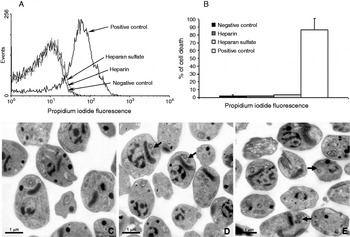
To ensure that the alterations observed in amastigote invasion were not related to toxic effects of soluble GAGs on amastigotes, we analysed the viability of heparin- and HS-treated amastigotes using propidium iodide (PI) labelling. The flow cytometry analysis demonstrated no significant alteration in the amastigote viability with GAGs treatment (Fig. 4). Membrane permeabilization was not observed after heparin or HS treatment, which exhibited a similar amount of PI uptake compared to control cells. Also, ultrastructural analysis revealed no evidence of morphological changes in amastigotes or signs of parasite death. In addition, amastigotes in the process of division were visualized after GAG treatment by transmission electron microscopy (Fig. 4).
Flow cytometry analysis of amastigotes treated with heparin and heparan sulfate (A). Note that treatment with GAGs did not induce cell death as compared to the positive control, i.e. parasites permeabilized with saponin, that reached 86·7% PI incorporation (B). Ultrastructural analysis of the glycosaminoglycan effect on amastigote forms of Trypanosoma cruzi. Extracellular amastigotes presented bar-shaped kinetoplasts positioned between the nucleus and flagellum (C). Heparin (D) and heparan sulfate (E) treatment neither altered parasite morphology nor their proliferation. Arrows show signs of a cellular division process in all conditions analysed. Scale bar=1 μm.

DISCUSSION
Intracellular pathogen-host cell interactions are dynamic and complex processes, in which parasite attachment is a primary requisite to establish infection. Glycosaminoglycans interact with cell surface molecules of a variety of intracellular pathogens, which enable attachment and trigger parasite invasion (Tossavainen et al. Reference Tossavainen, Pihlajamaa, Huttunen, Raulo, Rauvala, Permi and Kilpeläinen2006; Yokoyama et al. Reference Yokoyama, Okamura and Igarashi2006; Moelleken and Hegemann, Reference Moelleken and Hegemann2008; Schuksz et al. Reference Schuksz, Fuster, Brown, Crawford, Ditto, Lawrence, Glass, Wang, Tor and Esko2008). Therefore, efforts have been made to identify the parasite's heparin-binding proteins involved in the parasite-host cell recognition and their application as a prospective target for diagnostics or therapy.
In T. cruzi infection, both trypomastigotes and amastigotes are able to invade mammalian cells and sustain the intracellular cycle (Ley et al. Reference Ley, Andrews, Robbins and Nussenzweig1988). Although distinct mechanisms and signalling pathways involved in the invasion process have been reported between these infective forms (Mortara et al. Reference Mortara, Andreoli, Fernandes, da Silva, Fernandes, L'Abbate and da Silva2008; Alves and Mortara, Reference Alves and Mortara2009), the molecules required for amastigote invasion have been poorly investigated.
The fact that HSPG can activate signalling cascades involved in cytoskeleton remodelling (Dreyfuss et al. Reference Dreyfuss, Regatieri, Jarrouge, Cavalheiro, Sampaio and Nader2009) associated with the detection of glycosaminoglycan-binding proteins in amastigote forms of T. cruzi (Oliveira-Jr et al. Reference Oliveira, Alves, Calvet, Toma, Bouças, Nader, Castro, Côrtes, Krieger, Meirelles and Pereira2008) opened the question as to whether sulfated proteoglycans would also mediate amastigote invasion. Our results showed that treatment of amastigotes with heparin and heparan sulfate prevented cardiomyocyte invasion, achieving 82% and 65% inhibition, respectively. Similar to trypomastigotes, amastigote recognition is mediated by GAGs containing alpha-linked disaccharides, while the other negatively charged GAGs, such as chondroitin sulfate, dermatan sulfate and keratan sulfate, did not mediate amastigote invasion. Although the T. cruzi heparin-binding ligand has the ability to bind most sulfated glycosaminoglycan (Oliveira-Jr et al. Reference Oliveira, Alves, Calvet, Toma, Bouças, Nader, Castro, Côrtes, Krieger, Meirelles and Pereira2008), only heparan sulfate recognition triggers trypomastigote (Calvet et al. Reference Calvet, Toma, De Souza, Meirelles and Pereira2003; Oliveira-Jr et al. 2008) and amastigote invasion. The inability of GAGs to alter the uptake of Zy internalization by cardiomyocytes reinforces the important role of heparan sulfate in amastigote entry, suggesting that HS may be a key molecule in this process.
Few molecules have been demonstrated to mediate amastigote-host cell recognition. Mammalian lectins, such as mannose-binding proteins (MBP) and mannose receptor, are involved in the T. cruzi uptake by phagocytic cells (Kahn et al. Reference Kahn, Wleklinski, Aruffo, Farr, Coder and Kahn1995, Reference Kahn, Wleklinski, Ezekowitz, Coder, Aruffo and Farr1996). The protein SA85-1 expressed by amastigotes interacts strongly with MBP, but does not mediate MBP binding to trypomastigotes, suggesting a stage-specific recognition involved in amastigote attachment and invasion (Kahn et al. Reference Kahn, Wleklinski, Ezekowitz, Coder, Aruffo and Farr1996). In addition, it has recently been shown that membrane rafts, enriched in sphingolipids, cholesterol, ganglioside GM1, are implicated in the internalization of both extracellular amastigote and trypomastigote infective forms (Barrias et al. Reference Barrias, Dutra, De Souza and Carvalho2007; Fernandes et al. Reference Fernandes, Cortez, Geraldo Yoneyama, Straus, Yoshida and Mortara2007). Interestingly, evidence has demonstrated that heparan sulfate proteoglycans are localized in cholesterol-rich lipid raft domains (Chu et al. Reference Chu, Buczek-Thomas and Nugent2004) and modulate signal transduction events activated by fibroblast growth factor-2 at specialized membrane microdomains, favouring an effective sensitivity to diluted ligands (Fuki et al. Reference Fuki, Meyer and Williams2000).
Since it has also been demonstrated that RhoA GTPases play a key role in the invasion of extracellular amastigotes (Fernandes and Mortara, Reference Fernandes and Mortara2004; Mortara et al. Reference Mortara, Andreoli, Taniwaki, Fernandes, Silva, Fernandes, L'Abbate and Silva2005) and that HSPG-microbe interaction activates several signalling pathways, including PI3-kinase, focal adhesion kinase (FAK) and RhoA GTPases, triggering their internalization by endocytosis (Chen et al. Reference Chen, Goette, Liu and Park2008), we postulated that sulfated proteoglycans would mediate amastigote-cardiomyocyte invasion. Based on the fact that the cytoplasmic region of HSPG has a conserved domain, C1 domain, that binds to intracellular proteins, such as erzin, tubulin, Src kinase and cortactin (Granés et al. Reference Granés, Urena, Rocamora and Vilaró2000; Tehrani et al. Reference Tehrani, Tomasevic, Weed, Sakowicz and Cooper2007), it is possible that the binding of amastigote heparin-binding protein to host cell HS orchestrates different signalling transduction pathways, including (i) ezrin interaction eliciting actin remodelling by Rho GTPases, (ii) FAK/Src complex activation or (iii) phosphorylation of cortactin by Src activation, leading to actin polymerization and amastigote entry. Therefore, the investigation of signalling pathways triggered by binding of amastigotes to heparan sulfate may provide new insights into the mechanisms underlying T. cruzi amastigote infection.
ACKNOWLEDGMENTS
The authors thank Caroline Amaral for technical support and the Platform of Electron Microscopy of the Institute Oswaldo Cruz for use of its facilities. The anti-Ssp4 monoclonal antibody was kindly supplied by Dr Norma Andrews from the Department of Cell Biology, Yale University School of Medicine, New Haven, Connecticut, USA.
FINANCIAL SUPPORT
This work was supported by grants from Fundação Oswaldo Cruz (FIOCRUZ), Conselho Nacional de Desenvolvimento Científico e Tecnológico (M.C.S.P., Grant number 473087/2007-3), Programa Estratégico de Apoio à Pesquisa em Saúde (M.C.S.P., Grant number 403504/2008-2), Fundação de Amparo à Pesquisa do Estado do Rio de Janeiro (M.C.S.P., Grant number E-26/111.624/2008) and Fundação de Amparo à Pesquisa do Estado de São Paulo (L.T., Grant number 07/59801-1).