

Introduction
The length of pregnancy in the horse is nearly 11 months and, during this period, some animals exercise naturally while on pastureReference England1, Reference Vaala, Marr, Maxson, Sertich, Higgins and Wright2. Studies of pregnant equines to date have focused only on the mare and the reproductive and nutritional aspects of pregnancyReference England1, Reference Vaala, Marr, Maxson, Sertich, Higgins and Wright2. However, recent papers by the group in Cambridge, UK, have reported on metabolic changes in pregnant mares and the equine foetusReference Silver and Fowden3–Reference Taylor, Silver and Fowden5. Their research showed that the uptake of a variety of metabolites is influenced by the stage of pregnancy and by other physiological challengesReference Silver and Fowden3–Reference Taylor, Silver and Fowden5. Unfortunately, those published reports did not provide any information on the effects of acute exercise. To that end, a great deal of information has been published on the effect of acute exercise on the health and well-being of pregnant humansReference Pomerance, Gluck and Lynch6–Reference Rice and Fort11. Human studies have shown that moderate exercise during pregnancy is beneficial for maintaining maternal cardiovascular health, preventing excessive weight gain and providing a level of fitness that is a benefit in coping with the rigours of deliveryReference Pomerance, Gluck and Lynch6–Reference Rice and Fort11. Maternal benefits may come at a cost, and several studies suggest that maternal exercise results in foetal heart rate (HR) changes indicative of mild to moderate foetal stress due to alterations in uterine blood flowReference Carpenter, Sady, Hoegsberg, Sady, Haydon, Cullinane, Coustan and Thompson12–Reference Wolfe and Weissgerber16. The common conclusion from human experiments is that the effects of moderate maternal exercise on foetal cardiovascular and metabolic functions are transient, and do not appear to produce abnormalities in development or long-term changes in foetal healthReference Hall and Kaufmann9Reference Wolfe and Weissgerber16–Reference Carpenter, Sady, Sady, Haydon, Coustan and Thompson20.
For gestating horses, exercise is an important physiological challenge to consider because many pregnant horses are ridden and moderately trained up until parturition. The fact that moderate exercise appears to do no harm in humans leads to the speculation that natural exercise in the pasture, or even moderate training, should be beneficial to a mare for reasons similar to those suggested for humans, including avoidance of excessive weight gain during pregnancy as well as the maintenance of physical conditioning for parturition. Interestingly, the concern about the safety of exercise during pregnancy has another focus with many horse owners, trainers and veterinarians suggesting anecdotally that pregnancy modulates the behaviour of some difficult to handle athletic mares. Thus, it is not uncommon for some owners to keep training a mare during the early part of pregnancy. However, despite the intuitive beneficial effects of exercise during pregnancy, some equine health professionals are still leery of exercising a pregnant mare. As with humans, these questions are raised more often when it comes to the safety of moderately intense exercise performed during the last trimester of pregnancy. Those concerns apply both to the mare and to the developing foal. In the case of the latter, decreases in blood flow and oxygenation to the developing foetus can have devastating effects on the development of key organ systems as well as on in utero growth. Taken to the extreme, some believe exertion can increase the risk of miscarriage or lead to the premature birth of a foal. While this opinion is based only on anecdotal information, it has led some to limit exercise and even stall confine a mare in the later part of pregnancy. When one considers the overall detraining effect of extended stall confinement, one could suggest that such an overprotective practice may actually decrease the ease of parturition due to the loss of fitness. Decisions on the safety of exercise should be based on data from controlled hypothesis-driven experiments.
To that end, we are aware of only one study that has been published on the effect of mild exercise on maternal or foetal health in equinesReference Causey, Lehnhard, Finucane and Oliver21. One should note that, unlike exercise performed on a treadmill, the intensity of the exercise in that study could only be controlled in a subjective fashion. Furthermore, the logistics of the study limited the measurement of HR to before and after exertion. Finally, while that study provided some information suggesting that mild exercise is safe, there was no assessment of anaerobic intensity or overall metabolic stress. Therefore, the purpose of the present study was to utilise a standardised exercise test performed on a treadmill to test two hypotheses. First, that moderate exercise would alter maternal and foetal HR in the horse. Second, that pregnancy would alter the HR, plasma lactate and plasma cortisol responses to moderate exercise.
Materials and methods
Six unfit, pregnant Standardbred mares, aged 6–19 years were the subjects for this investigation. They were fed ad libitum Timothy hay and received supplementation with commercial pellets (15% crude protein and 12.6 MJ kg− 1 dry matter). The energy and protein supplementations were 10% over the NRC (1989) recommendations for gestation, and c. 55% of the energy during pregnancy came from pellets. Animals were fed at 07.00 and 15.00 hours inside individual stalls, but were otherwise maintained on pasture during daylight hours and returned to the stalls overnight. Water and salt were available ad libitum. All mares were on the anthelmintic, vaccination and hoof-trimming routine adopted at the Equine Science Center–Rutgers University, and all methods were approved by the Rutgers Institutional Animal Care and Facilities Committee.
Each mare completed two exercise tests. The first was performed at approximately 9 months of gestation, and the second graded exercise tests (GXT) took place at approximately 6 months post-parturition, at least one month post-weaning. Each GXT consisted of running on a treadmill up a fixed 6% incline, for three 1 min stages at velocities of 4, 6 and 7 m s− 1, respectivelyReference McKeever and Malinowski22.
Maternal and foetal HRs were obtained in the pre-parturition tests using a Schiller model AT-6 portable hard wire electrocardiograph (ECG). The use of the ECG for the measurement of HR allows for the measurement of both maternal and foetal HR but it can have a noisy signal and thus maternal HR was also recorded via ventricular waveform utilising a micromanometer catheter in both the pre- and post-parturition GXTs. This allowed for an internal check on data obtained from the ECG recordings and allowed the use of the noise-free pressure waveform for both GXTs for the mares. The standard four limb electrodes were placed on the mare at the following anatomical reference points: the right leg and left leg electrodes were placed mid-flank, 10 in (25.0 cm). from the centre line. The right limb (RL) electrode was placed mid-abdominally on the forward margin of the foetus. The left limb (LL) electrode was also placed mid-abdominally on the rear margin of the umbilicus.
By placing the limb electrodes in the above configuration, both the mare and the foetal ECGs appear in lead I (RA negative, LA positive). However, the amplitude of the foetal R wave is less than that of the mare; thus, the gain was turned up to a range of 40–60 Hz in order to obtain the foetal ECG signal. The significantly shorter foetal R wave was easily distinguishable from that of the mare on the resulting ECG tracing. Foetal and mare HR data were recorded at rest and during the last 10 s of each stage of the GXT. During recovery, HR was recorded immediately within the first 10 s after exercise and at 2, 3, 4 and 5 min post-GXT.
Plasma lactate concentrations (LA) were measured in triplicate using a lactate analyzer (YSI Sport 1500) and samples obtained at rest, at 4, 6 and 7 m s− 1 and at 5 min post-GXT. Plasma cortisol concentration was measured in duplicate using radioimmunoassay kits previously validated for use in horsesReference Freestone, Wolfsheimer, Kamerling, Church, Hamra and Bagwell23. Total plasma protein concentration was measured in duplicate via refractometry, using plasma samples collected before the GXT, immediately after exercise and after 5 min of recovery.
Data were analysed (SigmaStat, version 3, Jandel Scientific, San Rafeal, CA, USA) using ANOVA for repeated measures and t-tests for paired comparisons. Where appropriate, the post hoc analysis utilised the Student Newman–Keuls test for all pairwise comparisons. Null hypothesis was rejected when P < 0.05.
Results
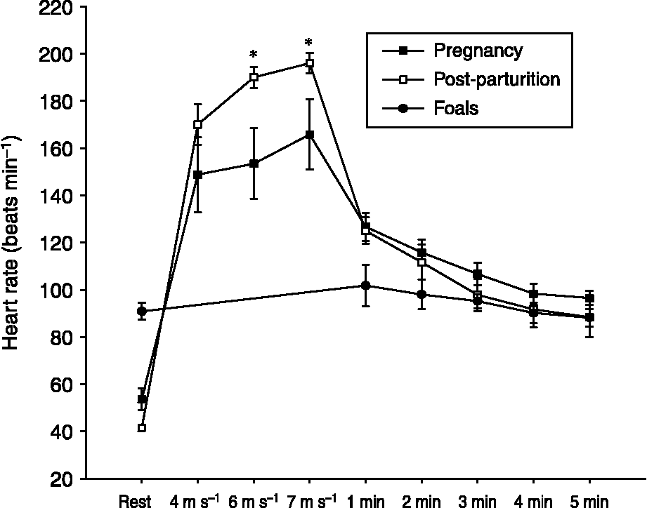
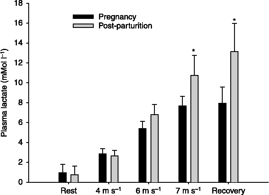
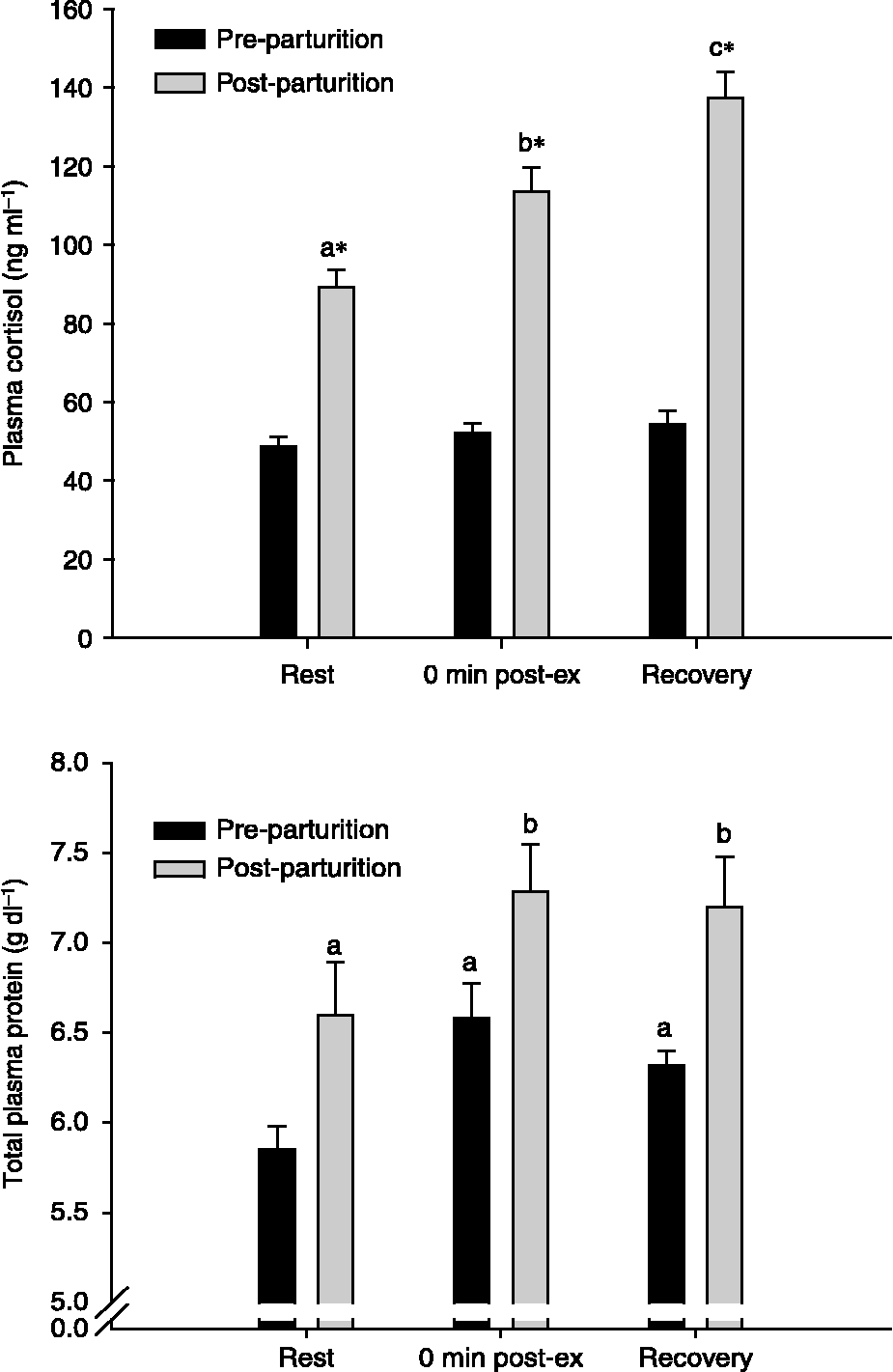
Data in the figures are expressed as means ± SE. Body weight (mean ± SE) was lower after parturition (570 ± 20 vs. 505 ± 22 kg; P < 0.05). Consequentially, there were differences (P < 0.05) in work rate (watts) at each step of the GXT (1557 ± 53 vs. 1381 ± 60; 2339 ± 80 vs. 2071 ± 90; and 2571 ± 1 21 vs. 2416 ± 105 W). Exercise caused no change (P>0.05) in foetal HR and their values were near 100 beats min− 1 or below (Fig. 1). Maternal HR (Fig. 1) and plasma LA (Fig. 2) were greater (P < 0.05) during the post-parturition GXT compared with the pre-parturition GXT. Interestingly, during pregnancy, plasma cortisol concentration was significantly lower at rest compared with post-parturition (Fig. 3). Furthermore, while there was a normal exercise-induced increase (P < 0.05) in plasma cortisol concentration during the post-parturition GXT, there was no similar exercise-induced increase (P>0.05) in plasma cortisol concentration during pregnancy.
Mean ( ± SE) HR response to incremental treadmill exercise measured in mares before (GXT 1) and after pregnancy (GXT 2). Foetal HR measured before, within 10 s immediately after exercise and at 2, 3, 4 and 5 min post-GXT 1. Bpm, beats per minute

Mean ( ± SE) plasma lactate response to incremental treadmill exercise measured before (pregnant) and after pregnancy (post-parturient)

Mean ( ± SE) plasma cortisol response to incremental treadmill exercise measured before (pre-parturition) and after pregnancy (post-parturition). Post-ex, post-exercise

Discussion
Mean gestational length in the horse is usually considered to be 340 days, and it is the longest in all domestic speciesReference England1, Reference Vaala, Marr, Maxson, Sertich, Higgins and Wright2. Because it is so long, the foetus is more at risk from the detrimental effects of stress than other domestic animals. The results obtained from the foetal HR measurements made in this experiment demonstrated that one acute bout of moderately intense exercise does appear to induce foetal cardiovascular stress during the late gestational period. However, one should note that, because of noise on the ECG signal, there were no recordings of foetal HR reported during exercise, and we cannot say for certain that there was no effect on foetal HR during the GXT. However, the immediate post-exercise recording was done early enough during recovery to reflect the effect of the GXT on foetal HR.
In common with most other species the HR in the foetal horse declines as term approaches, with a decrease in foetal HR from approximately 120 beats min− 1 during mid-gestation ( < 9 months of pregnancy) to approximately 90 beats min− 1 near parturitionReference Silver4. Foetal HR below 100 beats min− 1 recorded in the present study is in agreement with the results demonstrated by SilverReference Silver4. Interestingly, pregnancy appeared to affect the mares' HR response to exercise, suggesting a pregnancy-induced difference in their cardiovascular physiology. Post-parturient HR was higher during the GXT; however, the post-partum HR values were consistent with those observed in other studies of non-pregnant mares, including those performed in the laboratory at RutgersReference McKeever and Malinowski22. We do not have mechanistic data to explain this unexpected result. However, it is well documented that pregnant mammals have a greater circulating blood volume compared with non-pregnant animalsReference England1, Reference Vaala, Marr, Maxson, Sertich, Higgins and Wright2. It is possible that the extravascular fluid volume allows for greater venous return and cardiac filling. An increase in cardiac preload and end-diastolic volume would facilitate increased stoke volume. Such an increase in stroke volume would enhance cardiovascular stability and efficiency, allowing the generation of the same or even a greater cardiac output at a lower achieved HR. While purely speculative, this may explain the lower HR measured at each step of the GXT when the mares were pregnant vs. the HR measured in the post-partum exercise tests.
Plasma lactate concentrations were similar in both groups at rest; however, peak plasma LA concentrations were lower in pregnant mares compared with non-pregnant mares. Either the pregnant mares produced less LA or metabolised LA faster than non-pregnant mares. While purely speculative, this could have been due to greater cardiovascular efficiency when the horses were pregnant. However, the presence of the placenta in the pregnant animals could also be part of the reason for the modification of lactate metabolism. This is supported by the observations of Fowden et al. Reference Fowden, Taylor, White and Forhead24, who showed that LA delivered to the umbilical circulation in non-exercised mares was maximal between 281 and 300 days of gestation, a period similar to that when the pregnant mares performed the first GXT.
Finally, the effect of pregnancy on plasma cortisol concentration revealed some interesting responses in the horses of the present study. Resting plasma cortisol concentration increases over the course of gestation in humans and other mammalsReference Lindsay and Nieman25. This increase is mediated by pregnancy-induced alterations in several steps of the hypothalamic–pituitary–adrenal axis, with resulting alterations in production and release of corticotrophin-releasing factor, adrenocorticotropin hormone (ACTH), cortisol and cortisol-binding proteinReference Lindsay and Nieman25. Based on the human literature, we predicted an effect of pregnancy on resting plasma cortisol concentration as well as the response to the challenge of exercise. However, unlike pregnant humans, resting plasma cortisol concentration within the horses used in the present study was lower in samples collected during pregnancy when compared with samples collected post-partum. We have no explanation for this species-related difference. But, it should be noted that even though there were differences in pre-and post-pregnancy, the resting concentrations observed at both time points were similar to those reported in a range of papers on horses subjected to different experimental designs and perturbationsReference Sady, Carpenter, Thompson, Sady, Haydon and Coustan19Reference McCarthy, Jeffcott, Funder, Fullerton and Clarke26–Reference Malinowski, Shock, Roegner, Rochelle, Kearns, Guirnalda and McKeever31.
Another surprising observation made in the present study was the failure of exercise to induce an increase in plasma cortisol concentration when the mares were pregnant. This lack of a response during pregnancy contrasts with a normal increase in plasma cortisol concentration seen during and following exercise in non-pregnant horses subjected to a GXT Reference McCarthy, Jeffcott, Funder, Fullerton and Clarke26, Reference Malinowski, Shock, Roegner, Rochelle, Kearns, Guirnalda and McKeever31, Reference Gordon, McKeever, Betros and Manso Filho32. The cortisol response to exercise is affected both by the intensity and duration of the exercise challenge, providing a functionally significant boost to substrate mobilisation as well as a pre-potentiation of anti-inflammatory mechanismsReference Gordon, McKeever, Betros and Manso Filho32–Reference Marc, Parvizi, Ellendorff, Kallweit and Elsaesser37. We do not have mechanistic information to comment on the lack of a cortisol response during exertion in pregnancy; however, a similar lack of a response has been reported for humans performing an exercise test during the later part of pregnancyReference Artal, Platt, Sperling, Kammula, Jilek and Nakamura7, Reference Rauramo, Andersson, Laatikainen and Pettersson8. The authors of those papers showing a lack of a response in pregnant humans suggested that the altered plasma cortisol response to exercise may be related to either a disruption of feedback mechanisms affecting cortisol-releasing factor and/or ACTHReference Artal, Platt, Sperling, Kammula, Jilek and Nakamura7, Reference Rauramo, Andersson, Laatikainen and Pettersson8, Reference Lindsay and Nieman25. A similar lack of a response to exercise has also been reported in very old female horses subjected to an exercise testReference Malinowski, Shock, Roegner, Rochelle, Kearns, Guirnalda and McKeever31. The old horses also had a disruption of the normal glucose and insulin response to acute exercise. We do not know whether the same mechanisms play a role in the lack of a cortisol response to exercise in aged and pregnant horses, and we did not measure the glucose and insulin responses in our study; but one wonders whether there are similar or dissimilar reasons for this alteration in endocrine function.
Implications and comments
Foals from the pregnant mares used in this experiment were born healthy, with normal body weights and without complications associated with delivery. The data suggest that pregnant mares should be able to perform limited moderate exercise without any major deleterious effects on their unborn foals or themselves during late gestation.
Acknowledgements
Sponsored by the New Jersey State Initiative on Equine and New Jersey Agricultural Experiment Station Project # 99 566. Hélio C. Manso Filho was supported by a fellowship from CNPq/Brazil and University Federal Rural of Pernambuco, Brazil.