Introduction
Theileria annulata (T. annulata), an obligate intracellular protozoan parasite belonging to the phylum Apicomplexa, is economically important for domestic and wild bovids (Hayashida et al., Reference Hayashida, Hara, Abe, Yamasaki, Toyoda, Kosuge, Suzuki, Sato, Kawashima and Katayama2012; Rashid et al., Reference Rashid, Akbar, Rashid, Saeed, Ahmad, Ahmad, Shehzad, Islam and Farooqi2018, Reference Rashid, Rashid, Akbar, Ahmad, Hassan, Ashraf, Saeed and Gharbi2019b). It transforms macrophages, B-cells and dendritic cells (Moreau et al., Reference Moreau, Thibaud, Miled, Chaussepied, Baumgartner, Davis, Minoprio and Langsley1999; Stephens and Howard, Reference Stephens and Howard2002; Rashid et al., Reference Rashid, Guan, Luo, Zhao, Wang, Rashid, Hassan, Mukhtar, Liu and Yin2019a). These transformed cells constantly divide into daughter cells, which can be infinitely maintained in culture in fresh complete culture medium (Tretina et al., Reference Tretina, Gotia, Mann and Silva2015). The association of host cell mitotic apparatus with macroschizonts enables the parasite to divide at the same time as the host cell to ensure the partition of parasites into daughter cells. These infected cells competently express MHC class molecules on their surface (Sager et al., Reference Sager, Davis and Jungi1999) that play a role in antigen presentation and hence in the initiation and regulation of both innate and adaptive immune responses against target antigens (Sager et al., Reference Sager, Brunschwiler and Jungi1998; Kikuchi et al., Reference Kikuchi, Yanagawa and Onoé2005). Monocyte-derived dendritic cells (MoDCs) and T. annulata transformed dendritic cells (TaDCs) are potent antigen-presenting cells (APCs) capable of activating both T and B lymphocytes (Tretina et al., Reference Tretina, Gotia, Mann and Silva2015); and can also initiate strong primary and memory immune responses (Savina and Amigorena, Reference Savina and Amigorena2007). Hence, the current study was designed to compare the efficacy of antigen presentation by MoDCs and TaDCs for T lymphocyte proliferation. Efficient antigen presentation generates the development of effector cytotoxic T lymphocytes (CTL), while a less efficient presentation can lead to stimulation of CTL responses (Kennedy et al., Reference Kennedy, Undale, Kieper, Block, Pease and Celis2005; Umeshappa et al., Reference Umeshappa, Huang, Xie, Wei, Mulligan, Deng and Xiang2009). Three basic pathways are involved in antigen presentation. In one pathway, antigens derived from the extracellular milieu serve as substrates for presentation as peptide/class II MHC complexes to CD4+ T-cells consecutively activating dendritic cells (DCs) through CD40L signals; this activation can be reproduced by persistent antigen delivery. In the second pathway, intracellular antigens are synthesized and presented as peptide/class I MHC complexes to CD8+ T-cells. The third pathway of antigen presentation is termed as cross-presentation, where the exogenous antigen (Ag) peptides are presented to MHC class I complexes with the help of small GTPase of the Rab family (intracellular membrane trafficking proteins) and trans-locator (SEC61 and HSP90α) proteins. Rab family gene transcription in TaDCs was estimated to gain insight into the mechanism of antigen presentation by APCs. The above-mentioned pathways for antigen presentation trigger cellular immunity against the antigen (Savina et al., Reference Savina, Jancic, Hugues, Guermonprez, Vargas, Moura, Lennon-Duménil, Seabra, Raposo and Amigorena2006; Jancic et al., Reference Jancic, Savina, Wasmeier, Tolmachova, El-Benna, Dang, Pascolo, Gougerot-Pocidalo, Raposo and Seabra2007) and the extent of Ag internalization is a factor that affects the magnitude of cross-presentation (Weimershaus et al., Reference Weimershaus, Maschalidi, Sepulveda, Manoury, van Endert and Saveanu2012). Rab proteins are recruited to endosomes, phagosomes and the vacuole containing pathogen (Cebrian et al., Reference Cebrian, Croce, Guerrero, Blanchard and Mayorga2016). The APCs selectively process certain exogenous antigens for cross-presentation as peptide/MHC class, while degrading others to single amino acids (Schoenberger et al., Reference Schoenberger, Toes, van der Voort, Offringa and Melief1998; Obst et al., Reference Obst, van Santen, Melamed, Kamphorst, Benoist and Mathis2007; Kim et al., Reference Kim, Visser, Cruijsen, van der Velden and Boes2008). The antigen peptides presented with MHC-I to T cell receptors (TCR) activate the cytotoxic T-cell (CD8+) response, while presented with MHC-II activate helper T cells (CD4+) (Liu and Gao, Reference Liu and Gao2008; Huppa et al., Reference Huppa, Axmann, Mörtelmaier, Lillemeier, Newell, Brameshuber, Klein, Schütz and Davis2010; Hart et al., Reference Hart, MacHugh and Morrison2011). Adaptive immunity is initiated when APCs display antigenic peptides in the context of MHC I to CTL (Kim et al., Reference Kim, Visser, Cruijsen, van der Velden and Boes2008), an activated CTL can kill abnormal and deformed cells (Chiang et al., Reference Chiang, Theorell, Entesarian, Meeths, Mastafa, Al-Herz, Frisk, Gilmour, Ifversen and Langenskiöld2013).
The present study aimed to discern the impact of T. annulata on antigen presentation by infected DCs compared with non-infected MoDCs, as estimated by T lymphocyte proliferation. In addition, the transcription level of Rab genes was followed over multiple passages of TaDCs to gain insight into the mechanism/pathway of antigen presentation of these normal and transformed APCs.
Materials and methods
Materials and reagents
The details concerning the reagents and the antibodies (Abs) used in this study are as follows: OVA-FITC (lot# SF069; Solarbio Science and Technology Co, Ltd, Beijing, China), mouse anti-bovine CD14 antibody (MCA2678GA), mouse anti-bovine CD4 antibody (MCA1653PE), mouse anti-bovine CD8 antibodies (MCA837PE), bovine dendritic cell growth kit (PBP014KZZ), mouse anti-bovine MHC class I monomorphic antibody (MCA2444F) and mouse anti-bovine MHC class II DR antibody (MCA5656F) all from Bio-Rad (Hercules, California, USA). PrimeScript™ RT reagent Kit with gDNA Eraser (cat. RR047A) and SYBR® Premix Ex Tq™ II (Tli RNaseH Plus) (cat. RR820A) are from Takara, Co., Ltd (Dalian, China); carboxy-fluorescein succinyl ester (CFSE) (lot: 1938592) and TRIzol (cat. no. 15596-026) from Invitrogen (Carlsbad, CA, USA), anti-FITC microbeads (130-048-701), anti-PE microbeads (130-048-801) and LS column (order no. 130-042-401) from Miltenyi Biotec (Bergisch Gladbach, Germany), Gibco RPMI 1640 (lot: 1930005; Life Technologies, Carlsbad, California, USA).
Sample collection
Theileria annulata piroplasm-free cattle <1 year of age were maintained in the animal experimental unit of the Chinese Academy of Agricultural Sciences (CAAS), Lanzhou Veterinary Research Institute (LVRI), Lanzhou, Gansu, according to the instructions and guidelines of the Animal Ethics Committee (permit no. LVRIAEC-2018-001), approved by People's Republic of China. During the study period, blood samples were taken from these experimental animals for cell isolation.
Magnetic cell separation
The CD4+, CD8+ and CD14+ types of cells were isolated by magnetic separation according to published methods (Bull et al., Reference Bull, Lee, Stucky, Chiu, Rubin, Horton and McElrath2007; Zhao et al., Reference Zhao, Guan, Liu, Liu, Li, Yin and Luo2017) with minor modifications to attain high purity (>95%). Briefly, whole blood was collected by venepuncture of the jugular vein from piroplasm-free <1-year-old cattle into 9 mL tubes containing K3EDTA. Peripheral blood mononuclear cells (PBMCs) were isolated by density gradient centrifugation using Ficoll-Paque™ plus (product no. 17-1440-02; GE healthcare Uppsala, Sweden). Cells were labelled with mouse anti-bovine antibodies and anti-micro-beads for magnetic separation via LS column (Obermaier et al., Reference Obermaier, Dauer, Herten, Schad, Endres and Eigler2003). The percentage of specific cells in PBMCs and purity of isolated cells were analysed by flow cytometry. The pure isolated cells were seeded in 24-well plate at a concentration of 2 × 10⁶ cells per well and were cultured in complete culture medium containing Gibco RPMI 1640 (L-glutamine and 25 mm HEPES), 50 µg mL−1 gentamicin, 50 µ m 2-mercaptoethanol supplemented with 10% heat-inactivated foetal bovine serum (FBS) and incubated at 37 °C and 5% CO2 for further use. During the whole-cell isolation process, all solutions used were filtered through syringe supported Millex®GP filter of 0.22 µm (lot: R5SA76494; Merck Millipore Ltd, Ireland) to avoid any contamination.
MoDC generation and TaDC cell line maintenance
The culture medium of CD14+ cells was replaced with MoDC growth cocktail bovine dendritic cell growth kit containing granulocyte macrophages colony-stimulation factor (GM-CSF) and interleukin 4 (IL-4) at the rate of 1/20 with complete culture medium for 2 × 10⁶ cells per mL per well which was used to generate MoDCs according to the manufacture's instruction (Sallusto et al., Reference Sallusto, Cella, Danieli and Lanzavecchia1995; Kamphorst et al., Reference Kamphorst, Guermonprez, Dudziak and Nussenzweig2010). At day three, half of the medium was changed with fresh prepared MoDC growth cocktail medium and the cells were ready to use within 3 days. The established TaDC cell line was obtained from the Vector and Vector-Borne Diseases (VVBDs) Laboratory of LVRI. The cell line was maintained in complete culture medium at 37 °C and 5% CO2 and passaged twice a week according to the growth rate of the transformed cells.
Preparation of antigen-presenting cells
The MoDCs and TaDCs were seeded into 24-well plates at the rate of 2 × 10⁶ cells per mL per well with the addition of OVA-FITC (Ag) at the rate of 200 µg mL−1 in complete culture medium and incubated at 37 °C and 5% CO2. After 16–24 h, cells were washed three times with ice-cold PBS containing 0.2% Tween 20 to remove the rest of OVA-FITC. The endocytosis rate of antigen in both types of cells was measured by flow cytometry analysis. These normal and transformed APCs are ready to stimulate T lymphocyte proliferation.
T lymphocyte proliferation assay
To assess T lymphocyte proliferation, CD4+ and CD8+ T cells were isolated from non-immunized and not experimental antigen exposed cattle. The cells were incubated with carboxy-fluorescein succinyl ester (CFSE) at the rate of 10 µ m−1 for 2 × 10⁷ cells prior to culture with APCs (TaDCs and MoDCs) according to the previously used method (Hart et al., Reference Hart, MacHugh and Morrison2011). Briefly, CD4+ and CD8+ cells were washed with 1 × PBS containing 0.1% BSA followed by re-suspension into 10 µ m CFSE and incubated for 10 min at 37 °C in the presence of 5% CO2. The staining was stopped with the addition of five volumes of ice-cold complete culture medium followed by incubation on ice for 10 min and finally washed three times with ice-cold complete culture medium. In negative control assay, only TaDCs were used without the endocytosis of OVA-FITC. The CFSE stained cells and APCs (MoDCs and TaDCs) were poured into 6-well culture plates at the rate of 1:5 of APC to stained cells followed by incubation at 37 °C in the presence of 5% CO2. Cells were incubated 6–7 days to access T lymphocyte proliferation. Moreover, Rab genes were knockdown to evaluate their role for antigen presentation.
Sample preparation and flow cytometry analysis
The percentage of specific cells in PBMCs and isolated cell purity were performed after labelling with antibodies according to the method described in the previous section of magnetic cells separation. The MoDCs and TaDCs were labelled with MHC I and II antibodies and anti-FITC micro-beads for the percentage in both types of cells. Meanwhile, stained CD4+ and CD8+ cells cultured with APCs were washed and processed for flow cytometry analysis.
The fluorescence intensity for each assay was analysed by BD accuri C6 (Becton, Dickinson and Company 1 Becton Drive Franklin Lakes, NJ 07417-1880, USA). The stained, un-stained and antigen-free MoDCs and TaDCs were used as background fluorescence control (Monrad et al., Reference Monrad, Rea, Thacker and Kaplan2008) according to each assay. Stained cells were gated by side and forward scatter characteristics. For each sample, 5000– 20 000 events were collected by FACSCalibur (accuri C6) for flow cytometry analysis. Gating of live target cells was performed through the selection of main cell population in forward and side scatter profile. Normalized mean fluorescence was computed through the subtraction of geometric mean fluorescence of the cells that did not fluoresce (Wong et al., Reference Wong, Shenoi, Abbina, Chafeeva, Kizhakkedathu and Khan2017).
Primer optimization
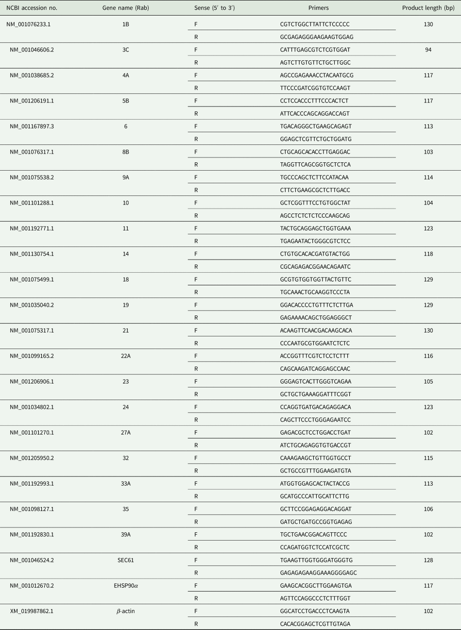
Bovine Rab genes (1B, 3C, 4A, 5B, 6, 8B, 9A, 10, 11, 14, 18, 19, 21, 22A, 23, 24, 27A, 32, 33, 35 and 39B) have been reported by others (Zou et al., Reference Zou, Zhou, Zhang, Li, Liu, Chai, Li, Liu, Li and Xie2009; Wang et al., Reference Wang, Liu and Huang2012). In addition, both heat shock protein 90α (Imai et al., Reference Imai, Kato, Kajiwara, Mizukami, Ishige, Ichiyanagi, Hikida, Wang and Udono2011) and endoplasmic reticulum (ER) membrane protein trans-locator (SEC61) genes (Zehner et al., Reference Zehner, Marschall, Bos, Schloetel, Kreer, Fehrenschild, Limmer, Ossendorp, Lang and Koster2015) were included in this study due to their roles in antigen cross-presentation. Primers for amplification of a short segment of these genes were designed through the online browser of GeneScript (https://www.genscript.com/tools/real-time-pcr-tagman-primer-design-tool) and NCBI (https://www.ncbi.nlm.nih.gov/tools/primer-blast/) and, melting temperature (difference of less than 2 °C) and self-dimer (less than 10 kcal mol−1) were double checked with the online browser (https://sg.idtdna.com/calc/analyzer). The primers which fulfil the criteria were selected and synthesized by Sangon (Biotech Co., Ltd, Shanghai, China). The target Rab genes were amplified from DNA and cDNA extracted from TaDCs, purified, ligated with pGEM™-T Easy vector (Promega, Madison, Wisconsin, USA) and transformed into DH5α. Briefly, polymerase chain reaction (PCR) products were analysed on 5% agarose gel and the desired bands were purified by gel/PCR extraction kit (Biomiga, Inc., San Diego, California, USA). The purified PCR products were then ligated into pGEM™-T Easy vector and transformed into competent cells (DH5α) (Hassan et al., Reference Hassan, Liu, Sajid, Mahmood, Zhao, Abbas, Guan, Yin and Luo2018). The bacterial culture samples were sent to Sangon (Biotech Co., Ltd, Shanghai, China) for sequencing. The optimized primers were used for Q-RT-PCR to check the transcription level of above-mentioned genes in the TaDC cell line (Table 1).
Details of Q-RT-PCR primers used during this study

F represents forward and R reverse primer sequences. The β-actin used was already optimized (Zhao et al., Reference Zhao, Liu, Guan, Liu, Li, Yin and Luo2018).
RNA isolation and quantitative real-time PCR
The MoDCs and TaDCs were used for RNA isolation. The cells were harvested by centrifugation at 12 000 g for 2 min followed by washing with 1 × PBS. For total RNA isolation, TRIzol method was used as described by others (Rio et al., Reference Rio, Ares, Hannon and Nilsen2010). The concentration and purity (260/280 nm ratio) of isolated RNA was determined through Nanodrop spectrophotometer (Thermo Scientific 2000/2001, Wilmington, DE 19810, USA). The cDNA from 1 µg RNA was synthesized with PrimeScript™ RT reagent Kit with gDNA Eraser according to the instruction of manufacturer and stored at −20 °C until processed.
The Q-RT-PCR was performed with SYBR® Premix Ex Tq™ II (Tli RNaseH Plus) according to the manufacture's instructions. Briefly, the volume of reaction mixture was 20 µL containing 10 µL of SYBR® Premix Ex Taq II, 0.8 µL (16 µ m) of each primer (forward and reverse), 0.4 µL of Rox reference dye II (50×), 2 µL of cDNA and 6 µL of RNase free water. The reaction conditions were set into thermocycler (Agilent Mx3005P; Agilent Technologies Santa Clara, California, USA) in three repeating steps/segments; consisting of one denaturation cycle at 95 °C for 30 s, 40 annealing cycles (95 °C for 5 s and 60 °C for 34 s) and one extension cycle at 95 °C for 15 s, 60 °C for 1 min and 95 °C for 0.15 s according to the instructions of the SYBR® Premix Ex Tq™ II kit. A serial dilution (1/10) of cDNA was performed followed by Q-RT-PCR to optimize the cytokines primers. The Q-RT-PCR data were analysed by regression (known as cDNA quantities) based on the threshold cycle (Ct) for each Rab gene to calculate R 2 values (Coussens et al., Reference Coussens, Verman, Coussens, Elftman and McNulty2004). All reactions were run in triplicate, and three samples of each gene were run in each Q-RT-PCR assay to analyse the expression level in TaDC cell line. The Ct values of β-actin and MoDCs were used as normalizer and calibrator, respectively. The fold change values were calculated by the formula of 2−ΔΔCt according to the described method (Rao et al., Reference Rao, Huang, Zhou and Lin2013).
Data analysis
Sequencing data were analysed by EditSeq and MegAlign (DNA Star, Madison, Wisconsin). Flow cytometry fluorescence data were analysed by FlowJo X (Tree Star; Version 10.0.7) of (BD Biosciences, Franklin Lakes, New Jersey, USA). Fold change values were calculated using 2−ΔΔCt in Microsoft Excel 2010 (Redmond, Washington, USA). Graphical analysis, Student's t test and one-way ANOVA were performed with GraphPad Prism Software version 7 (La Jolla, CA 92037, USA) for endocytosis and Rab genes expression, respectively. Significant values obtained are presented as *P ⩽ 0.05; **P < 0.01; ***P < 0.001 and ns represents non-significance (P > 0.05).
Results
Cell purity and MHC class molecules
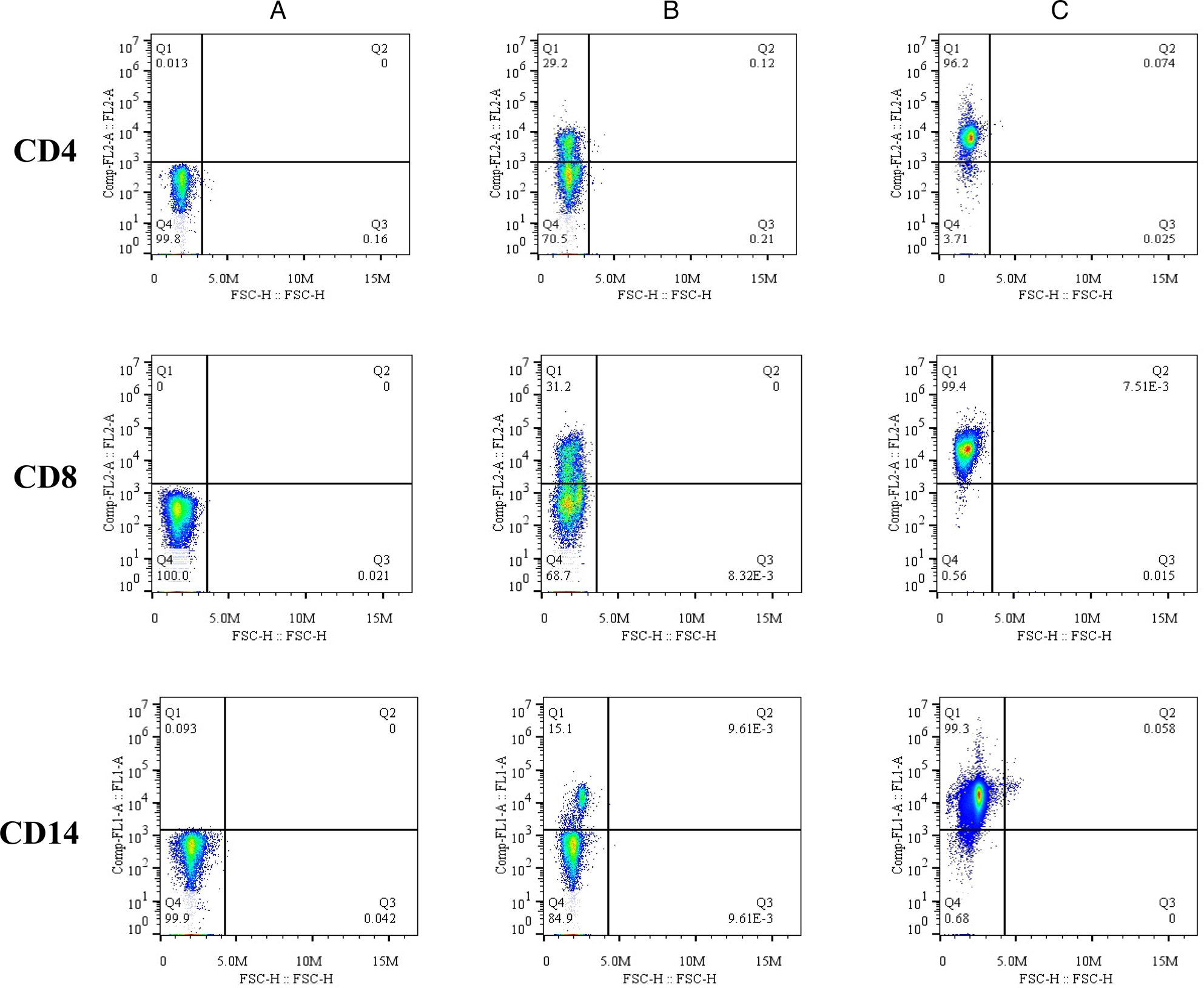
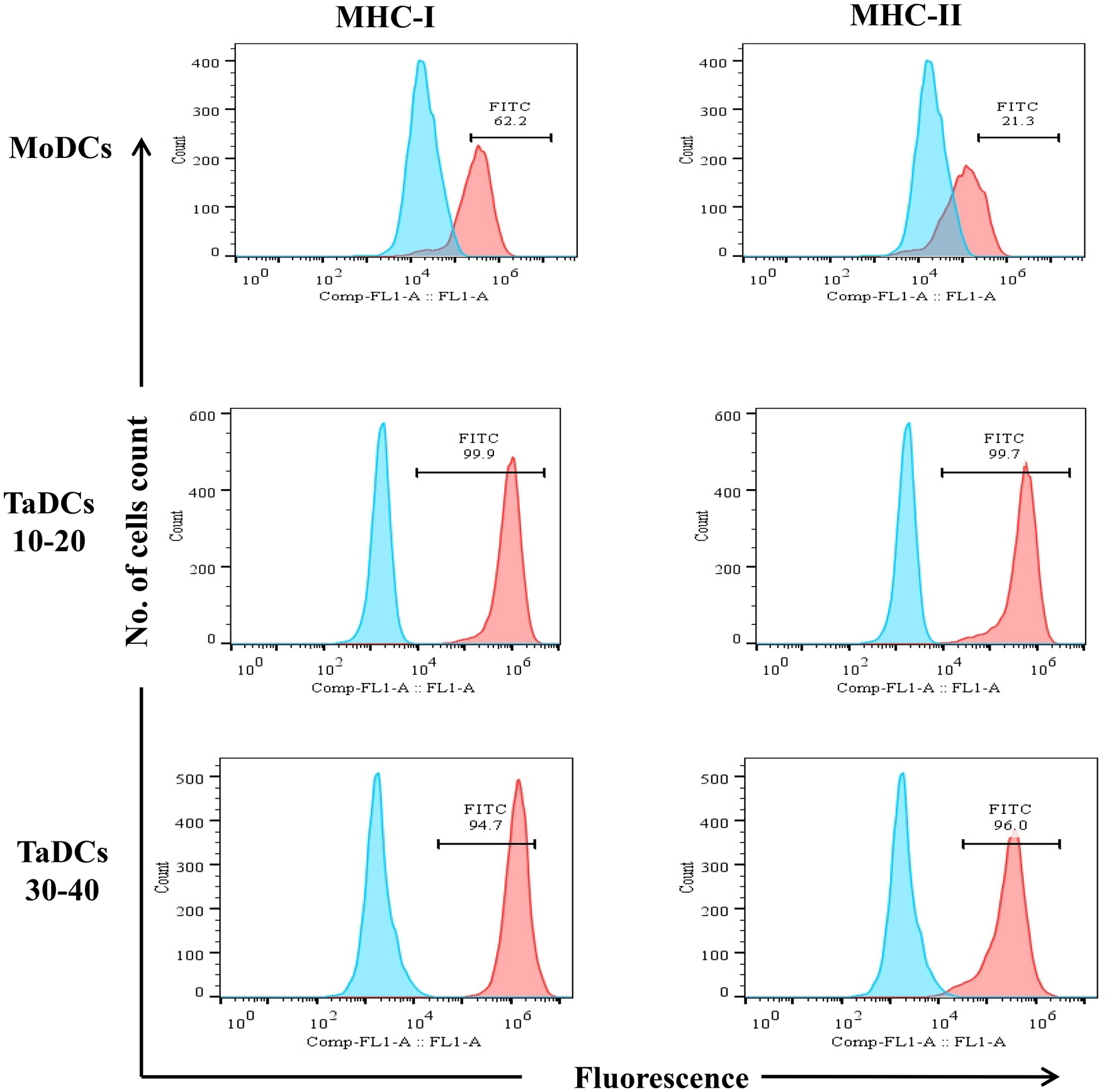
The isolated cells had >95% purity, whereas the percentage of CD4+, CD8+ and CD14+ cells in PBMCs were 29.20; 31.20 and 15.10%, respectively (Fig. 1). MHC-I (99.9 and 94.7%) and MHC-II (99.7 and 96%) were expressed by TaDCs between 10–20 and 30–40 passages, respectively. On the other hand, MHC-I and MHC-II were 62.20 and 21.30% in MoDCs, respectively (Fig. 2).
Purity analysis of magnetically isolated cells. Background control (A), percentage of specific cells in PBMCs (B) and purity analysis (C) of isolated CD4+, CD8+ and CD14+ cells are shown.

The flow cytometry analysis of MHC class molecules expression in MoDCs, and TaDC cell line at low (10–20) and higher (30–40) passages of the cell line.

Rate of endocytosis
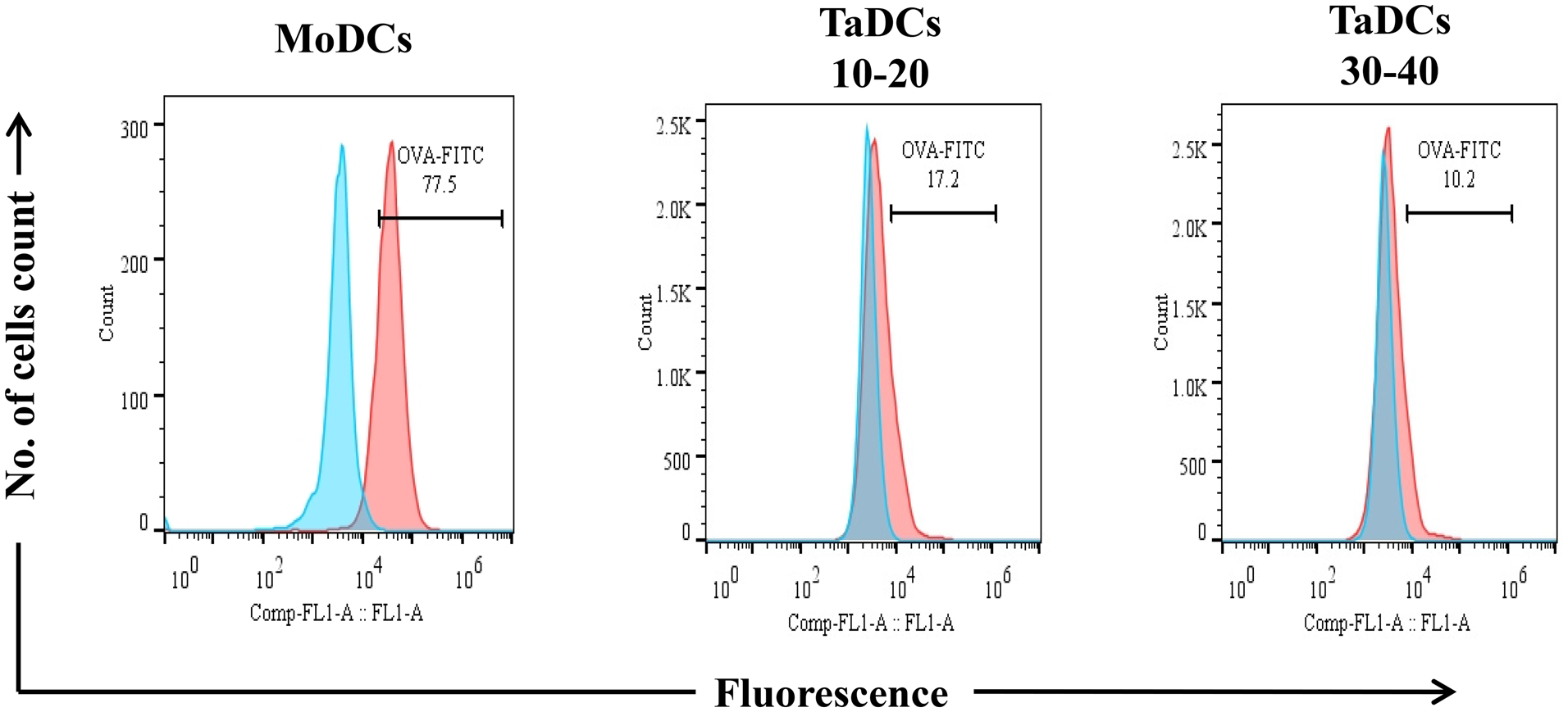
The endocytosis rate of OVA-FITC in MoDCs was 77.5%, while it was 17.2 and 10.2% in TaDCs at 10–20 and 30–40 passages, respectively. The endocytosis rate in TaDCs was significantly (P < 0.01) lower than MoDCs (Fig. 3).
The flow cytometry analysis of rate of endocytosis of OVA-FITC in MoDCs and TaDCs at 10–20 and 30–40 passages.

Rate of T lymphocyte proliferation
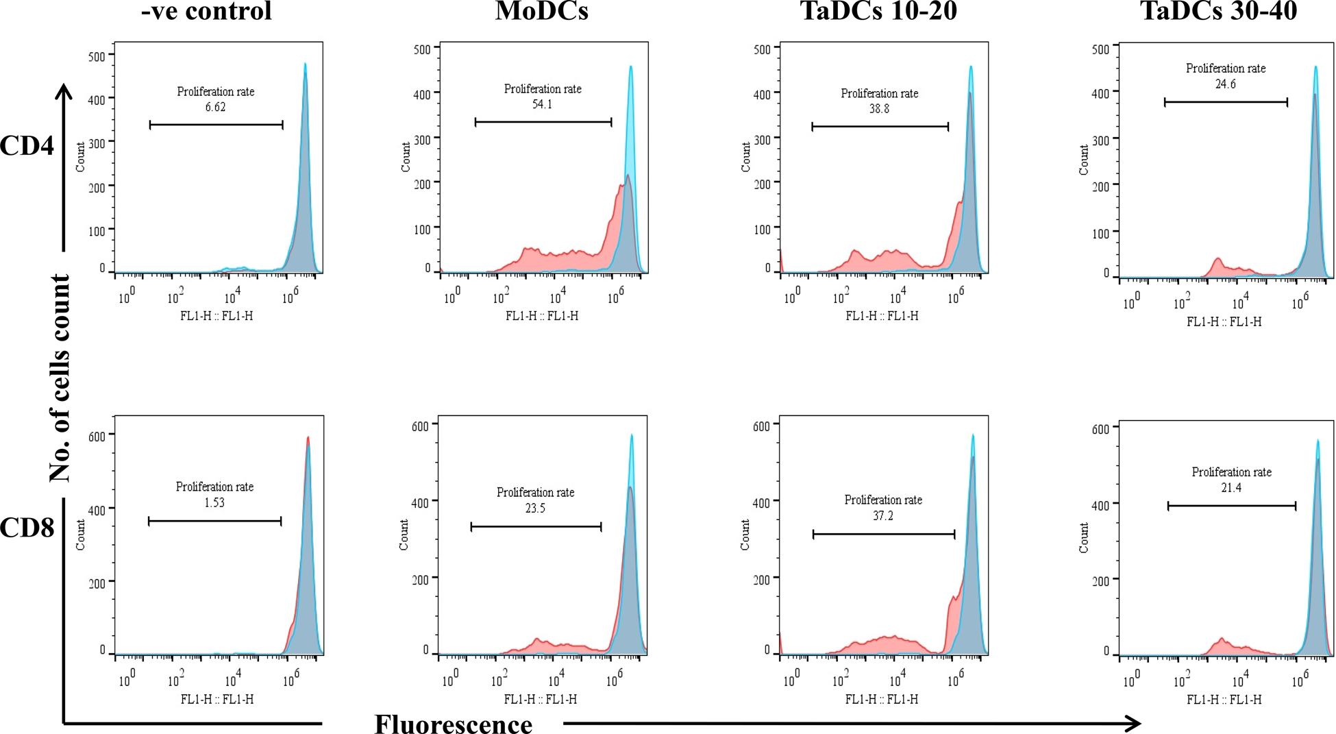
The antigen presentation assay revealed that both kinds of APCs have the ability to stimulate proliferation of purified T lymphocytes. MoDC-APC enhances the proliferation rate of CD4+ cells (54.1%) compared to CD8+ T cells (23.5%). Meanwhile, TaDC-APC also enhances the proliferation of both types of T lymphocytes, CD4 (38.8 and 24.6%) and CD8 (37.2 and 21.4%) at 10–20 and 30–40 passages, respectively. Nevertheless, the efficacy of antigen presentation as estimated by T lymphocyte proliferation declines with increases in a number of passages of the TaDC cell line. In the negative control group there was no significant proliferation of T lymphocytes (Fig. 4). There found a direct role of genes knockdown on T lymphocyte proliferation (Supporting information and Fig. S1).
The T lymphocyte proliferation upon co-culture of CFSE stained CD4+ and CD8+ cells with MoDC-APC and TaDC-APC (10–20 and 30–40 passages) in 6-well culture plate incubated for 6–7 days followed by flow cytometry analysis. For fluorescence analysis by flow cytometry, each type of purified stained cells was used as a background control (blue colour) while stained cells cultured with only TaDCs were taken as a negative control.

Rho small GTPase expression in TaDC cell line
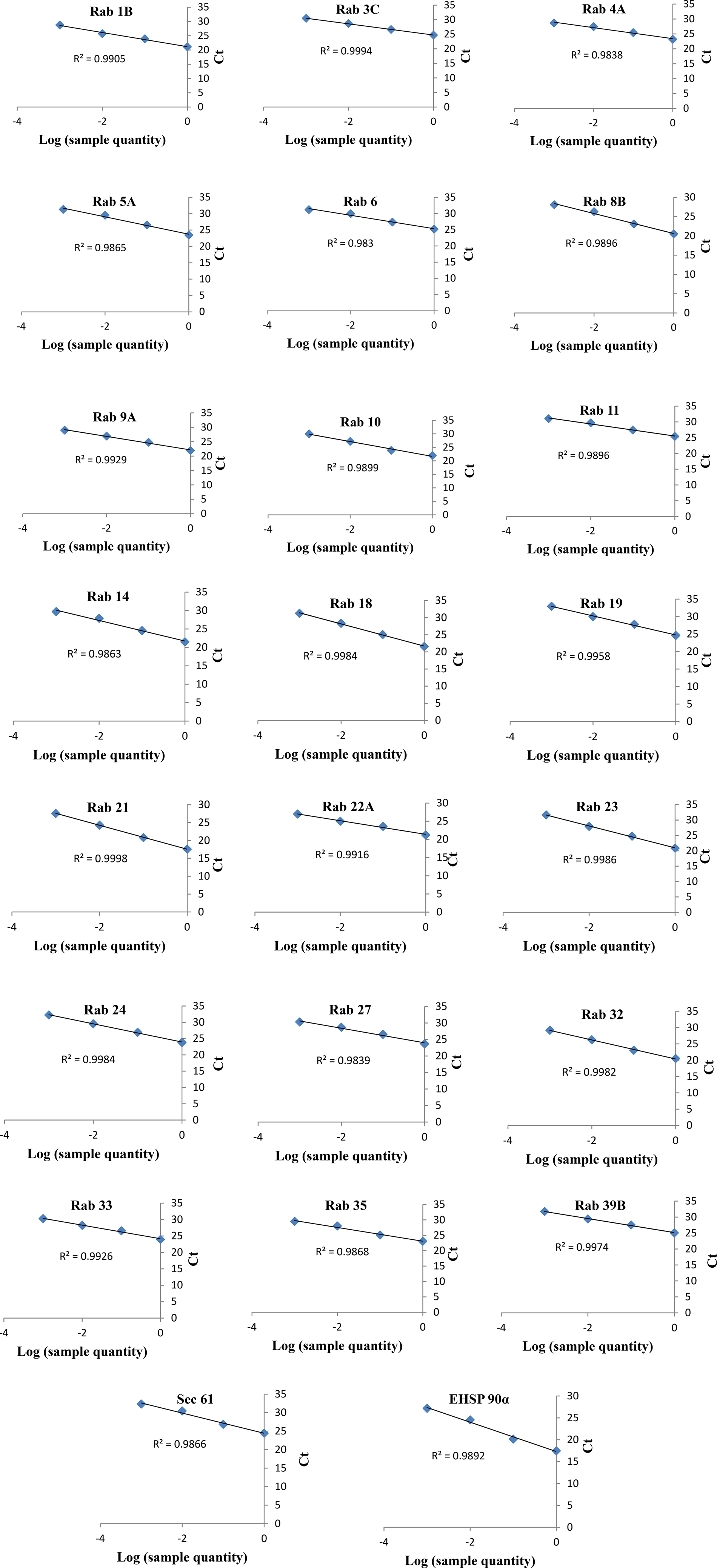
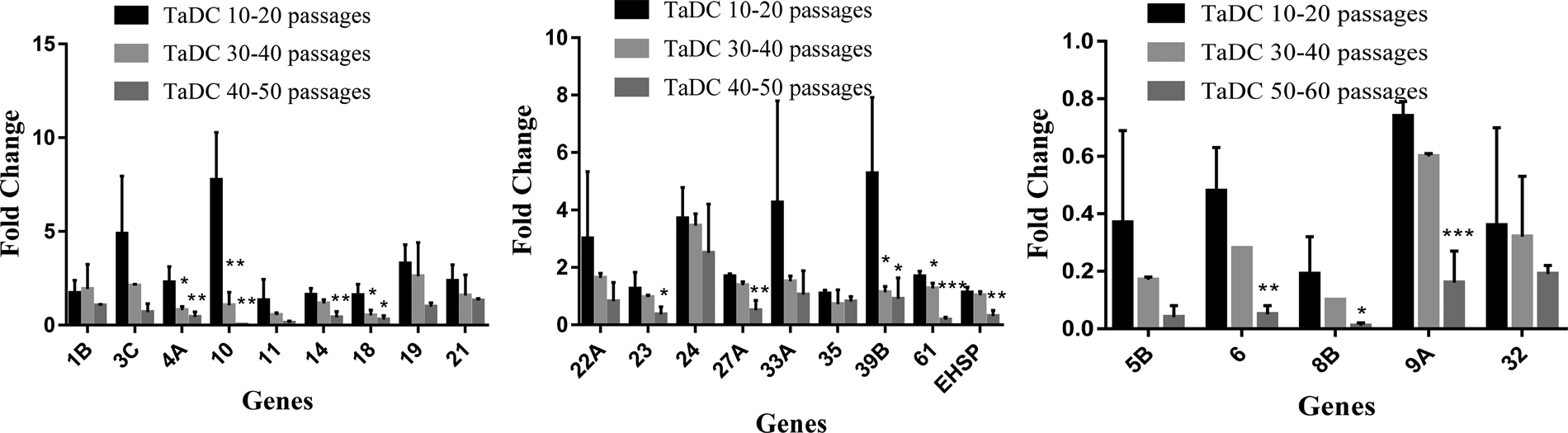
The optimized primers with correctly sequenced upon amplification and their R 2 values were used for Q-RT-PCR to evaluate the transcription levels of the abovementioned genes in TaDC cell line (Fig. 5). Q-RT-PCR was performed to estimate the level of transcription of the above mentioned 23 Rab genes. Out of these, 18 genes (1B, 3C, 4A, 10, 11, 14, 18, 19, 21, 22A, 23, 24, 27A, 33, 35, 39B, Sec61 and EHSP90α) were up-regulated, while the remaining five genes (5B, 6, 8B, 9 and 32) were down-regulated in TaDC cell line passage between 10–20 generations. Similarly, 13 genes (1B, 3C, 10, 14, 19, 21, 22A, 24, 27A, 33, 39B, Sec61 and EHSP90α) were up-regulated and 10 genes (4A, 5B, 6, 8B, 9, 11, 18, 23, 32 and 35) were down-regulated following 30–40 passages of TaDC cell line. Finally, four genes (1B, 21, 24 and 33) were up-regulated and 19 genes (3C, 4A, 5B, 6, 8B, 9A, 10, 11, 14, 18, 19, 22A, 23, 27A, 32, 35, 39B, Sec61 and EHSP90α) were down-regulated between 50 and 60 passages of TaDC cell line (Fig. 6). The up-regulated expression of these Rab genes likely contributes to antigen cross-presentation declines with the increased passage of transformed TaDCs.
The Q-RT-PCR linearity measurement of bovine cytokines in cDNA. The Q-RT-PCR was performed according to the method mentioned in quantitative PCR analysis in materials and methods section by serial dilution of 1/10 for detection of various cytokine transcripts. The Q-RT-PCR data were analysed by regression analysis between log values of samples quantity and cycles to the threshold (Ct) for each cytokine. The monitored cytokines and R 2 value of each regression line are shown.

Transcription level of small GTPase after analysis of Q-RT-PCR results. The cDNA of MoDCs was used as calibrator while β-actin was used as normalized. Transcription level was identified between 10–20, 30–40 and 50–60 passages of TaDC cell line. Significant values obtained are presented as *P ⩽ 0.05; **P < 0.01; ***P < 0.001 and ns represents non-significance (P > 0.05). The transcription level significantly (P > 0.001) decreased with increase in a number of passages.

Discussion
The current study compared the efficacy of TaDCs to that of MoDCs for antigen presentation and stimulation for the proliferation of T lymphocytes. The study confirmed that early passaged TaDCs act as APCs that stimulate the proliferation of T lymphocytes to promote an immune response against the presented antigen. Normally, endogenous antigen peptides are bound to MHC-I for presentation to CD8+ T-cells, whereas exogenous antigen peptides bind to MHC-II for presentation to CD4+ T-cells. However, a link exists between the two pathways when exogenous peptides are presented to CD8+ T-cells via MHC-I that is referred to as antigen cross-presentation. We observed that infection of DCs by T. annulata led to higher expressions of MHC class molecules on TaDCs compared to MoDCs. Previously, following activation with Ag DCs undergo numerous phenotypic changes that are responsible for their stronger stimulatory capacity like MHC class protein expression, up-regulation of co-stimulatory and adhesion molecules, and induction of cytokines and chemokine's secretions (Obst et al., Reference Obst, van Santen, Melamed, Kamphorst, Benoist and Mathis2007). The present study confirmed that endocytosis efficacy of TaDCs was significantly lower than MoDCs and this might be due to down-regulation of mannan receptors and continuous division phases by TaDCs (Mahnke et al., Reference Mahnke, Guo, Lee, Sepulveda, Swain, Nussenzweig and Steinman2000; Monrad et al., Reference Monrad, Rea, Thacker and Kaplan2008). In vitro studies revealed that immature and mature dendritic cells take up antigen by several endocytosis mechanisms, can retain antigen for 24 and 48 h, respectively, in its native form and can present processed peptides via MHC class to lymphocytes (Inaba et al., Reference Inaba, Metlay, Crowley and Steinman1990; Wykes et al., Reference Wykes, Pombo, Jenkins and MacPherson1998), or can excrete via exocytosis pathways (Wu et al., Reference Wu, Hamid, Shin and Chiang2014). Upon uptake antigen is partially degraded in phagosomes then access the lysosomes, where proteasomes degrade them into peptides (8–9 amino acids). For most protein antigens, only a few peptides are ultimately capable of binding to MHC class and presented to T lymphocytes (Jancic et al., Reference Jancic, Savina, Wasmeier, Tolmachova, El-Benna, Dang, Pascolo, Gougerot-Pocidalo, Raposo and Seabra2007). We observed that CD4+ T cell proliferation was higher than that of CD8+ T cells after co-culture with MoDC-APC. However, there was no difference in the proliferation rate of both types of T lymphocytes when they were cultured with TaDC-APC (Fig. 5). This confirms that TaDCs present processed antigen peptides in the context of both classes of highly expressed MHC molecules. T cell proliferation could be even higher if TaDCs endocytosed with greater efficiency. Previously it has been reported that T lymphocytes proliferate and rapidly expand following activation by antigen (Mannering et al., Reference Mannering, Zhong and Cheers2002; Karamitros et al., Reference Karamitros, Kotantaki, Lygerou, Kioussis and Taraviras2011). Proliferation was mainly due to activation and division of naïve and activated T lymphocytes following the formation of MHC/peptide–TCR complex (Liu and Gao, Reference Liu and Gao2008; Huppa et al., Reference Huppa, Axmann, Mörtelmaier, Lillemeier, Newell, Brameshuber, Klein, Schütz and Davis2010). Moreover, αCD40 treatment and persistent antigen exposure enhanced the cell proliferation rate (Obst et al., Reference Obst, van Santen, Melamed, Kamphorst, Benoist and Mathis2007). When transformed cells are used as APCs, the population of CD8+ instead of CD4+ cells increased (Nierkens et al., Reference Nierkens, Tel, Janssen and Adema2013). These cellular responses can be linked to the innate and adaptive immune responses depending on nature and presentation efficacies of antigen (Zhou et al., Reference Zhou, Wu, Liu, Guo, Zhu, Yao, Jiao, He, Han and Wu2016). Previously, T. annulata-transformed cells were shown to generate antigenic specificity to CD8 T-cells by presenting bovine herpes virus 1 as antigen (Hart et al., Reference Hart, MacHugh and Morrison2011). The capacity of the cells to generate antigen-specific CD8+ T-cell lines was initially validated using a recombinant canarypox virus expressing a defined immuno-dominant T. parva antigen (Tp1) (Hart et al., Reference Hart, MacHugh and Morrison2011). Furthermore, DCs capture and retain unprocessed antigen to transfer it to naïve lymphocytes or generation of immune responses (Inaba et al., Reference Inaba, Metlay, Crowley and Steinman1990; Wykes et al., Reference Wykes, Pombo, Jenkins and MacPherson1998).
The Rab family of GTPases regulate the transport of processed antigen peptides into the lumen of ER or phagosomes for loading to MHC-I (Jancic et al., Reference Jancic, Savina, Wasmeier, Tolmachova, El-Benna, Dang, Pascolo, Gougerot-Pocidalo, Raposo and Seabra2007; Zhou et al., Reference Zhou, Wu, Liu, Guo, Zhu, Yao, Jiao, He, Han and Wu2016). We observed that transcription of a number of Rab genes was up-regulated in early passaged TaDC cells, but became significantly (P > 0.001) down-regulated with multiple passages. The role of Rab proteins in antigen cross-presentation has been extensively studied (Zou et al., Reference Zou, Zhou, Zhang, Li, Liu, Chai, Li, Liu, Li and Xie2009; Cebrian et al., Reference Cebrian, Croce, Guerrero, Blanchard and Mayorga2016) and are confirmed from the current study (Supporting information). Twelve Rabs (Rab3b, Rab3c, Rab4a, Rab5b, Rab6, Rab8b, Rab10, Rab27a, Rab32, Rab33a, Rab34 and Rab35) were found to participate for antigen cross-presentation in dendritic cells (Zou et al., Reference Zou, Zhou, Zhang, Li, Liu, Chai, Li, Liu, Li and Xie2009). Rab5b, Rab8b and Rab10 are involved in phagosomes whereas, Rab3b, Rab3c, Rab6 regulate different exocytosis pathways. On the other hand, few Rabs (Rab4a, Rab27a, Rab32, Rab34 and Rab35) have both features (Zou et al., Reference Zou, Zhou, Zhang, Li, Liu, Chai, Li, Liu, Li and Xie2009). Progressive expression of Rab22A was found in tumour cells (Mayorga and Cebrian, Reference Mayorga and Cebrian2018) and in the absence of Rab27A phagosomes rapidly acidify at 6.5 pH causing acceleration of antigen degradation. Thus, delay in acidification of phagosomes due to up-regulation of Rab27A in dendritic cells (DCs) permits potentially antigenic peptides to escape degradation. In the absence of fusion between lysosome-related organelles and phagosomes, acidification happens promptly to degrade certain antigenic peptides and diminishes antigen cross-presentation efficacy (Jancic et al., Reference Jancic, Savina, Wasmeier, Tolmachova, El-Benna, Dang, Pascolo, Gougerot-Pocidalo, Raposo and Seabra2007). TaDC-APC equally promoted the proliferation of both types of T lymphocytes demonstrating that processed antigen was presented in the context of both MHC class I and II molecules.
Conclusions
This study demonstrated that MoDCs present antigen in the context of MHC-II, whereas TaDCs present antigen in the context of both MHC classes. The up-regulation of Rab gene expression in early passaged of TaDC cells suggests they are critical regulators of efficient and well-organized antigen presentation for the proliferation of both types of T lymphocytes. Thus, the current study furthers the understanding of molecular mechanisms that regulate antigen cross-presentation by T. annulata-infected transformed dendritic cells.
Further investigations are required to document the humoral immune response of TaDCs for a given antigen. Additionally, there need further investigations to find out the role of Rab genes for T lymphocyte proliferation induced by TaDC-APC upon knockdown of these genes by synthesized lentivirus-based siRNA for continuous genes silencing.
Supplementary material
The supplementary material for this article can be found at https://doi.org/10.1017/S0031182019001227.
Acknowledgement
We are thankful to Assistant Prof. Dr Haroon Akbar (Department of Parasitology, University of Veterinary and Animal Sciences, Lahore (54200), Pakistan) for revising this manuscript. We are also thankful to Mr Sahibzada Waheed Abdullah (Lanzhou Veterinary Research Institute) for guideline in gene knockdown.
Author contributions
Rashid, M., Guan, G., Liu, J. and Rashid, I. conceived and design experiment. Rashid, M. performed the experiment; Liu, J., Rashid, I., Li, Z. and Hassan, M.A. analysed the data; Wang J., Luo, J. and Yin, H. contributed reagents/materials/analysis tools. Rashid, M. and Mukhtar, U. wrote this paper. Liu, J. revised and did proofreading of this manuscript. All authors read and approved the final version of the manuscript for submission.
Financial support
This study was financially supported by the 973 Program (No. 2015CB150300); NSFC (No. 31402189); ASTIP (CAAS-ASTIP-2016-LVRI); NBCIS (CARS-37); Jiangsu Co-innovation Center programme for Prevention and Control of Important Animal Infectious Disease and Zoonosis, China.
Conflict of interest
The authors declare that they have no competing of interest.
Ethical standards
Samples were collected according to instruction and guideline of Animal Ethics Committee (Permit No. LVRIAEC-2018-001), approved by People's Republic of China.