




No CrossRef data available.
Article contents
Plasmodium chabaudi chabaudi chronic malaria and pathologies of the urogenital tract in male and female BALB/c mice
Published online by Cambridge University Press: 01 March 2004
Abstract
BALB/c mice of both sexes were infected with a non-virulent strain of Plasmodium chabaudi chabaudi and any pathologies occurring in the urogenital tract and its accessory glands were investigated. Organs and tracts were removed from infected and control mice at 15, 40 and 100 days post-injection, weighed and processed for macroscopical and histological analyses. The relative weights of preputial, clitoral glands and testes were modified in infected mice during the 40 days following infection. The preputial glands show a marked hypotrophy at 15 days post-infection. The bladders of half of the infected female mice and a few infected male mice displayed a conspicuous haemoglobinuria and frequent interstitial cystitis that worsened throughout the experiment. Also, several chronic inflammatory reactions were detected in the prostates, preputial and clitoral glands up to 100 days post-infection. A probable cause of such a divergence in the characteristics of the infection and in the nature of the pathologies identified in infected male and female mice is the interaction between the infection and the immune and endocrine systems of the host. The cause of the pathologies and their consequences on the host condition are discussed.
Information
- Type
- Research Article
- Information
- Copyright
- 2004 Cambridge University Press
INTRODUCTION
In rodents, both chronic and virulent erythrocytic malaria infections induce short-term, reversible, severe anatomical and physiological pathologies such as fever, pyrexia, anaemia, hepatomegaly and splenomegaly and also long-lasting or irreversible histopathological lesions in the liver, spleen, brain, heart and kidneys (Van Zon, Eling & Jerusalem, 1978; Cox, 1988; Andrade et al. 1991; Vuong et al. 1999). The pathologies are more or less specific to a host–parasite association and their relationship to the pathologies of human malaria is not evident (Landau & Gautret, 1998). However, it is noteworthy that whereas pyrexia, fever and anaemia intensities are more important in virulent infections, severe pathologies are found equally in virulent and chronic infections (Vuong et al. 1999).
In a virulent malaria–BALB/c mouse experimental model, the erythrocytic infection of Plasmodium vinckei petteri triggers consistent physiological and anatomical modifications in the genital tracts of female (Barthélémy et al. 2003) and male mice (unpublished observations). These pathologies were found to be reversible and led the authors to suggest, in agreement with a previous study on a virulent Plasmodium berghei infection (Akingbade et al. 1990), that an interaction between the parasite proliferation and the endocrine system of the host may allow for the alterations observed. Yet, considering the severity of the disease, such pathologies of the genital tract that cannot be implicated in the survival of the host may be of minor importance in the condition of the animal.
Surprisingly, data on the pathology of the genital tract during a chronic malarial infection are completely lacking in rodents and in humans. In lizards, malaria is always chronic, and numerous anatomical and physiological perturbations of the host are reported in both sexes (Schall, Bennett & Putnam, 1982; Schall, 1983; Schall & Dearing, 1987). Many other chronic parasitic diseases involving protozoa, such as trypanosomiasis and toxoplasmosis (Lamano Carvalho et al. 1992; Stahl, Kaneda & Noguchi, 1994; Stahl et al. 1995), have been associated with sexual dysfunctions of the rodent host. In all these host–parasite associations, the pathologies of the genital tract appear all the more important as, although the life of the host is not endangered, its reproductive success may be reduced.
The present study aimed to investigate the pathological effects of an infection with a non-virulent strain of Plasmodium chabaudi chabaudi on the urogenital tracts and accessory sex glands of male and female BALB/c mice. The analysis of the evolution of the occurrence and importance of the pathologies for up to 100 days following the infection is emphasized. The interplay of the immune and endocrine systems as a cause of the pathologies and their consequences on the host condition are discussed.
MATERIALS AND METHODS
Animals
Eight-week-old BALB/c male and female mice weighing 15–20 g were used in this study. The animals were provided free of specific pathogens by the Janvier breeding company (le Genest-Saint-Isle, France). Mice were maintained in the laboratory under constant conditions at 23±1 °C, 35–40% relative humidity and a 12:12 h artificial light-dark cycle.
Parasite strain
The subspecies Plasmodium chabaudi chabaudi Landau 1965 (864VD strain) was used. A frozen blood sample kept at −80 °C was thawed and then injected into BALB/c female mice before experimentation in order to prepare fresh inocula. A total of 35 male and 31 female mice were infected by intraperitoneal injection of 0·1 ml of a 10% heparinized fresh blood-saline buffer solution containing 30000 parasitized RBCs. For comparison, 30 male and 30 female mice injected with 0·1 ml of heparinized fresh blood from healthy mice were used as controls.
Follow-up of parasitaemia
In order to enumerate the parasitaemia, blood smears were taken using tail-blood, fixed in methanol and stained with Giemsa's stain. The parasitaemia was determined by counting the total number of parasitized RBCs in 2000 RBCs. Parasitaemias were monitored 3 times a week (on Mondays, Wednesdays, Fridays) throughout the experimentation.
Necropsy schedule and relative organ weights
Animals were killed by CO2 inhalation. Ten infected male mice, 8 infected female, 10 control male and 10 control female mice were killed at the end of the recovery phase following the peak parasitaemia (at 15 days post-infection (p.i.)), and at the time of the first recrudescence (at 40 days p.i.). Additionally, 15 infected and 15 control mice were killed at 100 days p.i. when they were supposed to have coped with the infection. Animals were weighed and autopsied: the gonads, genital tracts with the accessory sex glands (prostate, seminal vesicle, bulbourethral glands, preputial glands, clitoral glands) and the bladders with a small portion of the urethra were removed. After a minimum of 1 week in the fixative, the clitoral glands, preputial glands, gonads, epididimys and the uteri were carefully trimmed of extraneous tissue, placed on paper towelling to drain, then weighed on a Precisa model 410AM-FR analytic balance (with a sensivity of 0·001 mg). Relative organ weights were calculated in mg/100 g body weight. The spleens, livers, kidneys and the bone marrow extracted from the femurs of mice that showed gross haemoglobinuria at 100 days p.i. were removed, fixed in 10% formaldehyde and analysed in an attempt to estimate the overall health status of the animals.
Histology
Tissues were processed routinely and once dehydrated transferred to butanol before being oriented and embedded in paraffin. Three serial 7 μm thick histological transversal sections were cut from 3 different parts of each organ and stained with Haemalun and Eosin (H.E.) and analysed by 2 independent observers. Transversal sections of the spleen were stained by H.E. or by the Perls staining method. For preputial and clitoral glands, the size of the gland section and the proportion of the surface occupied by the whole acinous tissue were measured on 3 different sections by a complete morphometric study as previously described (Barthélémy et al. 2003). Necroses and/or inflammatory areas identified were also measured on 3 different sections.
Statistical analyses
For each set, Mann–Whitney U-tests and Fischer Exact tests were used to compare respectively data and frequencies between controls and surviving infected animals. Kruskall–Wallis tests were used to estimate any variations related to the time of autopsy within the groups of control mice and within the groups of infected mice. Spearman's rank correlation coefficient signification tests were used to establish any correlations between the parasitaemia (the value reached during the peak or the value of the strongest recrudescence), and morphological or histological modifications. P values [les ]0·05 were considered as significant. All tests were performed using STATVIEW v. 4.5 (SAS Institute: Cary, NC) statistic software package.
RESULTS
Parasitaemia
The parasitaemia of P. c. chabaudi-infected female and male mice increased quickly and steadily to a mean value of 19·3±8% and 18·6±8·5% of infected RBCs (iRBCs) respectively. Values reached at the time of the peak were not significantly different (U-test, P=0·19). One female out of the 65 animals studied did not survive the peak of parasitaemia. Recrudescences were observed after 30 days p.i. but never beyond 45 days p.i. (Fig. 1) and were significantly more frequent in females than in males: 16 out of the 22 infected female mice examined and 9 out of the 25 infected male mice examined showed recrudescences (Fischer exact test, P=0·01). Recrudescences lasted for 5–7 days in infected animals of both sexes but the maximal values of parasitaemia averaging 0·07±0·04% of iRBCs in female and 0·03±0·03% of iRBCs in male mice were found to be significantly different (U-test, P=0·016).

Fig. 1. Parasitaemia, body and relative organ weight changes in male and female mice infected with Plasmodium chabaudi chabaudi. Parasitaemia (males ([bull ]); females (○)) and body weights (males ([squf ]); females ([bull ]); infected (----); control (——)) are reported as means±S.D. A horizontal bar represents the phase of the infection where recrudescences were observed. Relative organ weights (mg/100 g body weight) (infected (----); control (——)) are reported as mean±S.E.M. The numbers of female mice (control, infected) examined at 15, 40 and 100 days p.i. were (10, 8), (10, 8) and (10, 14) respectively. The numbers of male mice (control, infected) examined at 15, 40 and 100 days p.i. were (10, 10), (10, 10) and (10, 15) respectively. *Significantly different from control value (P<0·05).
Body weight changes
The body weight of both controls and infected animals increased steadily throughout the experimentation period (Kruskall–Wallis, P<0·001). At 15 days p.i., the body weight of infected females was on the average 10% higher than that of controls (U-test, P=0·01), whereas the body weight of infected males was more than 6% lower than that of controls (U-test, P=0·01). At 40 and 100 days p.i., no significant difference was noticed in the body weight between infected animals and controls. However, the body weight of infected males at 40 days p.i. was negatively correlated to the maximum value of parasitaemia (r=−0·72; P=0·0017).
Organ weight changes
In females, the relative organ weight of the clitoral glands of infected females was significantly higher than that of the matched controls at 15 days p.i. (U-test, P=0·017) and 40 days p.i. (U-test, P=0·025), but was similar at 100 days p.i. The weight of the ovaries of both infected (Kruskall–Wallis, P<0·01) and control females (Kruskall–Wallis, P<0·001) increased throughout the experiment. Yet, no significant differences due to parasitism were recorded in the relative organ weight of the ovaries and uteri at 15, 40 and 100 days p.i.
In infected males, the relative organ weight of the preputial glands decreased at 40 days p.i. (Kruskall–Wallis, P=0·045) to a value 35% lower than that of the matched controls (U-test, P=0·011). No consistent variation was noted in the weight of the preputial glands of controls throughout the experimentation period. At 40 days p.i., the relative organ weight of the testes from infected males was more than 10% lower than that of matched controls (U-test, P=0·05). This decrease was still noted at 100 days p.i. but failed to reach significance (U-test, P=0·08). No significant differences due to parasitism were reported in the relative organ weight of the epididimys at 15, 40 and 100 days p.i.
Macroscopical and microscopical observations of the female urogenital tract
No consistent differences attributable to the parasitic infection were noted in the morphology and in the morpho-anatomy of the clitoral glands, ovaries, oviducts and uteri during the course of the experiments. Ovaries of both control and infected mice revealed follicles in all stages of development as well as numerous corpora lutea at 15, 40 and 100 days p.i.
In clitoral glands, the proportions of the area occupied by the glandular tissues relative to the entire sections were similar in control and infected mice throughout the infection. At high magnification, histological transverse sections of clitoral glands from infected mice showed regular functional acini at 15, 40 and 100 days p.i. and also a few lymphoid infiltrations into the glandular tissue from half of the animals at 100 days p.i. These infiltrations, mainly involving lymphocytes, were not associated with capillary thrombosis and were never accompanied by necrosis or fibrosis.
The bladders of half of the infected females presented a gross haemoglobinuria with casts at 15, 40 and 100 days p.i. At high magnification, histological transverse sections of bladders showed that haemoglobinuria was associated with urothelium thickening and frequently with interstitial cystitis with lymphoid hyperplasia (Fig. 2). Cystitis was also found in 4 infected females not presenting haemoglobinuria throughout the infection. Lymphoid infiltrations were mainly subacute (involving polynuclear cells) at 15 and 40 days p.i., and became mainly chronic (involving lymphocytes) and consistently larger (Kruskall–Wallis test, P=0·055) at 100 days p.i. (Fig. 2). In any case, no bacterial or fungal infection was observed and therefore was not suspected to be the cause of cystitis.

Fig. 2. Transverse sections of a bladder of a BALB/c female mouse infected with Plasmodium chabaudi chabaudi and autopsied at 100 days p.i. H.E. stained. At low magnification (A), the section shows a haemoglobinuria (H) with casts (Ca) and several lymphoid infiltrations (If) in the merge of the lamina propria (Lp) and in the muscularis (M). At high magnification (B, C), note in the muscularis the numerous vasodilatations (Vs) accompanying a lymphoid infiltration involving mainly lymphocytes (Lc) and a few polynuclear cells (Pc). Lymphoid infiltrations detected in the bladders of female mice autopsied at 15 and 40 days p.i. are smaller and involve mainly polynuclear cells. The acute, subacute and chronic inflammation occurring in the bladders of infected male at 15, 40 and 100 days p.i are smaller than those described in infected female mice at the same dates.
Macroscopical and microscopical observations of the male urogenital tract
No consistent differences attributable to the parasitic infection were noted in the morphology and in the morpho-anatomy of testes, epididymis, vas deferens, seminal vesicles, prostate and bulbourethral glands. Histological transverse sections of testes, epididimys and vas deferens of infected males showed a regular spermatogenesis and the presence of sperm in different parts of the tract throughout the infection.
At high magnification, histological transverse sections of the prostate from 5 and 3 infected males autopsied at 40 and 100 days p.i., respectively, showed several lymphoid infiltrations in the prostatic glands surrounded by vasodilatations. This chronic prostatitis, mainly involving lymphocytes, was not associated with either capillary thrombosis or tissue necrosis (Fig. 3). Interestingly, prostatitis was detected only in infected males that did not show any recrudescence.

Fig. 3. Transverse sections of a prostate of a BALB/c male mouse infected with Plasmodium chabaudi chabaudi and autopsied at 100 days p.i. H.E. stained. At low magnification (A), note the inflammation (If) of one of the prostatic glands (P) in the proximity of a blood vessel (Bv). At high magnification (B), this lymphoid infiltration appears to be mainly comprised of lymphocytes (Lc). The chronic inflammation detected in the prostates of infected male mice at 15 and 40 days p.i. are similar to that described in male mice at 100 days p.i.
Preputial glands of infected males displayed partially or entirely a transparent texture at 15 days p.i. This morphology of preputial glands was still observed in half of the infected males at 40 days p.i. and also in 5 infected and 3 control mice at 100 days p.i. At low magnification, histological transverse sections of preputial glands from infected mice autopsied at 15 days p.i. revealed a marked hypotrophy of the whole gland, i.e. a significant decrease in the glandular tissue accompanied by a thinning of the luminal epithelium (Fig. 4). The proportion of the area occupied by the glandular tissue relative to the entire section averaged 0·36 in controls, and 0·22 in infected males (U-test, P=0·021). At high magnification, it emerged that the global hypotrophy of the clitoral glands was supported by a global disorganization of the acinar cells. The accumulating fatty secretions in the acinar cell layer and the mature hypertrophic acinar cell layer were markedly reduced and the few remaining growing acinar cells were small and flattened (Fig. 4). In addition, several lymphoid infiltrations into the glandular tissue accompanied by vasodilatations were clearly identified in sections of preputial glands from infected males at 15, 40 and 100 days p.i. These infiltrations, mainly acute and subacute at 15 and 40 days p.i. and mainly chronic at 100 days p.i., were not associated with capillary thrombosis and were never accompanied by necrosis or fibrosis (Fig. 4).

Fig. 4. Transverse sections of the preputial glands of mice infected with Plasmodium chabaudi chabaudi at low and high magnification. H.E. stained. The preputial glands of control male mice (A, C) and infected male mice (B, D, E) autopsied at 15 and 40 days p.i. are examined. The acinous tissue (At) produced, via a holocrine process, a fatty secretion that filled the luminal ducts (Ld), although these products disappeared after the embedding process. The lymphoid infiltrations (If) detected at high magnification represent chronic (D) and subacute (E) inflammation, as lymphocytes (Lc) or polynuclear cells (Pc) were mainly present respectively. The lymphocyte infiltrations shown in preputial glands at 15 and 40 days p.i. are similar to those observed at 100 days p.i. and to those observed in clitoral glands of infected female mice at 15, 40 and 100 days p.i.
The bladders of 2, 1 and 5 infected males autopsied, respectively, at 15, 40 and 100 days p.i. presented gross haemoglobinuria with casts similar to that observed in infected females. However, in those cases, haemoglobinuria was associated merely with small lymphoid infiltrations and, thus, far less chronic cystitis was reported in bladders of infected male than in bladders of infected females (Fischer exact test, P=0·001). Also, it is noteworthy that the occurrence of cystitis in infected male mice was not related to the occurrence of prostatitis.
Bone marrow, spleen, liver and kidneys
The bone marrow of infected male and female mice showing gross haemoglobinuria at 100 days p.i. was found to be reactive (Fig. 5). Histological studies unveiled numerous histiocytes laden with haemozoin pigment that appeared dark blue when stained by the Perls staining method. The histiocytes were found to be frequently distributed in the red pulp of the spleen alongside sinusoids. A similar disposition was noted in the liver. No differences were detected in the macroscopical and microscopical morphology of the kidneys between infected and control mice of both sexes autopsied at 100 days p.i.

Fig. 5. High magnification of transverse sections of the bone marrow and the spleens of female mice infected with Plasmodium chabaudi chabaudi at 100 days p.i. H.E. stained (A–D) and Perls stained (E). Controls (A, C); infected (B, D, E). Note the density of the reactive bone marrow of the infected mouse displaying numerous megakaryocytes (M) and scarce adipocytes (Ad). Note the white pulp (Wp) and the red pulp (Rp) constituting the spleens, and the numerous dark staining haemozoin-laden histiocytes (H) distributed in the red pulp alongside the sinusoids (S) in the spleen of the infected mouse.
DISCUSSION
The present study shows that a chronic malarial infection triggers numerous morpho-anatomical perturbations of the urogenital tracts in inbred BALB/c mice of both sexes. Surprisingly, whereas pathologies occurring in the systemic organs are similar in infected male and female mice (Cox, 1988; Landau & Gautret, 1998; Vuong et al. 1999), the nature and the duration of the pathologies described in the urogenital tract appear to be markedly different depending on the sex of the infected animal examined.
The characteristics of the preputial gland transitory hypotrophy described in infected males autopsied at 15 days p.i. closely resembles the characteristics of the hypotrophy of clitoral glands described previously in an experimental model of acute P. v. petteri malarial infection (Barthélémy et al. 2003). The authors, in accordance with another study (Groot & Den Hartog, 1990), suggested that the global hypotrophy is the residual effect of an elevated activity occurring at the time of the peak of parasitaemia. Hence, the decrease in preputial gland weight of infected male mice at 40 days p.i. may reveal the slow restitution of the proliferative acinous tissue consecutive to the elimination of secretion. However, the development of preputial glands is known to be closely related to steroid signals (Huggins, Parsons & Jensen, 1955; Rudali, Roudier & Vives, 1974) and the hypotrophy observed in this study may be due to an impairment of steroid secretions as well. The decrease of testes weight observed at 40 days p.i. stresses the hypothesis of a reduction of androgen secretion due to Plasmodium proliferation in male mice. Indeed, a decrease of testis weight was previously described in association with a reduced plasma testosterone in Western fence lizards infected with P. mexicanum (Schall, 1983) and also in many other parasitic diseases of rodents (Ferreira & Rossi, 1973; Deleon, Zelinski-Wooten & Barkley, 1990; Hublart et al. 1990; Tavares et al. 1994; Larralde et al. 1995; Morales-Montor et al. 1999). Moreover, as androgens display immunosuppressive properties that greatly influence the malaria infection (Wunderlich et al. 1988; Benten et al. 1993, 1997; Zhang et al. 2000), a male mouse host may benefit from such a hormonal imbalance.
More surprising is the fact that the weight of clitoral glands of infected females is consistently higher than the weight of clitoral glands of controls at 15 and 40 days p.i. As clitoral glands (as well as preputial glands) are highly receptive to hormonal signals (Ozegovic & Milkovic, 1972; Mesquita-Guimaraes & Coimbra, 1981; Groot & Den Hartog, 1990), a possible explanation is that the hormonal imbalance associated with a virulent malarial infection in females (Akingbade et al. 1990) is not as important as or even different from a non-virulent malarial infection. In fact, a slight increase of steroid production might be sufficient to trigger a little enlargement of the clitoral glands but not enough to induce hyperactivity. Nevertheless, the occurrence of lymphoid infiltrations at day 100 p.i. suggests that clitoral glands of female mice infected by chronic malaria must have undergone several dysfunctions.
Obviously, the pathologies of the genital tract detected in malaria-infected mice in the present study do not interfere directly with the vital functions and the survival of the animal. Yet, considering the major role of preputial and clitoral glands in sex recognition (Bronson, 1971; Bronson & Marsden, 1973; Hayashi & Kimura, 1974; Lucas, Donohoe & Thody, 1982) and the paramount endocrine and germinal functions of testes, it can be suspected that these pathologies may greatly hinder the behaviour and the reproductive success of the animals. In lizards for instance, although the decrease of testes weight is not linked to a complete impairment of spermatogenesis, the consequences of the infection on social and sexual behaviour and thus on the reproductive success of infected males are severe (Schall & Dearing, 1987; Schall, 1990). Several studies based on social and sexual behaviours, mate choice tests and on reproductive capacity appear to be of particular interest in casting light on this situation.
Cystitis and prostatitis in rodents and humans can be caused by disordered immune responses or autoimmune responses (Mitra et al. 1999; Van De Merwe & Arendsen, 2000; Batstone, Doble & Gaston, 2002; Karlovsky & Pontari, 2002). As the cellular and humoral immune responses associated with erythrocytic malarial infection is considered to be the cause of several complications in malaria (Fell & Smith, 1998; De Souza & Riley, 2002; Langhorne, Quin & Sanni, 2002; Malaguarnera & Musumeci, 2002), they may, in the same way, trigger cystitis and prostatitis. The fact that infected females, more susceptible to autoimmune diseases (Olsen & Kovacs, 1996), more frequently show cystitis than males strongly supports this hypothesis. Nevertheless, the statement that cystitis or prostatitis seems not to have been reported in human malaria is somewhat puzzling.
Haemoglobinuria, an indisputable cue to severe haemolysis in P. falciparum infections (Ekvall et al. 2001), is detected in half of the infected female mice and in a few infected male mice at up to 100 days p.i., in spite of the low virulence and the low mortality rate characterizing the P. c. chabaudi infection. The fact that the bone marrow is reactive in infected animals at 100 days p.i. strongly suggests that erythropoiesis is increased to counterbalance an ongoing haemolysis that can account for haemoglobinuria. Indeed, chronic haemolysis may stem from irreversible alterations of blood vessel structure caused by the infection or from long-lasting autoimmune reactions triggered by the presence of circulating malarial antigens (Cox, 1988; Menendez, Fleming & Alonso, 2000). Another explanation is the persistence of a low and undetectable parasitaemia that can account for the occurrence of numerous haemozoin-laden histiocytes in the spleen at up to 100 days p.i. (Levesque, Sullivan & Meshnick, 1999). That is, whatever the cause of chronic haemolysis may be, such chronic physiopathology must greatly impair the host health and may result in a consistent reduction of the host life-span.
To conclude, it emerges from this study that a mouse infected with a given quantity of the parasite P. c. chabaudi displays different pathologies of its urogenital tract according to whether it is male or female. Numerous studies demonstrated that male and female mice were not equal in coping with a parasitic infection (Alexander & Stimson, 1988), and a consensus was that sex hormones interact largely with the immune system which, in turn, influences the endocrine system (Grossman, 1984, 1989; Olsen & Kovacs, 1996). In the present study, the divergence in the characteristics of the infection and in the nature of the pathologies detected in the urogenital tract is consistent with this statement. The erythrocytic proliferation of the parasite triggers an immune response that may cause a sex-specific hormonal imbalance more or less effective in inducing genital alterations and immune disorders that can lead to urinary inflammation. Finally, considering the divergence of the pathologies and the characteristics of the infection described in infected male and female mice, the nature and the duration of the consequences of the infection on the health of the host deserve attention. Indeed, whereas a female mouse might suffer more and longer from the parasitic infection, an infected male mouse may rather experience important transitory alterations in its reproductive success.
This work is dedicated to the late Dr Phat Nuoc Vuong. The authors gratefully acknowledge Touria Ben Assavy for careful technical assistance, Jacqueline Gabrion for her assistance during the preliminary phase of the study and Gloria Girton for reviewing the manuscript. This work was carried out under a grant to M. Barthélémy from the French Educational Ministry of Research and Technology (MENRT). The authors have adhered to the Guidelines for the Use and Care of Animals in Research and to the European recommendations (décret no. 87-848) for the care and use of laboratory animals adopted by the French government in October 1987.
References
REFERENCES
AKINGBADE, O.,
AINA, A. O.,
FOLASHADE, B. A. &
MODUPE, M. J.
(1990).
Malaria parasitization and hormonal imbalance in virgin mice.
Journal of the Medical Association of Thailand 73,
228–233.Google Scholar
ALEXANDER, J. &
STIMSON, W. H.
(1988).
Sex hormones and the course of parasitic infection.
Parasitology Today 4,
189–193.CrossRefGoogle Scholar
ANDRADE, H. F.,
CORBETT, C. E. P.,
LAURENT, M. D. &
DUARTE, M. I. S.
(1991).
Comparative and sequential histopathology of Plasmodium chabaudi-infected BALB/c mice.
Brazilian Journal of Medical and Biological Research 24,
1209–1218.Google Scholar
BARTHÉLÉMY, M.,
VUONG, P. N.,
GABRION, C. &
PETIT, G.
(2003).
Oestrus cycle perturbations and hypotrophy of clitoral glands in malaria-infected female BALB/c mice.
Parasitology Research 89,
134–140.CrossRefGoogle Scholar
BATSTONE, G. R.,
DOBLE, A. &
GASTON, J. S.
(2002).
Autoimmune T cell responses to seminal plasma in chronic pelvic pain syndrome (CPPS).
Clinical and Experimental Immunology 128,
302–307.CrossRefGoogle Scholar
BENTEN, W. P.,
WUNDERLICH, F.,
HERRMANN, R. &
KUHN-VELTEN, W. N.
(1993).
Testosterone-induced compared with oestradiol-induced immunosuppression against Plasmodium chabaudi malaria.
Journal of Endocrinology 139,
487–494.CrossRefGoogle Scholar
BENTEN, W. P.,
ULRICH, P.,
KUHN-VELTEN, W. N.,
VOHR, H. W. &
WUNDERLICH, F.
(1997).
Testosterone-induced susceptibility to Plasmodium chabaudi malaria: persistence after withdrawal of testosterone.
Journal of Endocrinology 153,
275–281.CrossRefGoogle Scholar
BRONSON, F. H. &
MARSDEN, H. M.
(1973).
The preputial gland as an indicator of social dominance in male mice.
Behavioural Biology 9,
625–628.CrossRefGoogle Scholar
COX, F. E. G.
(1988).
Major animal models in malaria research: rodent. In
Malaria: Principles and Practices of Malariology
(ed. Wersdorfer, W. H. &
Macgregor, I.),
pp. 1503–1543.
Churchill Livingston,
Edinburgh, UK.
DE SOUZA, J. B. &
RILEY, E. M.
(2002).
Cerebral malaria: the contribution of studies in animal models to our understanding of immunopathogenesis.
Microbes and Infection 4,
291–300.CrossRefGoogle Scholar
DELEON, D. D.,
ZELINSKI-WOOTEN, M. B. &
BARKLEY, M. S.
(1990).
Hormonal basis of variation in oestrous cyclicity in selected strains of mice.
Journal of Reproduction and Fertility 89,
117–126.CrossRefGoogle Scholar
EKVALL, H.,
ARESE, P.,
TURRINI, F.,
AYI, K.,
MANNU, F.,
PREMJI, Z. &
BJORKMAN, A.
(2001).
Acute haemolysis in childhood falciparum malaria.
Transactions of the Royal Society of Tropical Medicine and Hygiene 95,
611–617.CrossRefGoogle Scholar
FELL, A. H. &
SMITH, N. C.
(1998).
Immunity to asexual blood stages of Plasmodium: is resistance to acute malaria adaptative or innate?
Parasitology Today 14,
364–368.Google Scholar
FERREIRA, A. L. &
ROSSI, M. A.
(1973).
Pathology of the testis and epididymis in the late phase of experimental Chagas's disease.
American Journal of Tropical Medicine and Hygiene 22,
699–704.CrossRefGoogle Scholar
GROOT, M. J. &
DEN HARTOG, J. M.
(1990).
Histological changes in the genital tract of female veal calves implanted with naturally occurring anabolic steroids.
Zentralblatt für Veterinärmedizin, A 37,
775–786.CrossRefGoogle Scholar
GROSSMAN, C. J.
(1984).
Regulation of the immune system by sex steroids.
Endocrine Reviews 5,
435–455.CrossRefGoogle Scholar
GROSSMAN, C. J.
(1989).
Possible underlying mechanisms of sexual dimorphism in the immune response, fact and hypothesis.
Journal of Steroid Biochemistry 34,
241–251.CrossRefGoogle Scholar
HAYASHI, S. &
KIMURA, T.
(1974).
Sex-attractant emitted by female mice.
Physiology and Behavior 13,
563–567.CrossRefGoogle Scholar
HUBLART, M.,
TETAERT, D.,
CROIX, D.,
BOUTIGNON, F.,
DEGAND, P. &
BOERSMA, A.
(1990).
Gonadotropic dysfunction produced by Trypanosoma brucei brucei in the rat.
Acta Tropica 47,
177–184.CrossRefGoogle Scholar
HUGGINS, C.,
PARSONS, F. M. &
JENSEN, E. V.
(1955).
Promotion of growth of preputial glands by steroids and the pituitary growth hormone.
Endocrinology 57,
25–32.CrossRefGoogle Scholar
KARLOVSKY, M. E. &
PONTARI, M. A.
(2002).
Theories of prostatitis etiology.
Current Urology Reports 3,
307–312.CrossRefGoogle Scholar
LAMANO CARVALHO, T. L.,
CARRARO, A. A.,
LOPES, R. A. &
RIBEIRO, R. D.
(1992).
The male reproductive organs in experimental Chagas' disease. II. Morphometric study of the vas deferens in the chronic phase of the disease.
Experimental and Toxicologic Pathology 44,
147–149.CrossRefGoogle Scholar
LANDAU, I. &
GAUTRET, P.
(1998).
Animal models: rodents. In
Malaria: Parasite Biology, Pathogenesis, and Protection (ed. Sherman, I. W.),
pp. 401–417.
ASM Press,
Washington, D.C.
LANGHORNE, J.,
QUIN, S. J. &
SANNI, L. A.
(2002).
Mouse models of blood-stage malaria infections: immune responses and cytokines involved in protection and pathology.
Chemical Immunology 80,
204–228.CrossRefGoogle Scholar
LARRALDE, C.,
MORALES, J.,
TERRAZAS, I.,
GOVEZENSKY, T. &
ROMANO, M. C.
(1995).
Sex hormone changes induced by the parasite lead to feminization of the male host in murine Taenia crassiceps cysticercosis.
Journal of Steroid Biochemistry and Molecular Biology 52,
575–580.CrossRefGoogle Scholar
LEVESQUE, M. A.,
SULLIVAN, A. D. &
MESHNICK, S. R.
(1999).
Splenic and hepatic hemozoin in mice after malaria parasite clearance.
Journal of Parasitology 85,
570–573.CrossRefGoogle Scholar
LUCAS, P. D.,
DONOHOE, S. M. &
THODY, A. J.
(1982).
The role of estrogen and progesterone in the control of preputial gland sex attractant odors in the female rat.
Physiology and Behavior 28,
601–607.CrossRefGoogle Scholar
MALAGUARNERA, L. &
MUSUMECI, S.
(2002).
The immune response to Plasmodium falciparum malaria.
The Lancet Infectious Diseases 2,
472–478.CrossRefGoogle Scholar
MENENDEZ, C.,
FLEMING, A. F. &
ALONSO, P. L.
(2000).
Malaria-related anaemia.
Parasitology Today 16,
469–476.CrossRefGoogle Scholar
MESQUITA-GUIMARAES, J. &
COIMBRA, A.
(1981).
The effect of sexual hormones on the lipid and proteinaceous secretion of the rat preputial sebaceous gland.
Archives of Dermatological Research 270,
325–331.CrossRefGoogle Scholar
MITRA, S.,
DAGHER, A.,
KAGE, R.,
DAGHER, R. K. &
LUBER-NAROD, J.
(1999).
Experimental autoimmune cystitis: further characterization and serum autoantibodies.
Urological Research 27,
351–356.CrossRefGoogle Scholar
MORALES-MONTOR, J.,
GAMBOA-DOMINGUEZ, A.,
RODRIGUEZ-DORANTES, M. &
CERBON, M. A.
(1999).
Tissue damage in the male murine reproductive system during experimental Taenia crassiceps cysticercosis.
Journal of Parasitology 85,
887–890.CrossRefGoogle Scholar
OLSEN, N. J. &
KOVACS, W. J.
(1996).
Gonadal steroids and immunity.
Endocrine Reviews 17,
369–384.Google Scholar
OZEGOVIC, B. &
MILKOVIC, S.
(1972).
Effects of adrenocorticotrophic hormone, growth hormone, prolactin, adrenalectomy and corticoids upon the weight, protein and nucleic acid content of the female rat preputial glands.
Endocrinology 90,
903–908.CrossRefGoogle Scholar
RUDALI, G.,
ROUDIER, R. &
VIVES, C.
(1974).
The preputial gland of the male mouse.
Pathologie Biologie (Paris) 22,
895–899.Google Scholar
SCHALL, J. J.
(1983).
Lizard malaria: cost to the vertebrate host's reproductive success.
Parasitology 87,
1–6.CrossRefGoogle Scholar
SCHALL, J. J.
(1990).
Virulence of lizard malaria: the evolutionary ecology of an ancient parasite–host association.
Parasitology 100,
S35–S52.CrossRefGoogle Scholar
SCHALL, J. J.,
BENNETT, A. F. &
PUTNAM, R. W.
(1982).
Lizards infected with malaria: physiological and behavioral consequences.
Science 217,
1057–1059.CrossRefGoogle Scholar
SCHALL, J. J. &
DEARING, M. D.
(1987).
Malarial parasitism and male competition for mates in the western fence lizards, Sceloporus occidentalis.
Oecologia 73,
389–392.CrossRefGoogle Scholar
STAHL, W.,
KANEDA, Y. &
NOGUCHI, T.
(1994).
Reproductive failure in mice chronically infected with Toxoplasma gondii.
Parasitology Research 80,
22–28.CrossRefGoogle Scholar
STAHL, W.,
KANEDA, Y.,
TANABE, M. &
KUMAR, S. A.
(1995).
Uterine atrophy in chronic murine toxoplasmosis due to ovarian dysfunction.
Parasitology Research 81,
109–113.CrossRefGoogle Scholar
TAVARES, M. C.,
CARRARO, A. A.,
FAVARETTO, A. L.,
PETENUSCI, S. O.,
LOPES, R. A.,
RIBEIRO, R. D. &
CARVALHO, T. L.
(1994).
The male reproductive organs in experimental Chagas' disease. III. Plasma testosterone and accessory sex glands in the acute phase of the disease.
Experimental and Toxicologic Pathology 46,
243–246.CrossRefGoogle Scholar
VAN DE MERWE, J. P. &
ARENDSEN, H. J.
(2000).
Interstitial cystitis: a review of immunological aspects of the aetiology and pathogenesis, with a hypothesis.
British Journal of Urology International 85,
995–999.CrossRefGoogle Scholar
VAN ZON, A.,
ELING, W. &
JERUSALEM, C.
(1978).
Histo- and immunopathology of a malaria (Plasmodium berghei) infection in mice.
Israel Journal of Medical Sciences 14,
659–672.CrossRefGoogle Scholar
VUONG, P. N.,
RICHARD, F.,
SNOUNOU, G.,
COQUELIN, F.,
RENIA, L.,
GONNET, F.,
CHABAUD, A. G. &
LANDAU, I.
(1999).
Development of irreversible lesions in the brain, heart and kidney following acute and chronic murine malaria infection.
Parasitology 119,
543–553.CrossRefGoogle Scholar
WUNDERLICH, F.,
MOSSMANN, H.,
HELWIG, M. &
SCHILLINGER, G.
(1988).
Resistance to Plasmodium chabaudi in B10 mice: influence of the H-2 complex and testosterone.
Infection and Immunity 56,
2400–2406.Google Scholar
ZHANG, Z.,
CHEN, L.,
SAITO, S.,
KANAGAWA, O. &
SENDO, F.
(2000).
Possible modulation by male sex hormone of Th1/Th2 function in protection against Plasmodium chabaudi chabaudi AS infection in mice.
Experimental Parasitology 96,
121–129.CrossRefGoogle Scholar
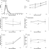
Fig. 1. Parasitaemia, body and relative organ weight changes in male and female mice infected with Plasmodium chabaudi chabaudi. Parasitaemia (males ([bull ]); females (○)) and body weights (males ([squf ]); females ([bull ]); infected (----); control (——)) are reported as means±S.D. A horizontal bar represents the phase of the infection where recrudescences were observed. Relative organ weights (mg/100 g body weight) (infected (----); control (——)) are reported as mean±S.E.M. The numbers of female mice (control, infected) examined at 15, 40 and 100 days p.i. were (10, 8), (10, 8) and (10, 14) respectively. The numbers of male mice (control, infected) examined at 15, 40 and 100 days p.i. were (10, 10), (10, 10) and (10, 15) respectively. *Significantly different from control value (P<0·05).

Fig. 2. Transverse sections of a bladder of a BALB/c female mouse infected with Plasmodium chabaudi chabaudi and autopsied at 100 days p.i. H.E. stained. At low magnification (A), the section shows a haemoglobinuria (H) with casts (Ca) and several lymphoid infiltrations (If) in the merge of the lamina propria (Lp) and in the muscularis (M). At high magnification (B, C), note in the muscularis the numerous vasodilatations (Vs) accompanying a lymphoid infiltration involving mainly lymphocytes (Lc) and a few polynuclear cells (Pc). Lymphoid infiltrations detected in the bladders of female mice autopsied at 15 and 40 days p.i. are smaller and involve mainly polynuclear cells. The acute, subacute and chronic inflammation occurring in the bladders of infected male at 15, 40 and 100 days p.i are smaller than those described in infected female mice at the same dates.

Fig. 3. Transverse sections of a prostate of a BALB/c male mouse infected with Plasmodium chabaudi chabaudi and autopsied at 100 days p.i. H.E. stained. At low magnification (A), note the inflammation (If) of one of the prostatic glands (P) in the proximity of a blood vessel (Bv). At high magnification (B), this lymphoid infiltration appears to be mainly comprised of lymphocytes (Lc). The chronic inflammation detected in the prostates of infected male mice at 15 and 40 days p.i. are similar to that described in male mice at 100 days p.i.
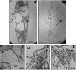
Fig. 4. Transverse sections of the preputial glands of mice infected with Plasmodium chabaudi chabaudi at low and high magnification. H.E. stained. The preputial glands of control male mice (A, C) and infected male mice (B, D, E) autopsied at 15 and 40 days p.i. are examined. The acinous tissue (At) produced, via a holocrine process, a fatty secretion that filled the luminal ducts (Ld), although these products disappeared after the embedding process. The lymphoid infiltrations (If) detected at high magnification represent chronic (D) and subacute (E) inflammation, as lymphocytes (Lc) or polynuclear cells (Pc) were mainly present respectively. The lymphocyte infiltrations shown in preputial glands at 15 and 40 days p.i. are similar to those observed at 100 days p.i. and to those observed in clitoral glands of infected female mice at 15, 40 and 100 days p.i.
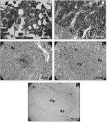
Fig. 5. High magnification of transverse sections of the bone marrow and the spleens of female mice infected with Plasmodium chabaudi chabaudi at 100 days p.i. H.E. stained (A–D) and Perls stained (E). Controls (A, C); infected (B, D, E). Note the density of the reactive bone marrow of the infected mouse displaying numerous megakaryocytes (M) and scarce adipocytes (Ad). Note the white pulp (Wp) and the red pulp (Rp) constituting the spleens, and the numerous dark staining haemozoin-laden histiocytes (H) distributed in the red pulp alongside the sinusoids (S) in the spleen of the infected mouse.
 You have
Access
- 7
- Cited by