Introduction
The Lavrion Mining District, Greece, is part of the Attic–Cycladic–Pelagonian ore belt, and includes – apart from Lavrion – significant base- and precious-metal deposits in south Evia, Sifnos, Tinos and Kythnos (Vavelidis, Reference Vavelidis and Papunen1997; Skarpelis, Reference Skarpelis2002; Tombros et al., Reference Tombros, Seymour and Spry2004; Neubauer, Reference Neubauer, Mao and Bierlein2005; Tombros et al., Reference Tombros, Seymour, Williams-Jones and Spry2007). Several styles of sulfide mineralisation, including skarns, skarn-free carbonate replacement and vein-type ores, are found in the area (Voudouris, Reference Voudouris, Mao and Bierlein2005). The Lavrion District is famous for its lead–zinc–silver ores that were mined in ancient times, most notably 600–400 B.C. and contributed to the wealth of Athens during the classical period. The district covers an area of ~150 km2 at the southern tip of the Attica peninsula. Conophagos (Reference Conophagos1980) estimated that, until the depletion and closure of the mines, 2.3 million metric tons of lead and 7.800 tons of silver were extracted from the ores.
The area is also well-known for the occurrence of (up to now) ~580 mineral species, and it is the type locality of ~20 new minerals (Rieck et al., Reference Rieck, Kolitsch, Voudouris, Giester and Tzeferis2018). The district has been the subject of intense investigation by the authors for more than two decades (Rieck, Reference Rieck1999; Rieck and Rieck, Reference Rieck and Rieck1999; Rieck and Wendel, Reference Rieck and Wendel1999; Wendel and Rieck, Reference Wendel and Rieck1999a,Reference Wendel and Rieckb; Giester et al., Reference Giester, Lengauer and Rieck2000; Giester et al., Reference Giester, Kolitsch, Leverett, Turner and Williams2007; Rieck, Reference Rieck2012; Giester and Rieck, Reference Giester and Rieck2014; Kolitsch et al., Reference Kolitsch, Rieck, Brandstätter, Schreiber, Fabritz, Blaß and Gröbner2014a,Reference Kolitsch, Rieck, Brandstätter, Schreiber, Fabritz, Blaß and Gröbnerb; Kolitsch et al., Reference Kolitsch, Rieck and Voudouris2015). Several new mineral species have been described by our research group, i.e. mereiterite, K2Fe2+(SO4)2·4H2O (Giester and Rieck, Reference Giester and Rieck1995), niedermayrite, Cu4Cd(SO4)2(OH)6·4H2O (Giester et al., Reference Giester, Rieck and Brandstätter1998), kapellasite, Cu3Zn(OH)6Cl2 (Krause et al., Reference Krause, Bernhardt, Braithwaite, Kolitsch and Pritchard2006), and a secondary zinc mineral phase with the formula Zn9(SO4)2(OH)12Cl2·6H2O (Kolitsch and Giester, Reference Kolitsch and Giester2013). In addition two minerals have been approved recently: prachařite, Ca ${\rm Sb}_2^{5 +} $(
 ${\rm As}_2^{3 +} $O3)4·10H2O, (IMA2018-081, Kolitsch et al., Reference Kolitsch, Sejkora, Topa, Kampf, Plášil, Rieck and Fabritz2018) and CaZn2(AsO4)2·4H2O, (IMA2018-051a, Rieck et al., Reference Rieck, Giester, Lengauer and Topa2019). The paragenesis of niedermayrite (at the Esperanza Mine, Kaminiza Area) was further studied in detail (Rieck, Reference Rieck2012) on freshly excavated rocks revealing colourless crusts of water-soluble minerals (Fig. 1), which previously had been eliminated on some old niedermayrite specimens due to ultrasonic cleaning in water. These colourless grains were studied by single-crystal and powder X-ray diffraction as well as electron microprobe analysis and are new minerals of chemically simple composition Cd(SO4)·H2O (voudourisite), and Cd3(SO4)3·8H2O (lazaridisite). The crystal structures of the respective isotypic synthetic sulfates have been described already by Brégeault and Herpin (Reference Brégeault and Herpin1970), Theppitak and Chainok (Reference Theppitak and Chainok2015), Lipson (Reference Lipson1936) and Caminiti and Johansson (Reference Caminiti and Johansson1981). A cadmium sulfate monohydrate was described from Radvanice, Czech Republic, by Sejkora and Kotrlý (Reference Sejkora and Kotrlý1998). The Radvanice occurrence, however, does not fulfil the criteria for acceptance as a new mineral as it was reported to be the efflorescence from a burning coal dump. Another new cadmium sulfate tetrahydrate, Cd(SO4)·4H2O (drobecite), also mentioned from the Esperanza Mine (Giester and Rieck, Reference Giester and Rieck2010), is unfortunately poorly characterised. Voudourisite and lazaridisite have since been observed in Lavrion at the #19 Mine in the Ano Sounio area and at the North Mine in the Villia area, with the finds at #19 Mine enabling a full description of the two new species.
Clear lazaridisite, white powdery voudourisite and blue–green niedermayrite from the #19 Mine, Ano Sounio, Lavrion, Field of view: 1.5 mm. Photo: H. Schillhammer. Private collection, B. Rieck, specimen no. S19108-0010.

Mineral names and type material
Voudourisite is named in honour of Professor Panagiotis Voudouris (born 1962), Department of Mineralogy and Petrology, Faculty of Geology and Geoenvironment, National and Kapodistrian University of Athens, in recognition of his groundbreaking work on the Lavrion deposits. Lazaridisite is named after Stathis Lazaridis (1953–2010), a mineral collector from Lavrion, who contributed significantly to our current understanding of the paragenetic sequences within the Lavrion deposits.
Both minerals and their names have been approved by the Commission on New Minerals, Nomenclature and Classification of the International Mineralogical Association under numbers IMA2012-042 (Rieck and Giester, Reference Rieck and Giester2012a) and IMA2012-043 (Rieck and Giester, Reference Rieck and Giester2012b), respectively. The type specimen for both minerals is deposited in the collection of the Institut für Mineralogie und Kristallographie, University of Vienna, Althanstraße 14, 1090 Wien, Austria, catalogue number HS13.077.
Occurrence
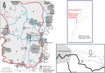
Both newly described minerals originate from the type locality of niedermayrite (Fig. 2) at the Esperanza Mine, Kaminiza Area, Lavrion Mining District, Attica, Greece. This small ancient mine is situated ~2.5 km NW of the town of Lavrion, close (<1 km) to the well-known ‘Km 3’ site along the road to Agios Konstantinos. The recent finds have been made at the #19 Mine (Ano Sounio area) and the North Mine (Villia area). At both localities, the two new minerals are associated with sphalerite, galena, niedermayrite, edwardsite, chalcanthite, gypsum and greenockite. Voudourisite and lazaridisite are secondary minerals formed as the result of the weathering of the primary cadmium sulfides hawleyite and greenockite (cf. Rieck, Reference Rieck2012) and are among the latest to form in this paragenetic sequence.
Type locality of cadmium-bearing minerals within the Esperanza Mine. Mapping by K.H. Fabritz.

Appearance, physical and optical properties
The two minerals form tiny clusters of stacked, short prismatic and rounded crystals on the sulfide minerals of the assemblage. Voudourisite also appears as powdery coatings covering areas of several square centimetres at the #19 Mine.
While neither cleavage nor preferred parting has been observed, the minerals exhibit conchoidal fracture. The Mohs hardness is ~3 and both have brittle tenacity. Densities were measured using micro-pycnometry (bromoform-filled capillary with an inner diameter of 0.5 mm and a length of 10 cm) to be 3.80(5) g/cm3 (voudourisite) and 3.10(5) g/cm3 (lazaridisite). These values compare well with the calculated densities of 3.838 and 3.089 g/cm3, respectively, based on the empirical chemical formula and the unit-cell dimensions obtained from single-crystal X-ray diffraction.
Both minerals are colourless or white, show transparent to translucent diaphaneity and have a vitreous lustre; the streak is white. No fluorescence was observed in either longwave or shortwave ultraviolet illumination.
Mineral fragments were mounted on glass fibres and inspected under a Leitz Ortholux polarising microscope equipped with a Trelle micro-refractometer spindle stage using Cargille liquids (Series A: 1.460–1.640, Δn: 0.002). The optical angle and orientation of the optical axis were based on extinction data evaluated with the software EXCALIBR (Gunter et al., Reference Gunter, Bandli, Bloss, Evans, Su and Weaver2004). Voudourisite is biaxial (–) with refractive indices at 589(1) nm of α = 1.580(2), β = 1.624(2), γ = 1.640(2), 2Vmeas. = 70(5)°, 2Vcalc. = 61° and β ~ || [010]. Lazaridisite is biaxial neutral with refractive indices at 589(1) nm of α = 1.552(2), β = 1.561(2), γ = 1.570(2), 2Vmeas. = 90(5)° and 2Vcalc. = 90°. Calculating K c for the empirical formulae using the Gladstone-Dale relationship (Gladstone and Dale, Reference Gladstone and Dale1863) with the values given by Mandarino (Reference Mandarino1976), combined with the optical data, resulted in a compatibility index for the empirical formula of 0.004 (voudourisite) and 0.008 (lazaridisite), which is rated as superior (Mandarino, Reference Mandarino1981). Both species show no discernible pleochroism.
Chemical composition
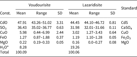
Chemical analyses (6 spots for voudourisite and 9 for lazaridisite) were done using an electron microprobe JEOL JXA-8500F (energy-dispersive spectroscopy mode, 20 kV, 20 nA, 5–6 μm beam diameter). The size and scarcity of the crystals did not allow direct measurement of H2O; it was calculated by stoichiometry from the results of the crystal-structure analysis. No elements other than those reported were detected. Analytical data are given in Table 1.
Chemical compositions (wt.%) for voudourisite and lazaridisite.

*Calculated on the basis of the crystal structure refinement and 5 (voudourisite) and 20 (lazaridisite) O atoms per formula unit.
Const. – constituent; SD – standard deviation.
The empirical formula for voudourisite (based on 5 O atoms per formula unit) is (Cd0.81Cu0.16Fe0.04Mg0.01)Σ1.02S0.99O4·H2O. The simplified formula is Cd(SO4)·H2O, which requires CdO 56.69, SO3 35.35, H2O 7.96, total 100 wt.%.
The empirical formula for lazaridisite (based on 20 O atoms per formula unit) is 3(Cd0.86Cu0.09Fe0.04Mg0.01)Σ1.00S1.00O4·8H2O. The simplified formula is Cd3(SO4)3·8H2O, which requires CdO 50.06, SO3 31.21, H2O 18.736, total 100 wt.%.
Powder X-ray diffraction
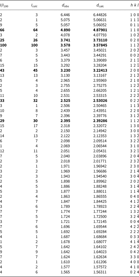
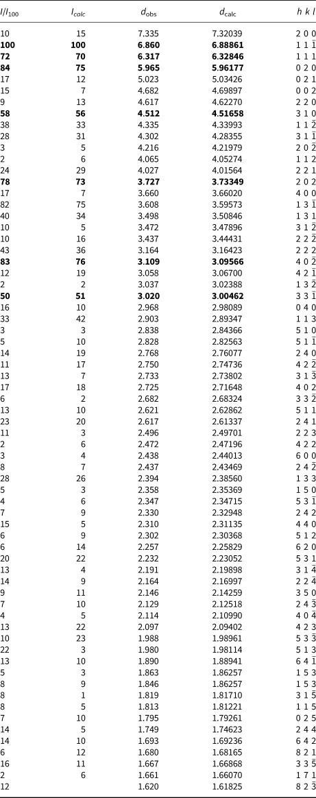
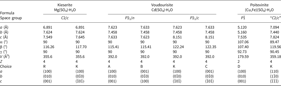
Measurements were carried out with a Philips PW3020 diffractometer using CuKα radiation. At the time of the IMA proposals, only lazaridisite had precise data (Table 3). However, the recent finds of voudourisite from the #19 mine in Sounion have allowed a new measurement of powder diffraction data (Table 2) thus completing the preceding Gandolfi examinations. The hkl assignments of the Gandolfi measurement for voudourisite were done initially according to the known data for the synthetic material from ICDD entry 20-1411 (powder diffraction files from the International Centre for Diffraction Data, http://www.icdd.com/). The correct indexing of the new powder X-ray diffraction data (Holland and Redfern, Reference Holland and Redfern1997) in Tables 2 and 3 is based on the consistent unit-cell choices given in Table 4. The refined unit-cell parameters from these powder data are a = 7.638(3), b = 7.462(3), c = 8.156(3) Å, β = 122.41(3)° and V = 392.4(2) Å3 for voudourisite, and a = 14.774(6), b = 11.924(5), c = 9.483(4) Å, β = 97.70(3)° and V = 1655.5(8) Å3 for lazaridisite, both in good agreement to the values obtained from single-crystal X-ray work. The strongest lines in the powder X-ray pattern [d in Å (I) (hkl)] are: 4.890 (66) (110); 3.741 (25) (020); 3.578 (100) (11 $\bar 2$); 3.230 (43) (200); 2.525 (33) (022); 2.395 (29) (112) for voudourisite and 6.860, (100) (200); 6.317 (72) (111); 5.965 (84) (020); 4.512 (58) (310); 3.727 (78) (202); 3.608 (82) (13
 $\bar 1$); 3.109 (83) (40
 $\bar 2 $); 3.020 (50) (33
 $\bar 1$) for lazaridisite, respectively. The d values for both minerals compare very well with the data for the synthetic analogues (ICDD entries 20-1411, 53-841 and 20-187 for observed powder X-ray data).
Powder X-ray diffraction data for voudourisite*.

* Intensities were calculated using VESTA 3 (Momma and Izumi, Reference Momma and Izumi2011); the strongest lines are given in bold.
Powder X-ray diffraction data for lazaridisite*.

* Intensities were calculated using VESTA 3 (Momma and Izumi, Reference Momma and Izumi2011); the strongest lines are given in bold.
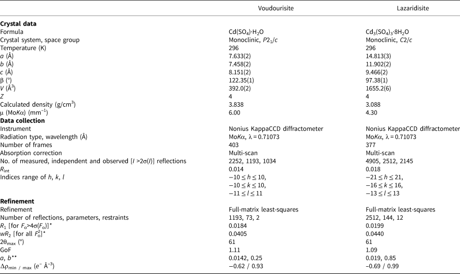
Crystal data and details of the single-crystal intensity measurement and structure refinement for voudourisite and lazaridisite.

* R 1= Σ F o – F c / Σ F o ; wR 2 = [Σw(F o2 – F c2)2 / Σw(F o2)2]½;
** w = 1 / [σ2(F o2) + (a × P)2+ b × P]; P = {[max of (0 or F o2)] + 2F c2}/3
Single-crystal X-ray diffraction data and crystal-structure refinements
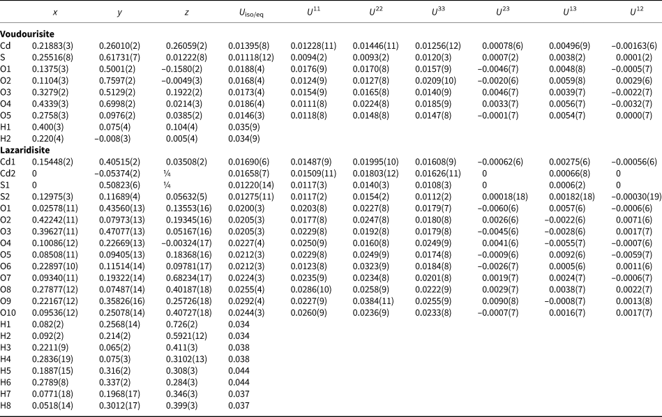
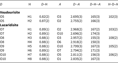
Small fragments of voudourisite and lazaridisite were separated carefully for collection of single crystal X-ray diffraction data (MoKα radiation, microcapillary optics) at room temperature on a Nonius KappaCCD diffractometer. Several sets of phi- and omega-scans with 2° scan width (total 403/377 frames, 100/220 s/frame for voudourisite and lazaridisite, respectively) were measured at a crystal-to-detector distance of 30 mm up to 61°2θ. The absorption was corrected by evaluation of partial multi-scans. Selected crystal parameters and a summary of data collection and structure refinements for both minerals are given in Table 4. The atomic coordinates reported for synthetic Cd3(SO4)3·8H2O (Caminiti and Johansson, Reference Caminiti and Johansson1981) were used as starting parameters for lazaridisite. For the description of voudourisite, the unit-cell setting in space group P21/c was chosen (cf. Table 5) and initial values for the atomic parameters according to Brégault and Herpin (Reference Brégeault and Herpin1970) and Theppitak and Chainok (Reference Theppitak and Chainok2015). The crystal structures were refined by full-matrix least-squares techniques using SHELXL97 (Sheldrick, Reference Sheldrick2008). A substitution of Cd by Cu could not be proven reliably by the structure refinements (Cu < 2%), and was therefore ignored. Final atom positions and important interatomic distances and angles are listed in Tables 6 and 7, respectively. Details on the hydrogen-bond schemes are summarised in Table 8.
Compilation of unit-cell choices from literature used for kieserite, compared with voudourisite and related compounds.

R: ‘Reduced’ monoclinic setting. Kieserite-type sulfates (Le Fur et al., Reference Le Fur, Coing-Boyat and Bassi1966)
K: Kieserite (Leonhardt and Weiss, Reference Leonhardt and Weiss1957; Brégault et al., Reference Brégeault and Herpin1970; Hawthorne et al., Reference Hawthorne, Groat, Raudsepp and Ercit1987), kieserite-type sulfates and selenates (Wildner and Giester, Reference Wildner and Giester1991; Giester and Wildner, Reference Giester and Wildner1992; Bechtold and Wildner, Reference Bechtold and Wildner2016) or atomic arrangement according to kieserite.
A: based on reduced unit cell with monoclinic setting
B: voudourisite (original submission: IMA2012-042)
C: voudourisite (present study); Cd(SO4)·H2O (Brégault and Herpin, Reference Brégeault and Herpin1970; Theppitak and Chainok, Reference Theppitak and Chainok2015); Hg(SeO4)·H2O and Cd(SeO4)·H2O (Stålhandske, Reference Ståhlhandske1978; Reference Ståhlhandske1981)
D: poitevinite (reduced unit cell, Giester et al., Reference Giester, Lengauer and Redhammer1994)
Atom coordinates and displacement parameters with estimated standard deviations in parentheses for voudourisite and lazaridisite.*

* Site symmetries (with Wyckoff positions in parentheses) are 1 (4e) resp. (8f) for all atoms except 2 (4e) for Cd2 and S1. For lazaridisite, the geometry of hydrogen atoms was refined with soft restrictions and U iso values fixed to 1.5 times U eq of the parent oxygen atom.
Selected interatomic distances (Å) and angles (°) for voudourisite and lazaridisite.

Hydrogen-bonding schemes (Å, °) in voudourisite and lazaridisite.

Structure descriptions
Voudourisite, Cd(SO4)·H2O, belongs to the kieserite group M(SO4)·H2O, with M = Mg (kieserite), Mn (szmikite), Fe (szomolnokite), Co (cobaltkieserite), Ni (dwornikite), Zn (gunningite) and Cu/Fe (poitevinite), listed as subdivision 7.CB of the Strunz Mineralogical Tables (Strunz and Nickel, Reference Strunz and Nickel2001). Except for poitevinite and voudourisite, these minerals crystallise in the monoclinic space group C2/c (Leonhardt and Weiss, Reference Leonhardt and Weiss1957; Brégeault and Herpin, Reference Brégeault and Herpin1970; Hawthorne et al., Reference Hawthorne, Groat, Raudsepp and Ercit1987; Wildner and Giester, Reference Wildner and Giester1991; Holtstam, Reference Holtstam2002), as do the synthetic compounds M(SeO4)·H2O where M = Mg, Mn, Co, Ni or Zn (Giester and Wildner, Reference Giester and Wildner1992). Poitevinite (Giester et al., Reference Giester, Lengauer and Redhammer1994) is triclinic P  $\overline 1$, as observed for Cu(XO4)·H2O with X = S or Se (Giester, Reference Giester1988). Voudourisite crystallises in the space group P21/c, a subgroup of C2/c, and is isotypic with Hg(SeO4)·H2O and Cd(SeO4)·H2O (Stålhandske, Reference Ståhlhandske1978; Reference Ståhlhandske1981). Several solid-solution series occur between end-members of the kieserite group, some of them (i.e. those incorporating copper) accompanied by a reduction of crystal symmetry (Cu/Fe: Giester et al., Reference Giester, Lengauer and Redhammer1994; Cu/Mg: Lengauer and Giester, Reference Lengauer and Giester1995; Mg/Co: Bechtold and Wildner, Reference Bechtold and Wildner2016; or Mg/Fe: Talla and Wildner, Reference Talla and Wildner2017). The kieserite structure type is also known for some phosphates and arsenates with trivalent cations (e.g. Fe3+, Mn3+ or V3+), including serrabrancaite, Mn(PO4)·H2O (Witzke et al., Reference Witzke, Wegner, Doering, Pöllmann and Schuckmann2000); close structure relations are further evident with amblygonite, lazulite and titanite.
The atomic arrangement in kieserite-group minerals is characterised by a framework structure, composed by a corner-linkage of MO4(H2O)2 octahedra and XO4 tetrahedra, further strengthened by hydrogen bonding. The characteristic structure motif are chains of corner-sharing MO6 octahedra (with H2O molecules acting as bridging ligands) parallel to the [001] direction for unit-cell choice K according to kieserite (Fig 3a), for details see the compilation in Table 5. In the present paper, we decided to describe voudourisite according to earlier literature on synthetic Cd(SO4)·H2O (Brégeault and Herpin, Reference Brégeault and Herpin1970; Theppitak and Chainok, Reference Theppitak and Chainok2015), and on isotypic Hg(SeO4)·H2O and Cd(SeO4)·H2O (Stålhandske, Reference Ståhlhandske1978; Reference Ståhlhandske1981), i.e. with respective chains arranged also along [001] but with a along [101] of kieserite (Fig. 3b). Differences in interatomic distances between synthetic Cd(SO4)·H2O (Theppitak and Chainok, Reference Theppitak and Chainok2015) and voudourisite are negligible with individual <Cd–O> and <S–O> distances ≤0.006 Å. In kieserite-type compounds with space group C2/c the atoms M (4b) and S, O3 (both 4e) are located on special positions with site symmetries of  $\bar 1$ and 2, respectively; in voudourisite (P21/c), all atoms have site symmetry 1, allowing a symmetry un-constrained variability of bond-length and bond-angle distortions. The Cd–O5 distances, i.e. those to the H2O molecule, are the longest observed for the corresponding M–O3 distances (cf. Wildner and Giester, Reference Wildner and Giester1991).
Atomic arrangement in (a) kieserite and (b) voudourisite in projections on (010). All crystal structure drawings are done with ATOMS (Dowty, Reference Dowty2016).

The crystal structure of lazaridisite (C2/c) is a three-dimensional, heteropolyhedral framework of CdO4(H2O)2 and SO4 groups sharing common corners (Fig. 4) and one free H2O molecule. Except Cd2 and S1 (both on 4e with site symmetry 2), all atoms are located on general sites. Again, differences of interatomic distances from the synthetic analogue Cd3(SO4)3·8H2O (Caminiti and Johansson, Reference Caminiti and Johansson1981) are small, with individual <Cd–O> and <S–O> distances ≤0.01 Å. Both CdO4(H2O)2 octahedra are moderately distorted, and contrary to voudourisite, the Cd–O(H2O) bond lengths are similar to Cd–O(SO4). For a detailed description of this structure type see Caminiti and Johansson (Reference Caminiti and Johansson1981).
Crystal structure of lazaridisite in a projection on (010).

The hydrogen bonding schemes (Table 8) in voudourisite and lazaridisite are characterised by O–H···O distances ranging from 2.695–2.755 Å and 2.698–2.967 Å, respectively. Note that the hydrogen bond O5–H2···O2 in voudourisite is different to the local arrangement observed in kieserite.
Acknowledgements
The authors are grateful to Peter Leverett and two anonymous reviewers for helpful comments.
Supplementary material
To view supplementary material for this article, please visit https://doi.org/10.1180/mgm.2018.157