Patent ductus arteriosus is associated with morbidity and mortality especially in preterm very low birth weight infants.Reference Sellmer, Bjerre and Schmidt 1 Treatment of patent ductus arteriosus remains a subject of tremendous controversy, and disagreement remains about the thresholds and strategies for patent ductus arteriosus closure among premature infants earlier in life.Reference Hamrick and Hansmann 2 Percutaneous closure of patent ductus arteriosus is well established in infants weighing >5 kg, but data regarding outcome of preterm especially very low birth weight infants is minimal. Although surgical ligation of patent ductus arteriosus is the preferred and well-accepted modality of treatment after failure of drug therapy in preterm infants, it has also got its own demerits in such a small and fragile subset. Device closure in infants weighing <1.5 kg is rarely attempted because of high chances of complications, especially acute arterial injury due to the arterial sheath.Reference Backes, Cheatham and Deyo 3 We present here an innovative technique in which successful patent ductus arteriosus device closure was done in a 1.4-kg infant without using arterial sheath.
Case report
A very low birth weight male preterm weighing 1.12 kg was born at gestational age of 28+6 weeks in a private hospital. He was intubated and ventilated just after the birth owing to respiratory distress. On the 3rd day of life, two-dimensional echocardiography was done, which showed a large patent ductus arteriosus of 3 mm left-to-right shunt, with severe pulmonary arterial hypertension. The infant was referred to us at an age of 40 days with a weight of 1.4 kg. Until then, several failed attempts were made to extubate the infant. Medical closure of patent ductus arteriosus was tried several times both with ibuprofen and paracetamol, but it was unsuccessful.
After receiving the infant, he was kept on a ventilator and endotracheal secretions were sent, which showed Acenatobacter baumanni. Appropriate antibiotics were started, andtwo-dimensional echocardiography showed large haemodynamically significant patent ductus arteriosus of 3.2 mm left-to-right shunt, and severe pulmonary arterial hypertension. The left atrium to aorta ratio was 2.1:1. After giving antibiotics for 7 days, it was decided to go for patent ductus arteriosus device closure.
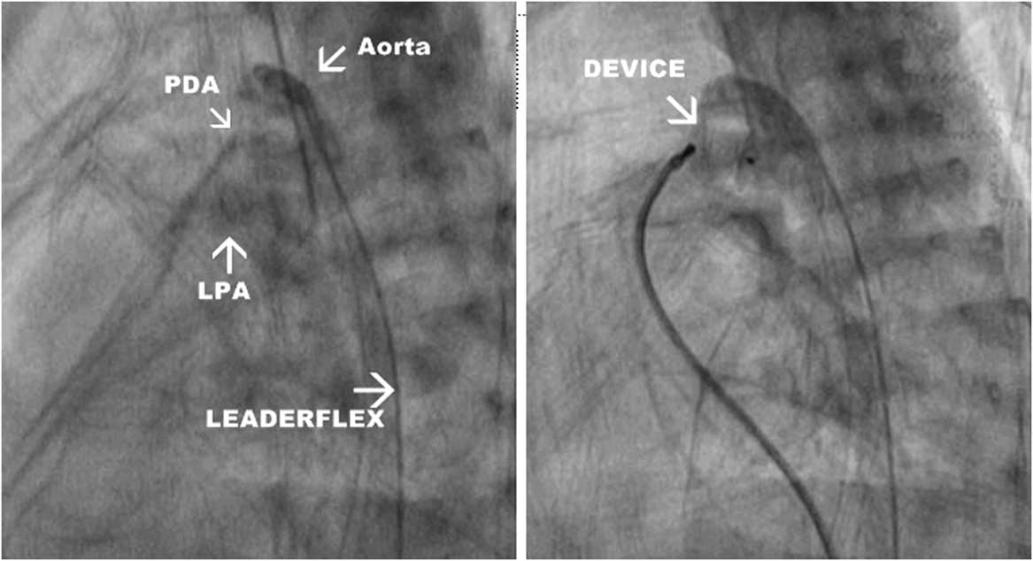
After taking written informed consent, the patient was taken to a cath lab, and under general anaesthesia right femoral venous access was taken with 4 French short sheath under ultrasound guidance. Right femoral artery access was taken and instead of using a short sheath we used 2 French, 22-gauze, 20-cm-long leaderflex catheter – radiopaque polyurethane catheter made by Vygon. The tip of the catheter could easily reach up to the transverse arch of the infant. A measure of 100 units/kg unfractionated heparin was given. Aortogram was performed in lateral view using leaderflex catheter (Fig 1), which showed large patent ductus arteriosus with roomy ampulla. It was decided to use 5×4 ADO 2 AS device (St. Jude Medical; Saint Paul, Minnesota, United States of America). The 4 F right coronary artery catheter was tracked from the venous side along with a 0.035-inch j tip terumo wire. Patent ductus arteriosus was crossed from the pulmonary artery end with the help of a wire, and the wire was parked in the descending aorta. The catheter was removed and the 4 F long sheath (Amplatzer) was tracked over the terumo wire and the tip of the long sheath was placed in the descending aorta. Next, 5×4 ADO 2 AS device – preloaded on cable – was taken and inserted inside the long sheath. The device was deployed across the defect, but the aortic end got pulled inside the ampulla because of very roomy ampulla. Constant push was maintained on the cable and long sheath to prevent dislodgement of the device in the pulmonary artery owing to force of delivery cable; the device is extremely soft and can be easily pulled with a little force. After keeping the device there for about 10 minutes, aortogram was done with leaderflex catheter, which showed good device position, small residual flow through the device, device well inside the ampulla, and no left pulmonary artery/arch obstruction (Fig 1). Next, two-dimensional echocardiography was done, which confirmed the above findings. There was a gradient of 5 mmHg in the left pulmonary artery, which was accepted. The device was released successfully. Aortogram done after the release of the device showed similar findings. Long sheath and short sheath were removed. The 20-cm leaderflex was exchanged with a 4-cm leaderflex, and the patient was shifted to the neonatal ICU.
Aortogram showing large patent ductus arteriosus (PDA). Leaderflex catheter can be seen in the arch and descending aorta (left side). Device in situ occluding the large PDA. Left pulmonary artery is seen filling with contrast, which signifies small residual flow (right side). LPA=left pulmonary artery.

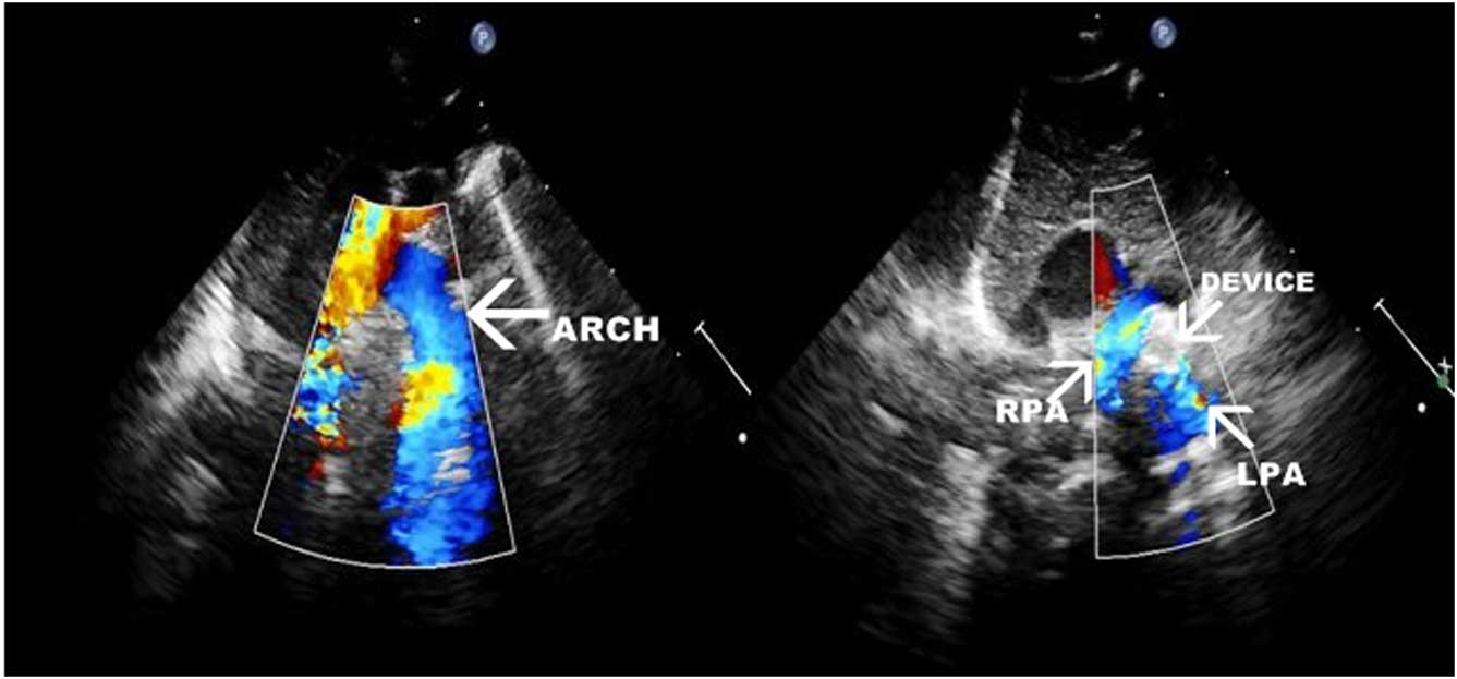
The patient was extubated the next day. Next, two-dimensional echocardiography with color Doppler was done after 24 hours, and it showed good device position, no residual flow, no arch obstruction, and mild flow turbulence in the left pulmonary artery with a peak gradient of 8 mmHg (Fig 2). He was kept on nasal continuous positive airway pressure for 6 days and slowly weaned off from oxygen in 20 days. Retinopathy of prematurity screening was normal. The child started gaining weight and was discharged from the hospital after 35 days with a weight of 1.82 kg. Post discharge, he is on regular follow-up and gaining adequate weight.
Color Doppler showing device in situ, no residual flow, and good flow in the arch and branch pulmonary arteries. LPA=left pulmonary artery; RPA=right pulmonary artery.
Discussion
Patent ductus arteriosus is a frequent congenital cardiac condition in low birth weight premature neonates. The consequences of a significant left-to-right shunting through the patent ductus arteriosus may present haemodynamic and respiratory problems – chronic lung disease, intraventricular haemorrhage, necrotising enterocolitis, retinopathy, and so on.Reference Raval, Laughon and Bose 4 , Reference Hammerman, Bin-Nun and Kaplan 5 Spontaneous closure of the patent ductus arteriosus in normal birth weight neonates occurs in 3 days, but it may persist longer in prematures.Reference Hsiao, Wung, Tsao and Chang 6 When drug therapy fails, surgical ligation is the preferred option for closure in preterm infants. Apart from complications such as residual shunt, aorta or left pulmonary artery obstruction, recurrent laryngeal nerve palsy, chylothorax, surgical intervention with thoracotomy incision itself is another factor for respiratory distress. Compression of the left lung may cause parenchymal bleeding and postoperative atelectasis. This risk increases manifolds if the patient is on ventilator for a long period and has got infection in the lungs like in this case. Evidence on the risks of surgical patent ductus arteriosus ligation has led to growing interest in the use of percutaneous closure in premature infants.Reference Dimas, Takao and Ing 7 , Reference Zahn, Nevin, Simmons and Garg 8
Performing device closure of patent ductus arteriosus in very low birth preterm infants is also a high-risk procedure owing to complications such as device embolisation, residual shunt, aorta, or left pulmonary artery obstruction. Risk of acute arterial injury is the highest.Reference Backes, Cheatham and Deyo 3
In our case, we used a 2 F leaderflex catheter instead of arterial sheath for angiography, which is routinely used in these infants in the neonatal ICU. By using this technique, we could avoid chances of acute arterial injury, and the same catheter with shorter length was used for post-procedure blood pressure monitoring.
Conclusion
In conclusion, we state that device closure of patent ductus arteriosus in very low birth weight infants is feasible, and complications such as acute arterial injury can be avoided by using 2 F leaderflex catheter. Further studies are required to compare this procedure with surgical ligation so that it can be used as an alternative to ligation in future.
Acknowledgements
The authors thank Dr Poonam Sidana and Dr Gaurav Mandhan, Department of NICU, for pre- and post-procedure intensive care.
Financial Support
This research received no specific grant from any funding agency, commercial or not-for-profit sectors.
Conflicts of Interest
None.