Fontan palliation is used for single-ventricle physiology to direct systemic venous blood flow from the inferior caval vein to the pulmonary arteries. The conduits used for Fontan palliation generally are single baffle. However, Y-shaped conduit grafts are chosen in selected cases. There are theoretical benefits in reducing the energy loss and achieving better hepatic flow distribution.
We report a case of a patient who underwent Fontan palliation with a bifurcated Y-shaped Dacron conduit. Three years later, the patient was noted to have significant conduit stenosis with complete obliteration of one arm of the Y-shaped conduit. This is the first case report that describes the clinical course of a patient with a severely stenotic Y-shape Dacron conduit for Fontan palliation.
Case report
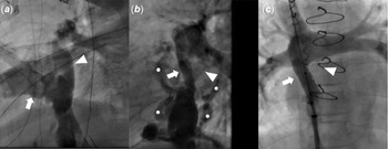
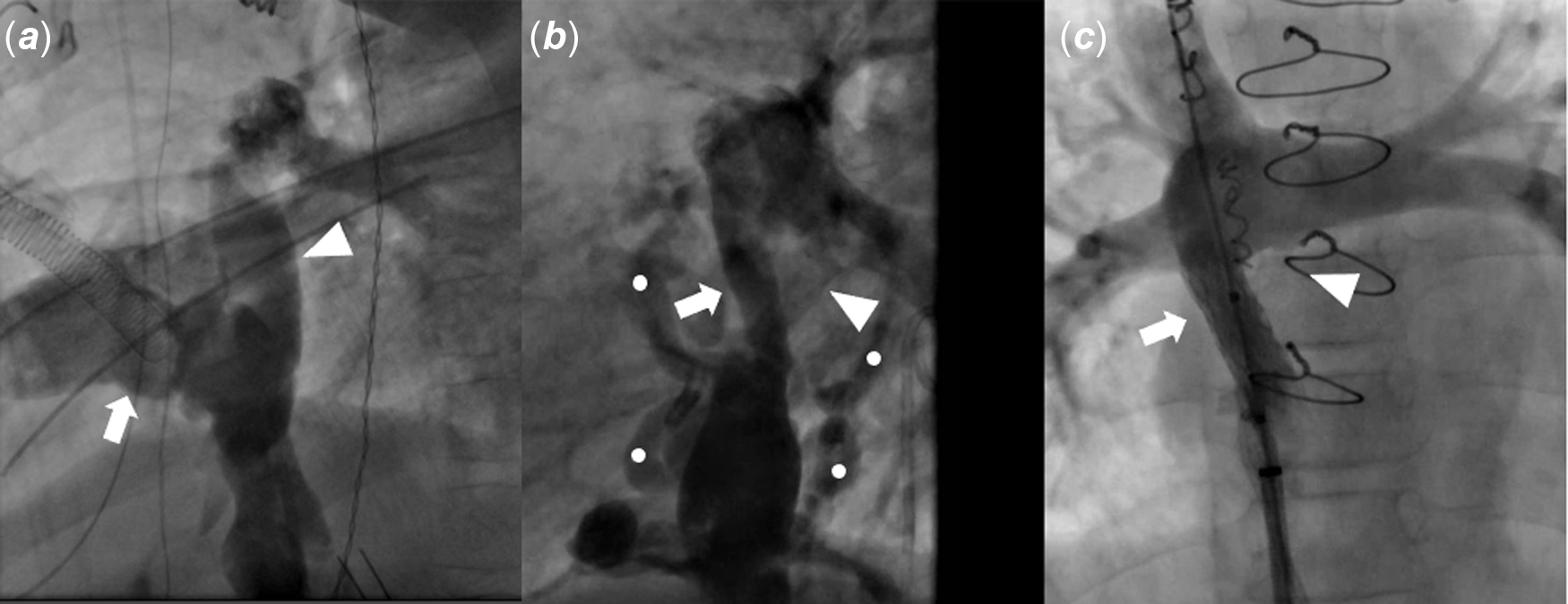
A 6-year-old female with heterotaxy syndrome with asplenia who had undergone Fontan palliation was found to have severe stenosis of the Fontan conduit. She was born with a double outlet right ventricle, pulmonary valve stenosis, D-transposition of the great arteries, total anomalous pulmonary venous connection of the cardiac type with moderate obstruction, and right-sided aortic arch with mirror image branching. She underwent the modified Blalock–Taussig shunt with the repair of obstructed total anomalous pulmonary venous connection at 11 days of life. She subsequently underwent a right-sided bidirectional cavopulmonary anastomosis at the age of 12 months. Her Fontan palliation was completed at the age of 3. Her intraoperative course was very complicated. During the operation, a standard 18 mm diameter polytetrafluoroethylene tube graft was initially attempted. The bulk and stiffness of the polytetrafluoroethylene tube graft sutured to the anterior, augmented aspect of the right pulmonary artery distorted the distal right pulmonary artery unacceptably. The second attempt to attach the distal end of the polytetrafluoroethylene tube graft to the inferior aspect of the right pulmonary artery failed due to the unacceptable amount of compression on the anterior aspect of the right-sided pulmonary vein. Notably, the right-sided pulmonary veins were represented by approximately four very thin branches that entered the right atrium. They coursed more anteriorly than usual. The surgeons felt that they would need to abandon the use of polytetrafluoroethylene tube graft and to devise a mechanism to create an extracardiac Fontan that they would not compress the multiple, quite small and delicate right-sided pulmonary veins. Therefore, the decision was made to use an 18 mm diameter, bifurcated, Gelweave Dacron graft (Terumo Cardiovascular Systems Inc., Ann Arbor, MI, USA) with 9 mm limbs rather than to try a smaller sized polytetrafluoroethylene tube graft. The attempt with bifurcated, Gelweave Dacron graft was successful. A cardiac catheterisation performed on post-operative day 9 showed a widely patent right and left arm of the Y-shaped conduit (Fig 1a).
Right arm of the conduit (arrow) and left arm of the conduit (arrowhead) in the angiogram. ( a ) Pre-stenotic condition. Catheterisation was performed on POD 9 of Fontan completion while the patient was on ECMO support. ( b ) Stenotic right arm of the conduit (arrow). Left arm of the conduit (arrowhead) is completely obstructed and not visualised and a large collateral is seen from the right side of conduit just below the stenotic portion. There are moderate-sized venovenous collaterals superior to the hepatic veins and inferior to the baffle obstruction (dot). ( c ) Post-stent placement showing the improvement in the flow to pulmonary arteries.
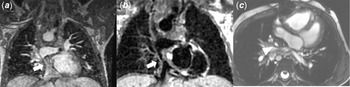
At 3 years after Fontan palliation, the Fontan conduit and bifurcation could not be visualised with an echocardiogram. She underwent a surveillance cardiac MRI to further delineate her Fontan conduit and pulmonary veins. Cardiac MRI revealed obstruction of the conduit (Fig 2a,b,c). There was a narrowing just below the junction of the right pulmonary artery and the right arm of the conduit and complete obstruction of the left arm of the Y-shaped conduit.
Cardiac MRI imaging with gadolinium enhancement showing the stenotic right arm of the conduit (arrow). The left arm of the conduit is completely obstructed and not visualised. ( a and b ) Coronal imaging. ( c ) Axial imaging showing intimal thickening of right arm of the conduit (arrowhead).

A cardiac catheterisation was performed and showed an elevated inferior caval vein pressure of 19–20 mmHg with a pulmonary artery and superior caval vein pressure of 16–17 mmHg. The left arm of the conduit was completely obstructed, and the right arm was severely obstructed (50% obstruction) (Fig 1a and b). There was no right pulmonary venous obstruction or branch pulmonary artery stenosis. A Genesis XD 24 mm stent was placed on an 8 mm balloon catheter across the right arm of the stenotic conduit (Fig 1c). No stent was placed in the left arm of the conduit due to the complete obstruction. After the stent placement, there was a significant improvement in flow with no further gradient across the conduit.
Over the next 5 months after cardiac catheterisation, she has been doing well. The cardiology team discussed the case, and the decision was made to perform reoperation in 6–12 months with a plan for Fontan revision.
Discussion
The Fontan technique has been used for over half a century for palliation of single-ventricle physiology. A single-baffle polytetrafluoroethylene conduit is the most commonly used material for the extracardiac conduit. A Dacron conduit might be chosen for its flexibility and adaptability in specific anatomic situations.Reference Careddu, Petridis and Angeli1 However, the use of Dacron conduit may have a higher complication rate. Careddu et al. reported that 17/25 patients (68%) who underwent cavopulmonary anastomosis with a Dacron conduit had conduit occlusion or severe stenosis requiring intervention, while only 1 out of 80 patients (1.25%) with polytetrafluoroethylene conduit had the same after a mean 46.9 ± 35 months after the operation. They suggest that the use of Dacron is an independent risk factor for midterm stenosis or obstruction, which requires early reintervention.Reference Careddu, Petridis and Angeli1 Generally, a single-baffle conduit is used for Fontan operation because the placement of a Y-shaped conduit is technically more complex. There are other concerns, including the risk of thrombosis in the graft arms, kinking, and impingement of the graft arms on surrounding structures.Reference Martin, Feinstein and Chan2
However, there are some cases in which a Y-shaped conduit option has been selectively implemented. Theoretically, a Y-shaped conduit in Fontan palliation has the advantage of reducing energy losses and a more balanced hepatic flow distribution by directing hepatic flow through two Y-graft branches towards the left pulmonary artery and right pulmonary artery. It is believed that a balanced hepatic flow distribution is an important factor to prevent the development of pulmonary arteriovenous malformation, although its clinical significance is still unknown.Reference Trusty, Wei and Sales3 In our case, a Y-shaped Dacron conduit was not the first choice in the operation. We initially attempted polytetrafluoroethylene tube graft, which resulted in failure due to compression of the right-sided pulmonary veins. A commercially available Y-shaped Dacron conduit was the only alternative option available.
Identifying conduit stenosis by echocardiogram is challenging and even more challenging in the setting of a Y-shaped conduit. Slesnick et al reported that the Y-arm connections to the branch pulmonary arteries were not visualised in all 45 patients who underwent a Y-graft Fontan palliation. Cardiac MRI was performed in 39 patients a median of 9 days after Fontan (range: 4–295 days) and CT angiography was performed in 6 patients due to pacemakers. One patient was found to have thrombosis in the baffle, and 40 patients had some degree of stenosis in a Fontan baffle or branch pulmonary artery. Twenty-seven patients showed mild stenosis, while 13 patients had moderate or severe stenosis. The authors conclude that cross-sectional imaging is essential to assess the bifurcated Y-graft Fontan.Reference Slesnick, Parks and Sallee4
Acknowledgement
None.
Financial support
None.
Conflicts of interest
None.
Ethical standards
There is no research involving this patient.
Author contributions
Daiji Takajo wrote the manuscript; all authors: critically reviewed the manuscript.