




No CrossRef data available.
Article contents
Testing key predictions of the associative account of mirror neurons in humans using multivariate pattern analysis
Published online by Cambridge University Press: 29 April 2014
Abstract
Cook et al. overstate the evidence supporting their associative account of mirror neurons in humans: most studies do not address a key property, action-specificity that generalizes across the visual and motor domains. Multivariate pattern analysis (MVPA) of neuroimaging data can address this concern, and we illustrate how MVPA can be used to test key predictions of their account.
Information
- Type
- Open Peer Commentary
- Information
- Copyright
- Copyright © Cambridge University Press 2014
References
Catmur, C., Walsh, V. & Heyes, C. M. (2007) Sensorimotor learning configures the human mirror system. Current Biology
17(17):1527–31.CrossRefGoogle ScholarPubMed
Edelman, S., Grill-Spector, K., Kushnir, T. & Malach, R. (1998) Toward direct visualization of the internal shape representation space by fMRI. Psychobiology
26:309–21.CrossRefGoogle Scholar
Haxby, J. V., Gobbini, M. I., Furey, M. L., Ishai, A., Schouten, J. L. & Pietrini, P. (2001) Distributed and overlapping representations of faces and objects in ventral temporal cortex. Science
293(5539):2425–30.CrossRefGoogle ScholarPubMed
Haynes, J. D. & Rees, G. (2005) Predicting the orientation of invisible stimuli from activity in human primary visual cortex. Nature Neuroscience
8:686–91.CrossRefGoogle ScholarPubMed
Kriegeskorte, N. (2009) Relating population-code representations between man, monkey, and computational models. Frontiers in Neuroscience
3:363–73.CrossRefGoogle ScholarPubMed
Kriegeskorte, N., Goebel, R. & Bandettini, P. (2006) Information-based functional brain mapping. Proceedings of the National Academy of Sciences USA
103:3863–68.CrossRefGoogle ScholarPubMed
Kriegeskorte, N., Mur, M. & Bandettini, P. (2008) Representational similarity analysis – Connecting the branches of systems neuroscience. Frontiers in Systems Neuroscience
2:3863–68.Google Scholar
Mukamel, R., Ekstrom, A. D., Kaplan, J., Iacoboni, M. & Fried, I. (2010) Single-neuron responses in humans during execution and observation of actions. Current Biology
20(8):750–56.CrossRefGoogle ScholarPubMed
Norman, K., Polyn, S., Detre, G. & Haxby, J. V. (2006) Beyond mind-reading: Multi-voxel pattern analysis of fMRI data. Trends in Cognitive Sciences
10:424–30.CrossRefGoogle ScholarPubMed
Oosterhof, N. N., Tipper, S. P. & Downing, P. E. (2012) Viewpoint (In)dependence of action representations: An MVPA study. Journal of Cognitive Neuroscience
24:975–89.CrossRefGoogle ScholarPubMed
Oosterhof, N. N., Tipper, S. P. & Downing, P. E. (2013) Cross-modal and action-specific: Neuroimaging the human mirror neuron system. Trends in Cognitive Sciences
17:211–318.CrossRefGoogle Scholar
Oosterhof, N. N., Wiestler, T., Downing, P. E. & Diedrichsen, J. (2011) A comparison of volume-based and surface-based multi-voxel pattern analysis. Neuroimage
56:593–600.CrossRefGoogle ScholarPubMed
Oosterhof, N. N., Wiggett, A. J., Diedrichsen, J., Tipper, S. P. & Downing, P. E. (2010) Surface-based information mapping reveals crossmodal vision-action representations in human parietal and occipitotemporal cortex. Journal of Neurophysiology
104:1077–89.CrossRefGoogle ScholarPubMed
Peelen, M. V. & Downing, P. E. (2007) Using multi-voxel pattern analysis of fMRI data to interpret overlapping functional activations. Trends in Cognitive Sciences
11:4.CrossRefGoogle ScholarPubMed
Press, C., Gillmeister, H. & Heyes, C. (2007) Sensorimotor experience enhances automatic imitation of robotic action. Proceedings of the Royal Society of London B: Biological Sciences
274(1625):2509–14.Google ScholarPubMed
Sereno, M. I. & Tootell, R. B. (2005) From monkeys to humans: What do we now know about brain homologies?
Current Opinion in Neurobiology
15:135–44.CrossRefGoogle ScholarPubMed
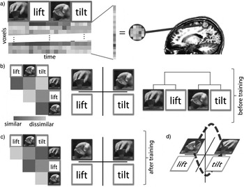
Figure 1. Hypothetical example illustrating neural effects of visuomotor “counter-mirror” training. (a) Neural responses are measured in a region of interest using fMRI while participants observe (red boxes) or are instructed to perform (green boxes) two actions (lifting or tilting a cup-shaped object). Each trial is associated with a spatially distributed pattern of responses over voxels. (b) Before training, congruent actions across the visual and motor domain are represented more similarly, as illustrated by a similarity matrix (higher pattern similarity indicated in dark orange; left), multi-dimensional scaling in two dimensions (more similar patterns depicted nearer in space; centre), and a dendrogram (“leaves” representing patterns are connected by shorter paths if patterns are more similar; right). (c) After counter-mirror training, where participants learn to lift (or tilt) the object after observing a tilt (or lift, respectively) action, the neural similarity structure might have changed, where non-matching actions are represented similarly across the visual and motor domain. (d) When visualized using multi-dimensional scaling, here in three dimensions, the similarity “trajectories” (dark purple arrows) during learning and unlearning of the associations can be visualized, allowing for assessment of the temporal dynamics of changes in visuomotor associations. A color version of this image is available at http://dx.doi.org/10.1017/S0140525X13002434.
The discovery of mirror neurons (MNs) in macaques has undoubtedly had a major impact on the field of social neuroscience by providing a potential mechanism for associating visual and motor aspects of actions, which could be a core component in action understanding. The timely article by Cook et al. addresses one of the fundamental questions on this topic, namely, how such visuomotor associations emerge in the first place.
While we agree with much in Cook et al.'s article, our concern is that some of the interpretations and conclusions the authors draw about mirror neurons in humans generally, and in favour of the associative account specifically, risk being overstated based on the evidence reviewed. Specifically, most inferences (but see Mukamel et al. Reference Mukamel, Ekstrom, Kaplan, Iacoboni and Fried2010) about MNs in humans are based on a combination of proposed homologies between macaques and humans, and on studies employing less invasive methods to study the brain, including transcranial magnetic stimulation, magneto- and electroencephalography, and most notably, functional magnetic resonance imaging (fMRI). The former approach assumes a mapping between brain regions across monkey and human species, but this homology is imperfect (Sereno & Tootell Reference Sereno and Tootell2005). The latter ostensibly provides a wealth of evidence for human MNs; for example, numerous fMRI studies have shown common areas demonstrating an increased response during observation and execution of actions (compared to a baseline condition), but such effects could be explained by task engagement or attention processes (Oosterhof et al. Reference Oosterhof, Tipper and Downing2013).
In contrast, we would argue that stricter requirements are needed to infer that a brain region may contain mirror neurons. In macaques, MNs have been shown to code specific, individual manual actions that generalize across the visual and motor domains. In contrast, most fMRI studies with humans do not test for this key property: action-specificity. To demonstrate action-specificity in the human brain, the same action, whether observed or executed, should elicit more similar neural responses than dissimilar actions (Oosterhof et al. Reference Oosterhof, Tipper and Downing2013).
In our view, the most promising approach addressing this limitation is the application of multivariate pattern analysis (Edelman et al. Reference Edelman, Grill-Spector, Kushnir and Malach1998; Haxby et al. Reference Haxby, Gobbini, Furey, Ishai, Schouten and Pietrini2001; Haynes & Rees Reference Haynes and Rees2005; Norman et al. Reference Norman, Polyn, Detre and Haxby2006), which considers neural responses across a group of voxels. The logic behind this method is that the spatially distributed pattern, across voxels, of a specific observed (or executed) action should be more similar to the pattern associated with executing (or observing, respectively) the same action than a different action. Although fMRI cannot be used to measure individual neurons, MVPA complements single-cell recording approaches by considering spatially distributed responses at a system level. Importantly, this sensitive approach allows for dissociating responses of spatially overlapping neural populations (Peelen & Downing Reference Peelen and Downing2007).
Relevant to the topic of the present article, the human mirror system, recent MVPA studies have provided evidence for cross-modal action-specific representations of manual actions in anterior parietal and lateral occipito-temporal cortex (Oosterhof et al. Reference Oosterhof, Wiggett, Diedrichsen, Tipper and Downing2010; Reference Oosterhof, Tipper and Downing2012). These findings, together with evidence from single-cell recordings in humans (Mukamel et al. Reference Mukamel, Ekstrom, Kaplan, Iacoboni and Fried2010), indicate that regions consistent with MN properties can also be found outside the canonical fronto-parietal network, consistent with Cook et al.'s associative account.
Particularly relevant for the target article is that MVPA – in particular, representational similarity analysis (RSA; Kriegeskorte Reference Kriegeskorte2009; Kriegeskorte et al. Reference Kriegeskorte, Mur and Bandettini2008) – can be used to test key predictions, at a neural population level, of the associative account of the human mirror system. Cook et al. write “the properties of MNs can be changed in radical ways by relatively brief periods of sensorimotor experience” (sect. 3.4, para. 4), based on evidence from several behavioural studies showing that priming effects (e.g., automatic imitation effects) can be reduced or even reversed after counter-mirror training. Although we agree that these findings are interesting and consistent with an associative learning account, we do not believe that results from such studies alone provide strong evidence to support the authors' claim, as the neural correlates of these effects were not measured in fine detail. We believe that MVPA enables such detailed measurement and can be used to characterize where and when such changes occur at a neural population level.
To illustrate this, we provide a hypothetical example based on associative account predictions brought forward by Cook et al. of how counter-mirror learning can be adapted to study the changes in neural representations using MVPA (cf. Catmur et al. Reference Catmur, Walsh and Heyes2007; Press et al. Reference Press, Gillmeister and Heyes2007). Patterns of responses can be measured in the brain when participants observe or execute two different manual actions (Fig. 1a). Before training, observing and executing actions congruent across the visual and motor domain are represented similarly (Fig. 1b). After counter-mirror training, where actions incongruent across the visual and motor domain are associated with each other, this situation is reversed: manual movements incongruent across the visual and motor domain might now be represented similarly (Fig. 1c).
Hypothetical example illustrating neural effects of visuomotor “counter-mirror” training. (a) Neural responses are measured in a region of interest using fMRI while participants observe (red boxes) or are instructed to perform (green boxes) two actions (lifting or tilting a cup-shaped object). Each trial is associated with a spatially distributed pattern of responses over voxels. (b) Before training, congruent actions across the visual and motor domain are represented more similarly, as illustrated by a similarity matrix (higher pattern similarity indicated in dark orange; left), multi-dimensional scaling in two dimensions (more similar patterns depicted nearer in space; centre), and a dendrogram (“leaves” representing patterns are connected by shorter paths if patterns are more similar; right). (c) After counter-mirror training, where participants learn to lift (or tilt) the object after observing a tilt (or lift, respectively) action, the neural similarity structure might have changed, where non-matching actions are represented similarly across the visual and motor domain. (d) When visualized using multi-dimensional scaling, here in three dimensions, the similarity “trajectories” (dark purple arrows) during learning and unlearning of the associations can be visualized, allowing for assessment of the temporal dynamics of changes in visuomotor associations. A color version of this image is available at http://dx.doi.org/10.1017/S0140525X13002434.
The application of MVPA provides other advantages. First, unlike TMS studies, MVPA does not require defining regions of interest a priori through the use of “searchlight” analyses (Kriegeskorte et al. Reference Kriegeskorte, Goebel and Bandettini2006; Oosterhof et al. Reference Oosterhof, Wiggett, Diedrichsen, Tipper and Downing2010; Reference Oosterhof, Wiestler, Downing and Diedrichsen2011). Second, MVPA enables the study of temporal dynamics of learning and unlearning new visuomotor associations of specific actions across the brain (Fig. 1d). Third, MVPA can be used to test generalization to other experimental factors such as viewpoint and different grasps. Fourth, MVPA allows for comparisons of neural representations across species (macaques and humans) and brain measurement methods (fMRI and neurophysiology), allowing for more detailed comparisons of (dis)similarities across species (Kriegeskorte Reference Kriegeskorte2009).
In conclusion, we agree that the existing evidence of a human mirror system is compatible with an associative learning account. However, we argue that the current evidence is not strong enough to fully support all the claims made by Cook et al. in the target article. We believe that the application of MVPA, in particular RSA and information mapping techniques, offers a promising avenue to more fully characterize the human mirror system, and thus provide evidence to support or falsify the associative learning hypothesis. We predict that these methods will be crucial for future fMRI studies if they are to advance our understanding of the human mirror system.