Haemorrhagic shock is the most common and preventable cause of mortality in trauma due to excessive bleeding. According to the statistics, ∼30–40% of global trauma-related deaths annually are attributed to severe haemorrhage (Bao et al., Reference Bao, Gao, Cau, Ali-Mohamad, Strong and Jiang2022). Effectively controlling bleeding during the critical ‘golden hour’ post-injury represents both a pivotal challenge and a key opportunity for improving survival rates, which also constitutes a crucial approach to improving military/civilian medical services and emergency response capabilities globally.
Medical gauzes have been widely used to protect wounds from bleeding for thousands of years due to the advantages of their low-cost, facile production, their ease of storage and the large pores that they contain, which are beneficial for exudate management and air permeability that can accelerate wound healing (Rezvani Ghomi et al., Reference Rezvani Ghomi, Niazi and Ramakrishna2023; Zhang et al., Reference Zhang, Zhang, Lin, Yang, Jiang, Chen and Ma2023b). An effective approach to improve the haemostatic performance of dressings involves integrating haemostats with medical gauze samples. Therefore, significant effort has been devoted to the development of highly efficient haemostatic gauzes, especially through the incorporation of natural clays or clay minerals with excellent haemostatic properties.
QuikClot Combat Gauze, a fabric knitted with rayon/polyester yarns loaded with kaolin particles (Littlejohn et al., Reference Littlejohn, Devlin, Kircher, Lueken, Melia and Johnson2011), can effectively increase survival rates by speeding up the blood-clotting reaction due to the active effect of kaolin, while the composite fibres of zeolite/cotton also produce greater haemostatic efficiency (Yu et al., Reference Yu, Shang, Chen, Xiao, Zhu and Fan2019). Furthermore, a composite gauze formulation prepared by coating zeolite gauze with kaolin particles using 0.75% styrene–butadiene latex as a binder demonstrated superior haemostatic efficacy with a minimal powder shedding ratio compared with both kaolin gauzes and zeolite gauzes currently employed in clinical practice (Wang et al., Reference Wang, Shi, Huang, Wei, Shi, Xiao and Fan2024a). The goal of this research is to make full use of the haemostatic properties of clay minerals whilst improving their biocompatibility.
Similarly to most clay mineral-based haemostatic agents, such as kaolinite, montmorillonite, halloysite, sepiolite, palygorskite and rectorite (Zhang et al., Reference Zhang, Xue, Zhu, Zhao, Chen and Tong2019, Reference Zhang, Pan, Dong, Yang, Huang, Yan and Gao2023a; Serati-Nouri et al., Reference Serati-Nouri, Jafari, Roshangar, Dadashpour, Pilehvar-Soltanahmadi and Zarghami2020; Feng et al., Reference Feng, Luo, Wu, Liu, Liang, He and Liu2022; Jiang et al., Reference Jiang, Yang, Peng, Li, Peng and Zhang2023; Song et al., Reference Song, Li, Chen, Han, Duns and Dessie2023; Yang et al., Reference Yang, Wang, Li, Mu, Yang, Wang and Liu2024), mixed-dimensional palygorskite clay (MDPal) composed of palygorskite, illite, chlorite, kaolinite and illite–smectite mixed-layer clays also demonstrated excellent haemostatic performance, with its performance even being superior to that of Yunnan Baiyao (Wang et al., Reference Wang, Yang, Yang, Mu and Wang2024b). However, there are no reports on the combination of MDPal and gauzes for haemostatic applications, and the problem of clay mineral detachment remains a common key issue to resolve.
The surface functional modification of gauzes has long been of great importance. Amongst the various modification approaches used, polydopamine (PDA) modification possesses obvious advantages of promoting blood coagulation, improving biocompatibility and biodegradability and providing excellent antioxidant properties to improve wound healing (Zhao et al., Reference Zhao, Zhang, Guo and Ma2016; Bao et al., Reference Bao, Zhao, Sun, Hu and Yang2018; Shin et al., Reference Shin, Ryu, Kim, Kim, Jo and Lee2018), and it is also a good option for the construction of high-performance gauze composites, in which PDA serves as a crucial bridge to simultaneously form a solid layer on the surface of the gauze and as a clay mineral to increase the loaded amount of clay mineral and strengthen the bonding interaction between the clay mineral and gauze.
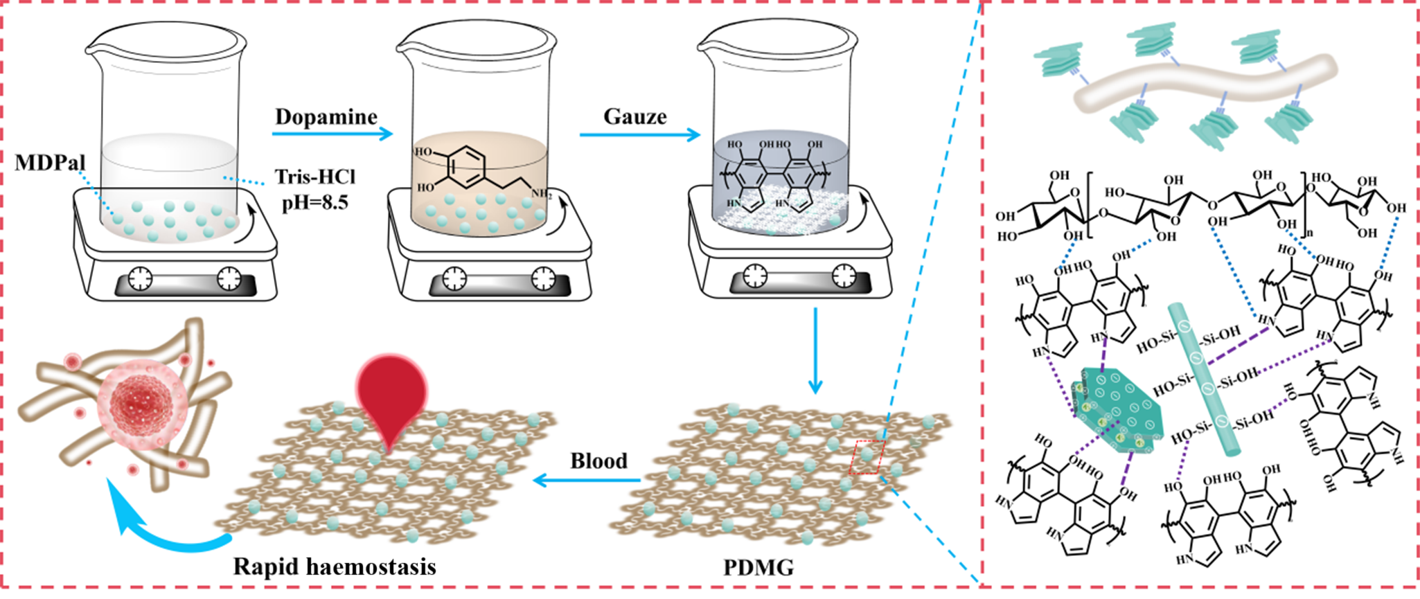
Thus, gauze composites were fabricated by loading MDPal onto gauzes with PDA surface modification for wound haemostasis (Scheme 1). First, a medical gauze – one of the most commonly used medical dressings – was modified with MDPal and PDA. The reaction duration and dopamine dosage were optimized based on haemostatic performance. Next, the physicochemical properties of the gauze composites were evaluated, including water vapour transmission, moisture absorption, hydrophilicity and hydrophobicity. In addition, haemostasis-related characteristics were tested, such as the blood-clotting index (BCI), coagulation kinetics of platelet-poor plasma (PPP), thrombogenic capacity and erythrocyte adhesion behaviour. Subsequently, the biocompatibility and haemocompatibility of the as-prepared gauze composites were investigated, and their procoagulant mechanism was explored. In addition, the in vivo haemostatic performance was further verified via animal models.
Preparation schematic of haemostatic gauze composites for wound haemostasis.

Scheme 1 Long description
The schematic illustrates the process of creating PDMG gauze using dopamine and MDPaI. On the left, three containers are shown sequentially. The first container holds MDPaI, with a label indicating its contents. An arrow points to the second container, which contains dopamine mixed with Tris-HCl at a pH of 8.5. Another arrow leads to the third container, labeled 'Gauze,' which shows the gauze being treated with the mixture. Below these containers, an arrow points to a depiction of rapid hemostasis, where blood interacts with the PDMG gauze, suggesting its effectiveness in stopping bleeding. On the right side, a detailed molecular structure is shown, highlighting the chemical interactions and bonds within the PDMG gauze. Various chemical groups and bonds are labeled, illustrating the complex molecular arrangement. The diagram emphasizes the transformation from raw materials to a functional gauze with rapid hemostatic properties.
Experimental
Materials
MDPal was purchased from Gansu Rongwan Technology Co., Ltd (Gansu, China), and pretreated with oxalic acid dihydrate (Chengdu Chron Chemicals Co., Ltd, Sichuan, China) at 80°C for 1 h. The treated MDPal was then sieved through a 200 mesh sieve for later use. Tris-(hydroxymethyl)aminomethane (Tris) was supplied by Aladdin Industrial Co., Ltd (China). Dopamine and sodium hydroxide were obtained from Macklin Biochemical Technology Co., Ltd, and Chengdu Chron Chemicals Co., Ltd, respectively. Medical degreased gauze (abbreviated as ‘gauze’; yarn density: 21 s × 21 s, 115 × 105; specifications: 80 mm × 80 mm × 8 P, ethylene oxide-sterilized) was purchased from Henan Yadu Industrial Co., Ltd (Henan, China). All water used in the experiments was ultrapure water. Sodium citrate-anticoagulated whole blood from healthy volunteers was provided by the Laboratory Medicine Center of Lanzhou University Second Hospital (Gansu, China). This study was approved by the Clinical Research Professional Ethics Committee (approval no.: 2023A-053).
Preparation of the gauze composites
Some 0.5 g of MDPal powder was dispersed in 100 mL of Tris solution (10 mM) and stirred for 0.5 h at 400 rpm to form a uniform solution. Various amounts of dopamine powder were added to this dispersion solution. The medical skimming gauze (80 mm × 80 mm × 4 P) was immersed into this solution after dopamine was completely dissolved, then 2 M sodium hydroxide solution was used to adjust the pH of the reaction solution to 8.5–9.5. The gauze composites were obtained by optimizing the two key factors of reaction duration (1.5, 4.0, 6.5, 9.0 h) and the added amount of dopamine (0, 50, 100, 200, 400 mg) at a stirring rate of 200 rpm. After the reaction, the gauze composites were obtained by washing them several times with ultrapure water and drying them to a constant weight in a vacuum oven at 60°C. The corresponding gauze composites were marked as PDMG1.5–9.0 and PDMG50–400, respectively. The loaded amounts of MDPal on the composite gauze samples are provided in Table S1. Subsequently, the unmodified gauzes and the gauzes modified with PDA or MDPal were designated as G, P-G and M-G, respectively, whereas the commercially available kaolin-impregnated QuikClot Combat Gauze was labelled as K-G and used as a control.
Characterizations
The main components and interactions of the gauze composites were recorded using attenuated total reflectance Fourier-transform infrared (ATR-FTIR) spectrometry (FTIR-Nicolet iS20, Thermo Fisher, Wilmington, DE, USA) from 4000 to 400 cm–1 at a resolution of 4 cm–1. The morphology, energy-dispersive X-ray spectroscopy (EDS) mapping and adherent erythrocytes of the gauze samples were observed via field-emission scanning electron microscopy (SEM; JSM-6701F, JEOL, Ltd, Japan). The contact angle (CA) of the gauze samples towards water was measured using a CA system (OCA20, DataPhysics Instruments, Filderstadt, Germany).
The gauze samples were cut to a size of 3.0 cm × 2.5 cm and evenly fixed on a glass slide, then 20 μL of water was dropped to the gauze surfaces. The CA was automatically recorded, and three parallel recordings for each sample were collected (n = 3). The thermal behaviour of the gauze samples was assessed using an integrated thermal analyser (STA 800, PerkinElmer Co., Groningen, The Netherlands) under an oxygen atmosphere (20 mL min–1) from 30°C to 600°C at 20°C min–1.
All experimental animals were obtained from Lanzhou Veterinary Research Institute, Chinese Academy of Agricultural Sciences (Beijing, China; license number: SCXK (Gan) 2020-0002), and the experiment protocols have been approved by our animal ethics committee.
Water vapour transmission rate and water absorption rate
To test water vapour permeation, the experimental procedures were derived from the literature with slight modification (Jia et al., Reference Jia, Hua, Yang, Li, Zhao and Zhou2023). A 15 mL tube filled with 10 mL of ultrapure water was covered with the gauze composites, and then the circumferential border was sealed firmly to prevent any penetration of water vapour through it. After being stored at 37°C for 24 h, the water vapour transmission rate (WVTR) was measured by weighing the mass loss of water from the tube. All of the results are presented as mean ± standard deviation (SD; n = 3), and the WVTR (g m–2 day–1) was calculated using Equation 1:
 \begin{equation}\text{WVTR} =\left(m_\text{b}-m_\text{a}\right)/A\end{equation}
where m b and m a (g) are the tube masses before and after the evaporation of water and A (m2) is the area of the tube mouth.
For the water absorption experiments (Yang et al., Reference Yang, Shi, Liu, Cheng, Li and Lyu2023), gauze composites (1.5 cm long, 1.5 cm wide and 1.0 mm thick) at a dry weight (m b) were soaked in water and then removed from the water at various time points to measure the weight of the gauze (m a), and the absorbed mass of water (m a – m b) was scaled to the dry weight of the gauze sample to determine the level of water absorption. The water absorption rates (W abs; %) of the gauze samples was determined (Equation 2) after 3 and 20 min. All of the results are presented at mean ± SD (n = 3).
 \begin{equation}W_\text{abs}\left(\text{\%}\right)=\left(m_\text{a}-m_\text{b}\right)/m_\text{b}\times100\text{\%}\end{equation}
In vitro blood coagulation test
In vitro haemostasis was evaluated according to the dynamic coagulation effects, plasma-clotting assay, thrombogenesis amount and erythrocyte adhesion assay.
Dynamic coagulation effects
Three layers of gauze of 1 cm length and 1 cm width were placed into tubes, and the coagulation results at 150 and 180 s were obtained following the process presented in our previous report (Wang et al., Reference Wang, Yang, Yang, Mu and Wang2024b). Then, 1 mL of ultrapure water was gently added to stop the coagulation reaction without damaging the blood clot. The erythrocytes not adhering to the clot would thus be destroyed and release haemoglobin. Subsequently, 200 μL of the supernatant was taken to 96 well plate, and the relative haemoglobin absorbance at 540 nm was measured using a microplate reader (Agilent Epoch 2 microplate spectrophotometer, Agilent, CA USA; n = 4). A lower absorbance value indicated a stronger blood clot and a greater coagulation efficiency.
Plasma-clotting assay
Some 100 μL of PPP was added to a 96 well plate followed by 10 μL of the sample suspension (10 mg mL–1), and 100 μL CaCl2 (0.025 M) was added to trigger coagulation. The coagulation reaction was recorded at 0, 2, 4, 6 and 8 min by measuring the absorbance at 405 nm (multi-point scan 4 rows × 4 columns) using the microplate reader (Agilent Epoch 2 microplate spectrophotometer). The kinetics of fibrin formation are indirectly expressed by subtracting the absorbance at 0 min from that of each specific time point (n = 3; Liu et al., Reference Liu, Liu, Liu, Wang, Chen and Yao2019).
Thrombogenesis amount
The in vitro thrombosis ability (i.e. blood coagulability to form a haemostatic plug) of the gauze samples was assessed according to processes described previously in the literature (Liu et al., Reference Liu, Zhou, Zhang, Zhu, Wang, Xu and Zhuang2022). Typically, gauze composites were first weighed (m i) were mixed with 1 mL of citrated whole blood in a culture plate at 37°C for 30 min. Deionized water (10 mL) was added to stop the reaction, and the thrombus was fixed with 2.5% glutaraldehyde solution for 10 min. The gauze samples were dried overnight to obtain constant-weight gauze composites, recorded as m t. All sample groups were analysed at least three times. The thrombogenesis amount was measured using Equation 3:
 \begin{equation}{\text{Thrombogenesis amount}}\left( {{\text{mg}}} \right) = {{\textit{m}}_{\text{t}}} - {{\textit{m}}_{\text{i}}}\end{equation}
In vitro haemocompatibility and cytocompatibility
The in vitro haemocompatibility was determined by measuring the extent of red blood cell (RBCs) damage following co-culture with the gauze. In more detail, 100 μL of the sample suspension (1 cm × 1 cm gauze leached in phosphate-buffered saline (PBS)) was diluted by 700 μL of PBS, and then 200 μL of 5% (v/v) RBC solution was added to obtain the mixed suspension. Here, double-distilled H2O (+ RBCs) and PBS (+ RBC) served as the positive control and negative control, respectively. After centrifugating at 1000 rpm for 5 min, 200 μL of the suspension was transferred to a 96 well plate to measure the absorbance at 540 nm using a microplate reader (Agilent Epoch 2 microplate spectrophotometer). The haemolysis rate (HR; %) was calculated using Equation 4 (n = 3):
 \begin{equation}\text{HR}\left(\text{\%}\right)=\frac{OD_\text{sample}-OD_\text{negative}}{OD_\text{positive}-OD_\text{negative}}\times100\%\end{equation}
where OD sample, OD negative and OD positive refer to the absorbance values of the sample group, negative group and positive group, respectively.
The cytotoxicity was evaluated using a Cell Counting Kit-8 (CCK-8; APExBio, TX, USA) assay used with slight modification to a previously described process (Chen et al., Reference Chen, Jin, Kamiya, Liu, Tian and Xia2025). At the predetermined time, 10 μL of CCK-8 solution was added to each well and then incubated for another 2 h at 37°C, and the absorbance was measured at 450 nm using a microplate reader (Agilent Epoch 2 microplate spectrophotometer; n = 3). The cell viability (%) was calculated using Equation 5 (n = 3), in which OD sample and OD blank represent the absorbance values of the sample and control groups, respectively.
 \begin{equation}\text{Cell viability}\left(\text{\%}\right)=\frac{OD_\text{sample}}{OD_\text{blank}}\times100\text{\%}\end{equation}
Animal experiments
Sprague-Dawley rats (male, weight range 220–250 g) were obtained from Lanzhou Veterinary Research Institute, Chinese Academy of Agricultural Sciences (license number: SCXK (Gan) 2020-0002) and were used as a liver haemorrhage model. The rats were anesthetized and fixed on an operating table, the abdominal cavity was exposed and a wound with a length of ∼20 mm was made in the liver. All experiment protocols were approved by our animal ethics committee. The gauze samples (G, P-G, M-G, K-G and PDMG) were weighed and recorded before covering the wound and weighed and recorded again after removal from the wound, where PDMG here refers to the PDMG200 sample, which exhibited the best in vitro haemostatic properties. The haemostatic time and blood loss were recorded and the injury model without any treatment was set as the control group (n = 3). It is worth noting that the small sample size and the single model are shortcomings of this study, and the validation of the promising findings presented here will be investigated in our future research using larger sample sizes (n ≥ 6 per group) and other haemorrhage models (e.g. arterial spurting haemorrhage in large animals).
Statistical analysis
Average values and SDs were calculated from at least three different measurements. The in vivo performance, in vitro cytocompatibility and haemocompatibility evaluation are averaged from three different measurements.
Results
Compositional and structural analysis of the gauze composites
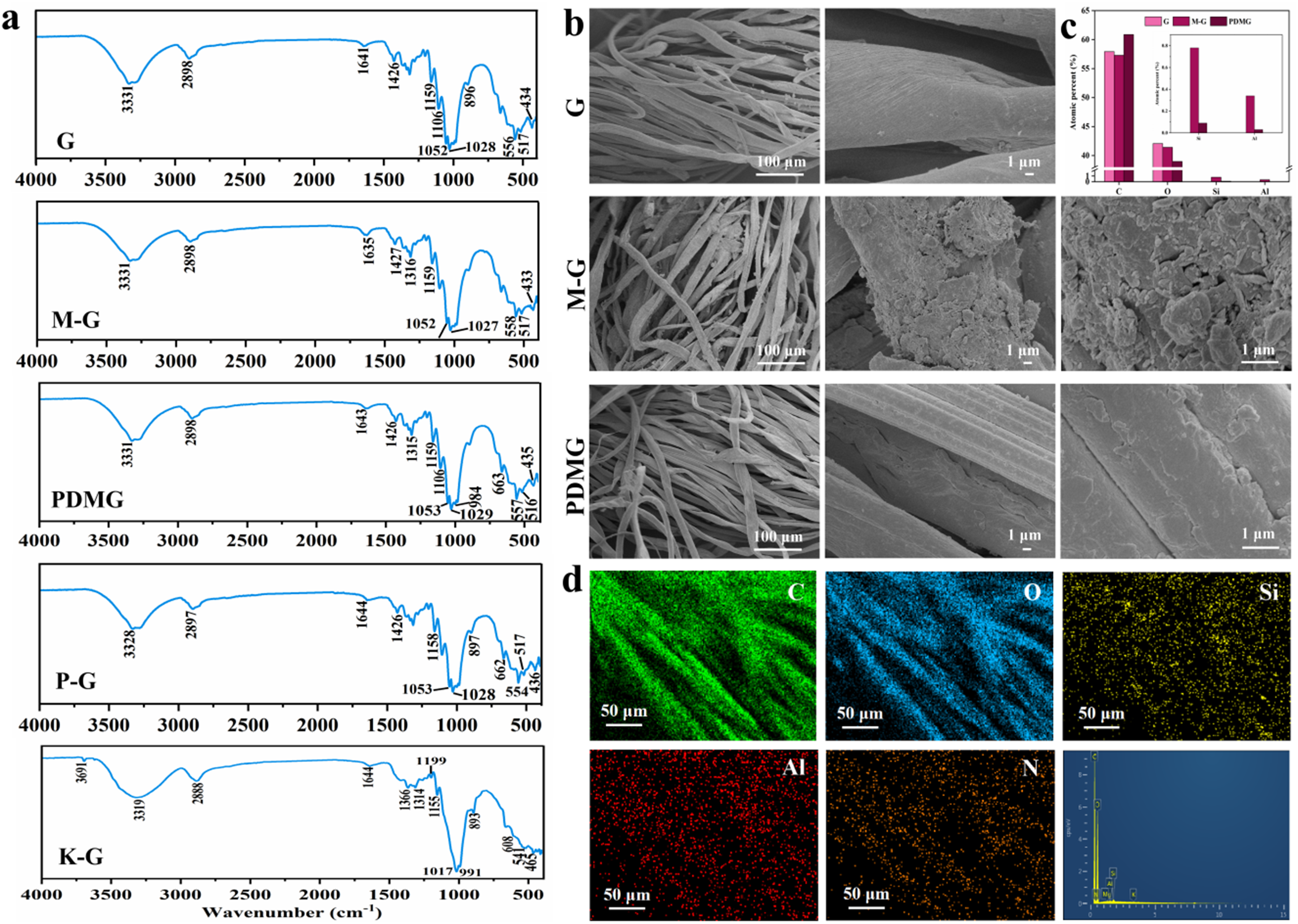
FTIR characterization was carried out to analyse the PDA, MDPal, gauze and composite gauze samples. As shown in Fig. 1a, the characteristic peaks of cellulose were found in the gauze, in which the peaks at 3331 cm–1 corresponded to the stretching vibration of hydroxyl groups (–OH) on the cellulose molecular chain, and the region of 1200–1000 cm–1 was attributed to the strong and complex C–O stretching vibration and C–O–C vibration regions. Moreover, the absorption bands at ∼900 and 2898 cm–1 were attributed to the characteristic peaks of β-glycosidic bonds and C–H stretching vibrations, respectively. In the gauze composites, dopamine monomers began to polymerize, and the primary –NH2 peaks disappeared and turned into secondary amine peaks. In addition, the strong C–O stretching vibration (catechol) decreased significantly at ∼1288 cm–1 (Fig. S1a). Thus, the catechol structure in the monomer can be assumed to have been consumed during oxidative polymerization and transformed into quinone or other structures.
(a) ATR-FTIR spectra of the gauze samples, (b) SEM images of G, M-G and PDMG, (c) main elemental contents of the gauze samples detected by SEM-EDS and (d) elemental mappings of PDMG.

Figure 1 Long description
The figure contains four panels labeled a, b, c and d. Panel a shows ATR-FTIR line spectra for five gauze samples: G, M-G, PDMG, P-G and K-G. The x-axis is labeled Wavenumber in cm superscript minus 1, ranging from 4000 to 500. The y-axis label is not legible in the image. Each sample is represented by a separate line spectrum stacked vertically. All five spectra share common peak positions. A broad peak appears near 3331 cm superscript minus 1 in G, M-G, PDMG and P-G; the K-G spectrum shows a corresponding peak near 3046 cm superscript minus 1. A peak near 2898 cm superscript minus 1 is visible in G, M-G, PDMG and P-G. Peaks near 1641 to 1645 cm superscript minus 1 appear across all samples. The region between 1200 and 1000 cm superscript minus 1 shows complex absorption features in all spectra, with labeled values including 1052, 1053, 1027, 1028 and 1029 cm superscript minus 1. Additional peaks near 900 and 435 cm superscript minus 1 are marked in several spectra. The K-G spectrum shows a distinct peak near 1199 cm superscript minus 1 and labeled values at 1017 and 601 cm superscript minus 1, differing from the other four samples. Panel b shows SEM micrographs for G, M-G and PDMG. Each sample row contains two images at different magnifications, with scale bars of 100 micrometers and 1 micrometer. The G images show smooth, elongated fiber bundles. The M-G images show fibers with visible surface texture differences compared to G. The PDMG images show fibers with a rougher surface and visible particulate deposits on fiber surfaces compared to G and M-G. Panel c shows a bar chart of main elemental contents for gauze samples detected by SEM-EDS. The x-axis shows element labels: C, O, N and Si. The y-axis label is not legible. Two bar series are shown per element, corresponding to two sample types indicated in the legend. C values appear highest across all elements for both sample types. Si bars are visibly elevated for one sample type compared to the other. Panel d shows elemental distribution maps for PDMG. Five maps are shown for elements C, O, Si, Al and N. Each map has a scale bar of 50 micrometers. An EDS spectrum plot is also present; the x-axis units are not legible and the y-axis label is not legible.
Generally, the FTIR spectra of the various samples indicated highly similar characteristic absorption peaks, suggesting that the gauze samples presented similar main chemical structures and functional groups. This might be due to the low content of the target components (e.g. MDPal) or due to their peaks being masked by the strong peaks of the gauze matrix. Furthermore, MDPal and PDA were not easy to be measure as they resided in the gauzes’ gaps, resulting in their detection being primarily related to the fibrous structure of the base gauze. However, the intensity of some characteristic peaks increased, such as at 1158, 1052 and 558 cm–1, indicating that there were interactions among the various components. For the K-G group, the main characteristic absorption bands of the matrix, the Si–O–Si vibration, the Si–OH vibration and the surface adsorbed water were detected at 465, 1155, 991 and 1644 cm–1, respectively (Xiao et al., Reference Xiao, Li, Huang, Gong, Diao, Wang and Bian2024), indicating the successful impregnation of the clay mineral on the gauze surface. The X-ray diffraction (XRD) trace of MDPal (Fig. S1b) suggested that MDPal mainly consisted of palygorskite (8.51°2θ, 19.89°2θ and 35.98°2θ; JCPDS card no. 88-1950), illite (8.9°2θ and 17.9°2θ; JCPDS card no. 26-0911), quartz (20.9°2θ, 26.60°2θ and 50.1°2θ; JCPDS card no. 78-1253) and a new whewellite phase (14.92°2θ, 15.30°2θ and 30.11°2θ; JCPDS card no. 75-1313), representing the mixed-dimensional structural properties of one-dimensional and two-dimensional clay minerals.
The presence of MDPal on the gauzes was determined using SEM and EDS. As shown in Fig. 1b, the SEM images show obvious differences between the basic gauze, M-G and PDMG gauze samples, where clay minerals were immobilized on the cotton fibres. In particular, the typical one-dimensional rod-like and two-dimensional lamellar morphologies were clearly observed on the surfaces of or gaps in the cotton fibres. After the incorporation of PDA, the surface of PDMG became smooth compared with M-G, and the morphology of the one- or two-dimensional clay minerals was not clearly visible, which was mainly due to the effect of the encapsulation of PDA. In addition, the chemical compositions of PDMG were analysed using SEM-EDS (Fig. 1d), and the main elements of Si and Al from MDPal and N from PDA were observed on the coated gauze, while only C and O were present in the basic gauze. By contrast, PDMG exhibited a higher atomic ratio (Fig. 1c).
Physicochemical properties of the gauze samples
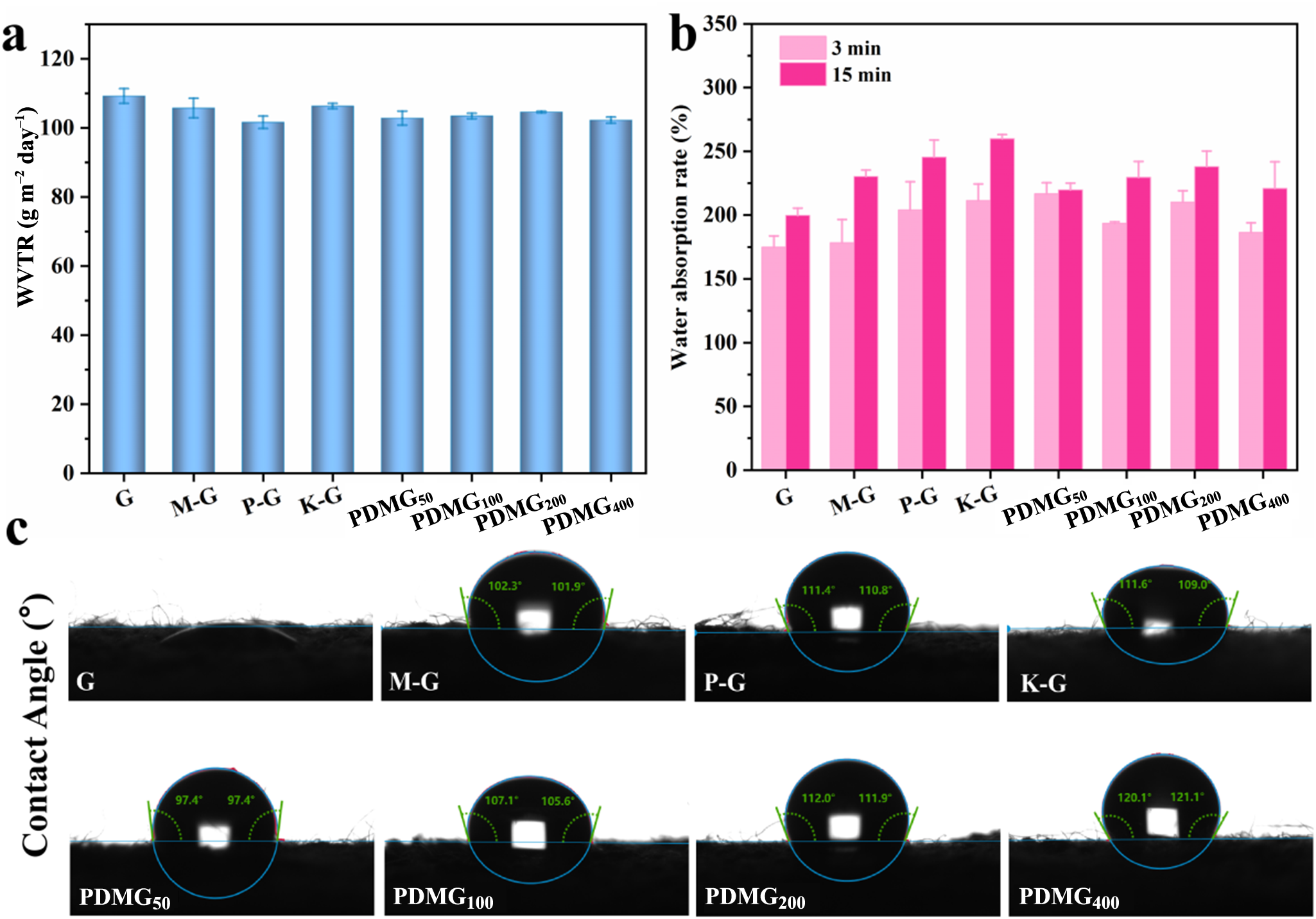
Important characteristics for the use of haemostatic gauzes in wound dressing are suitable water vapour transmission and moisture absorption properties (Khaliq et al., Reference Khaliq, Sohail, Shah, Mahmood, Kousar and Jabeen2022). We found that the WVTRs of the gauze composites were not influenced by the incorporation of MDPal, kaolin and PDA (Fig. 2a).
Physicochemical properties of the gauze samples. (a) Histogram of WVTR, (b) water absorption rate and (c) CAs of the various gauze samples to water.

Figure 2 Long description
The image A showing a bar chart of WVTR for eight gauze sample groups. The y-axis is labeled WVTR (g m superscript -2 day superscript -1) and runs from 0 to 120 with ticks every 20. The x-axis categories are G, M-G, P-G, K-G, PDMG, PDMG plus kaolin, PDMG plus PDA and PDMG plus kaolin plus PDA. Each category has one bar with an error bar. Bar heights are clustered near the 100 to 110 range on the y-axis. The image B showing a grouped bar chart of water absorption rate for the same eight gauze sample groups. The y-axis is labeled Water absorption rate (percent) and runs from 0 to 350 with ticks every 50. A legend shows two series: 3 min and 15 min. Each x-axis category has two bars with error bars. Across categories, the 15 min bars are higher than the 3 min bars. The tallest bars appear around the K-G category, with the 15 min bar near the upper 200s. The image C showing eight contact-angle images arranged in two rows of four, with a vertical label at left reading Contact Angle (degree). Each sub-image shows a droplet profile on a flat surface with an overlaid circular guide and numeric angle readouts near the droplet edges. The eight labels under the images match the x-axis categories: G, M-G, P-G, K-G, PDMG, PDMG plus kaolin, PDMG plus PDA and PDMG plus kaolin plus PDA.
Water absorption experiments were also conducted on the gauze samples at various times (Fig. 2b). With increasing time, the water absorption rate showed an increasing trend for all of the gauze samples. Among them, K-G displayed the greatest water absorption rate at 15 min. The dose of dopamine had an impact on their wettability, showing a trend of first increasing and then decreasing. These results suggest that the loaded clay minerals with inherently good water absorption properties improved the gauzes’ water absorption, complementing the role of PDA in bridging the gauze and clay minerals. Thus, PDMG demonstrated a synergistic effect that was related to the dosage of PDA and MDPal. It was shown that the amount of the loaded MDPal (Table S1) affected performance, with no benefit observed if the dosage was too high or too low.
Moreover, the hydrophilicity and hydrophobicity of the gauze surfaces were measured to gain further understanding of the surface contributions to haemostasis. As shown in Fig. 2c, the hydrophilicity of the gauze samples was assessed using CA tests. Compared with highly hydrophilic G due to its instant absorption (CA = 0°), the other gauze samples exhibited CA values in the range of 90–120° after contact with water molecules, and the gauze samples possessed obvious hydrophobic surfaces derived from PDA and MDPal. In particular, the CA values of the gauze samples increased linearly with dopamine content. It is reasonable to suggest that PDA and MDPal could interact with the fibrous structures (mainly hydroxyl functional groups) of the gauze, which would alter the spatial arrangement of surface functional groups, thereby affecting the wettability. Furthermore, the coating of MDPal might construct specific rough micro/nano-structures on the surface that would endow the gauze composites with hydrophobicity. This low-wetting property could contribute to keeping blood within wounds for forming clots, covering damaged vessels and reducing blood loss (Zhu et al., Reference Zhu, Wu, Zhao, Cai, Qian and Si2018; Li et al., Reference Li, Milionis, Zheng, Yee, Codispoti and Tan2019b). In addition, the highly hydrophobic surface of the modified gauze was revealed to have favourable anti-adhesion performance with respect to bacteria.
In vitro blood coagulation tests
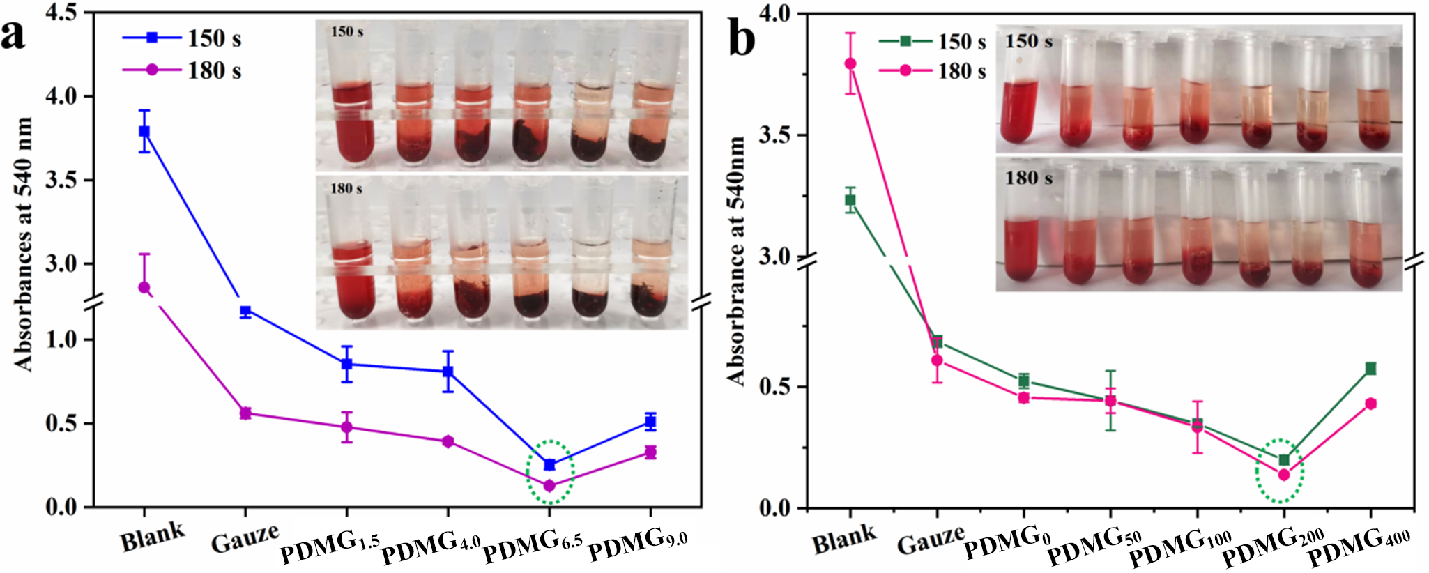
The excellent procoagulant potential of MDPal has been recognized in previous work (Wang et al., Reference Wang, Yang, Yang, Mu and Wang2024b). However, MDPal is easily detached from the substrate, which is the key factor affecting the haemostatic properties of gauze composites. PDA exhibits robust interfacial affinity and diverse functional groups that act as bridges to solve this problem. The two key factors of reaction duration and amount of dopamine were optimized by evaluating the dynamic coagulation effects of the gauze samples, where the lower absorbance of the released haemoglobin from the unbonded RBCs led to improved coagulation performance. The coagulation effects of the gauze samples first increased and then decreased with the prolonging in the reaction duration to both 150 and 180 s. Significantly, when the reaction duration was increased to 6.5 h, the colour of the supernatant became lighter, which was consistent with the findings from quantitative measurements, demonstrating the best coagulation activity (Fig. 3a). Compared with the blank group and basic gauze, the supernatant indicated a gradual lightening in colour with reaction duration, indicating an improvement in coagulation activity, which could also be determined quantitative from the absorbance value. A crucial contribution came from PDA, which increased the loaded amount of MDPal. However, the coagulation activity became weaker when the added amount of dopamine was over 200 mg. This phenomen on might be related to the rapid formation of PDA, which might hinder the increase in the loaded amount of MDPal on the surface of the gauze.
Optimization of the preparation conditions based on the blood coagulation test. (a) Reaction duration and (b) added amount of dopamine. The two dashed green circles on the graphs highlight that the PDMG6.5 and PDMG200 samples had the lowest absorbance values, which reflected the best clotting performances of the conditions of pH 6.5 and 200 mg of dopamine.

Figure 3 Long description
The image A showing a line graph and a set of sample tube images above the graph. The vertical axis label reads Absorbance at 540 nanometer. The vertical axis range shows 0.0 to 4.5. The horizontal axis categories read Blank, Gauze, PDMG subscript 1, PDMG subscript 2, PDMG subscript 3, PDMG subscript 4, PDMG subscript 5, PDMG subscript 6. The legend shows 150 s and 180 s. For 150 s, the plotted values by category are approximately: Blank about 3.0, Gauze about 1.2, PDMG subscript 1 about 0.9, PDMG subscript 2 about 0.8, PDMG subscript 3 about 0.8, PDMG subscript 4 about 0.3, PDMG subscript 5 about 0.4, PDMG subscript 6 about 0.5. For 180 s, the plotted values by category are approximately: Blank about 1.3, Gauze about 0.6, PDMG subscript 1 about 0.5, PDMG subscript 2 about 0.45, PDMG subscript 3 about 0.45, PDMG subscript 4 about 0.25, PDMG subscript 5 about 0.2, PDMG subscript 6 about 0.3. Error bars are drawn on the points. Above the graph are two rows of tube images labeled 150 s and 180 s. The image B showing a line graph and a set of sample tube images above the graph. The vertical axis label reads Absorbance at 540 nanometer. The vertical axis range shows 0.0 to 4.0, with a break mark on the axis. The horizontal axis categories read Blank, Gauze, PDMG subscript 1, PDMG subscript 2, PDMG subscript 3, PDMG subscript 4, PDMG subscript 5, PDMG subscript 6. The legend shows 150 s and 180 s. For 150 s, the plotted values by category are approximately: Blank about 3.9, Gauze about 0.6, PDMG subscript 1 about 0.5, PDMG subscript 2 about 0.45, PDMG subscript 3 about 0.45, PDMG subscript 4 about 0.35, PDMG subscript 5 about 0.2, PDMG subscript 6 about 0.45. For 180 s, the plotted values by category are approximately: Blank about 3.2, Gauze about 0.65, PDMG subscript 1 about 0.55, PDMG subscript 2 about 0.5, PDMG subscript 3 about 0.45, PDMG subscript 4 about 0.35, PDMG subscript 5 about 0.25, PDMG subscript 6 about 0.55. Error bars are drawn on the points. Above the graph are two rows of tube images labeled 150 s and 180 s.
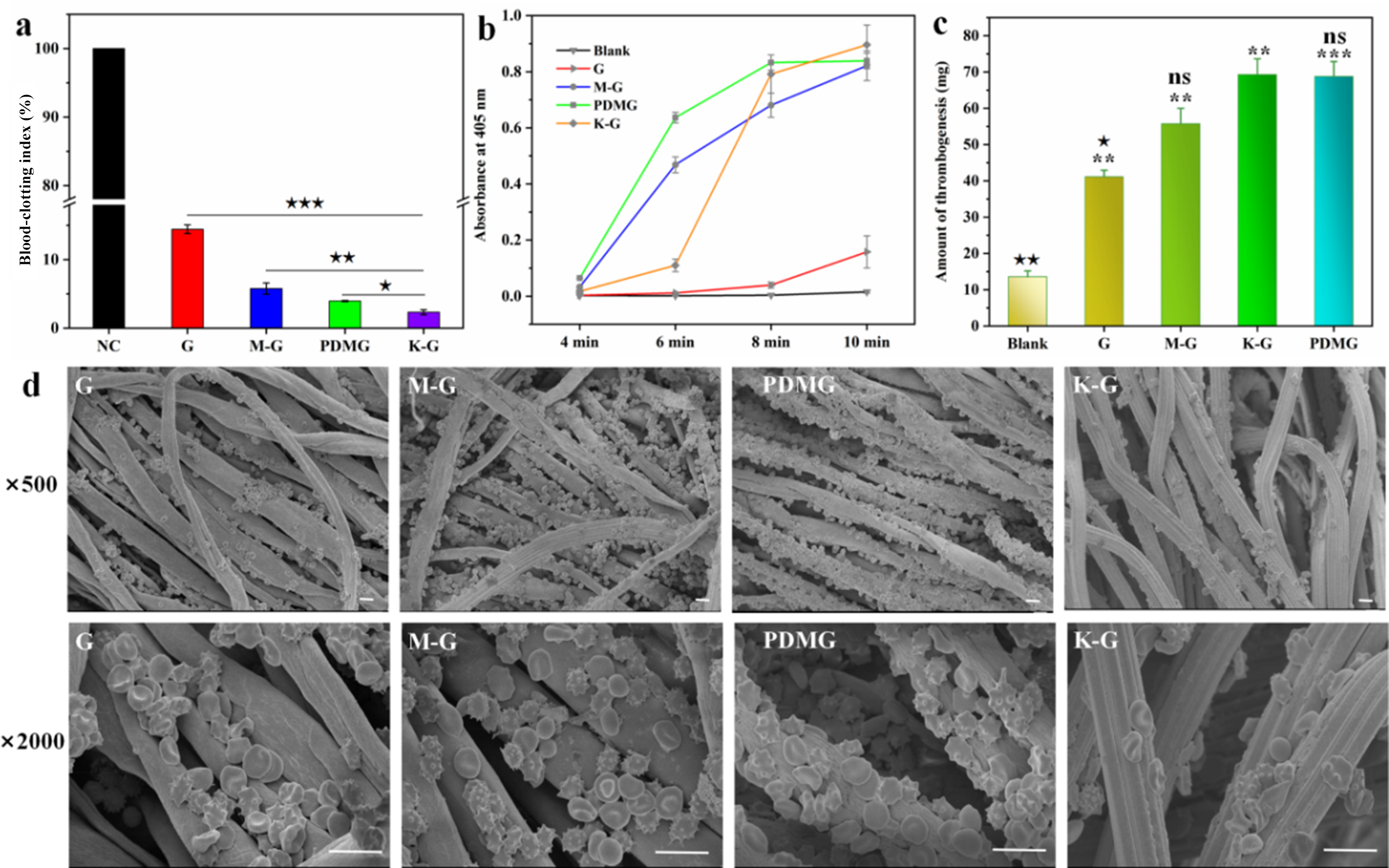
The BCI was employed to evaluate the haemostatic efficacy of the gauze samples. Erythrocytes that failed to be incorporated into the clot liberated free haemoglobin into the water. Therefore, a reduction in haemoglobin concentration directly correlates with a lower BCI, reflecting improved clotting performance. Compared with the negative control, all of the gauze samples showed lower BCI values. Among them, the commercial K-G gauze presented the lowest BCI value and the fastest coagulation, which were slightly better than those of the modified gauze, and it exhibited significant differences in these values compared with the M-G and G gauze samples (Fig. 4a).
In vitro blood coagulation tests of the gauze samples. (a) BCI (n = 3), (b) PPP coagulation (n = 3), (c) amount of thrombogenesis produced on the gauze samples after co-culturing with blood and (d) SEM images of adherent erythrocytes on the gauze surfaces at different magnifications (scale bar = 10 μm). Compared with the K-G group: ★p < 0.05, ★★p <0.01, ★★★p < 0.001; compared with the G or blank group: *p < 0.05, **p < 0.01, ***p < 0.001. NC = negative control; ns = non-significant.

Figure 4 Long description
The composite image consists of four parts: (a) A bar graph showing blood-clotting index percentages for different gauze samples: NC, G, M-G, PDMG and K-G. The K-G sample shows the lowest index. (b) A line graph depicting absorbance at 405 nm over time (4, 6, 8, 10 minutes) for Blank, G, M-G, PDMG and K-G samples. The K-G sample shows the highest absorbance. (c) A bar graph illustrating the amount of thrombogenesis in mg for Blank, G, M-G, K-G and PDMG samples. The K-G and PDMG samples show similar high values. (d) SEM images of gauze samples at two magnifications: 500 times and 2000 times. The images show the surface structure and adherent erythrocytes on M-G, PDMG and K-G samples.
Moreover, the presence of a large amount of plasma in the blood also plays a critical role in the coagulation process, and so it is essential to assess the plasma coagulation properties. The maximum fibrin level suggested plasma coagulation, and a shorter time to reach this maximum indicated a faster rate of plasma coagulation, and in this way the coagulation kinetics of PPP were evaluated. As shown in Fig. 4b, PDMG demonstrated a very rapid speed to triggering plasma coagulation, followed by the M-G group. After 8 min, the K-G group displayed the fastest rate of plasma coagulation, possibly illustrating the poor sensitivity of kaolinite to coagulation proteins and coagulation factors in the plasma coagulation process. Conversely, the MDPal loaded onto the gauze with PDA modification could improve wound haemostasis.
The thrombogenic capacity of the gauze samples was tested and compared with that of the blank sample (Fig. 4c). The incorporation of clays or clay minerals (kaolinite and MDPal) significantly improved the thrombogenesis of the gauze, but PDMG demonstrated greatly more significant differences from the blank group compared with the K-G group. This phenomenon might be due to the larger surface area of PDMG increased the contact between blood cells and silanol groups, thereby forming a stable haemostatic clot (Li et al., Reference Li, Lu, Shang, Ye, Yu and Lu2019a). Moreover, it was notable that the thrombogenic capacity of the M-G gauze was poor, which suggested the critical role of PDA in stabilizing MDPal on the gauze through electrostatic interaction.
During the process of controlling a haemorrhage, the adherent and aggregated erythrocytes are crucial components of the blood clot. Erythrocytes possess a biconcave discoid morphology with a central indentation on each face, and the adherent behaviour of erythrocytes was investigated for all of the gauze samples (Fig. 4d). Among the samples, PDMG demonstrated the highest number of adherent erythrocytes on its surface or gaps, which might be due to the physical absorption of MDPal and PDA, the latter of which not only acted as a bridge to increase MDPal loading but also modified the surface charge of the erythrocytes. These phenomena demonstrated the potential procoagulant effect of the PDMG gauze.
Biocompatibility
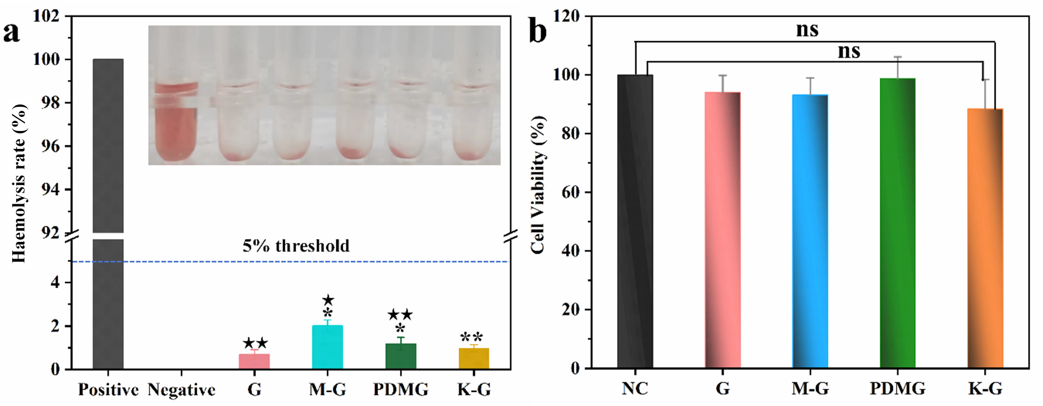
Before contact with the wound in the rat liver, the haemolytic potentials and cell viabilities of the gauze samples were evaluated. The haemolysis values of all of the gauze samples were less than 5% (Fig. 5a). Compared with the G group, M-G and PDMG displayed significant differences, and the K-G group demonstrated highly statistically significant differences, illustrating a less destructive nature to RBCs. Furthermore, the cytotoxicity of the gauze samples on NIH-3T3 cells during a 24 h incubation period was investigated (Fig. 5b). The cell viabilities of the extracts of the gauze samples were comparable to that of the blank sample, and the values ranged from 88.5% to 98.7%, demonstrating the good cytocompatibility and great potential for large wound management.
(a) Haemocompatibility and (b) cytocompatibility of the gauze samples. Compared with the K-G group: ★p < 0.05, ★★p <0.01, ★★★p < 0.001; compared with the G or blank group: *p < 0.05, **p < 0.01, ***p < 0.001. NC = negative control; ns = non-significant.

Figure 5 Long description
The image A showing a bar graph and a row of six sample containers. The vertical axis label is Haemolysis rate (percent). The vertical axis range is 0 to 102. The horizontal axis categories are Positive, Negative, G, M-G, PDMG and K-G. A horizontal line is labeled 5 percent threshold. Bar values: Positive 100; Negative 0; G 0; M-G 2; PDMG 1; K-G 0. The row of sample containers shows six containers aligned horizontally. The image B showing a bar graph. The vertical axis label is Cell viability (percent). The vertical axis range is 0 to 120. The horizontal axis categories are NC, G, M-G, PDMG and K-G. Bar values: NC 100; G 95; M-G 92; PDMG 98; K-G 88. Three bracket annotations above the bars are labeled ns, ns and ns.
Haemostatic experiments on animals
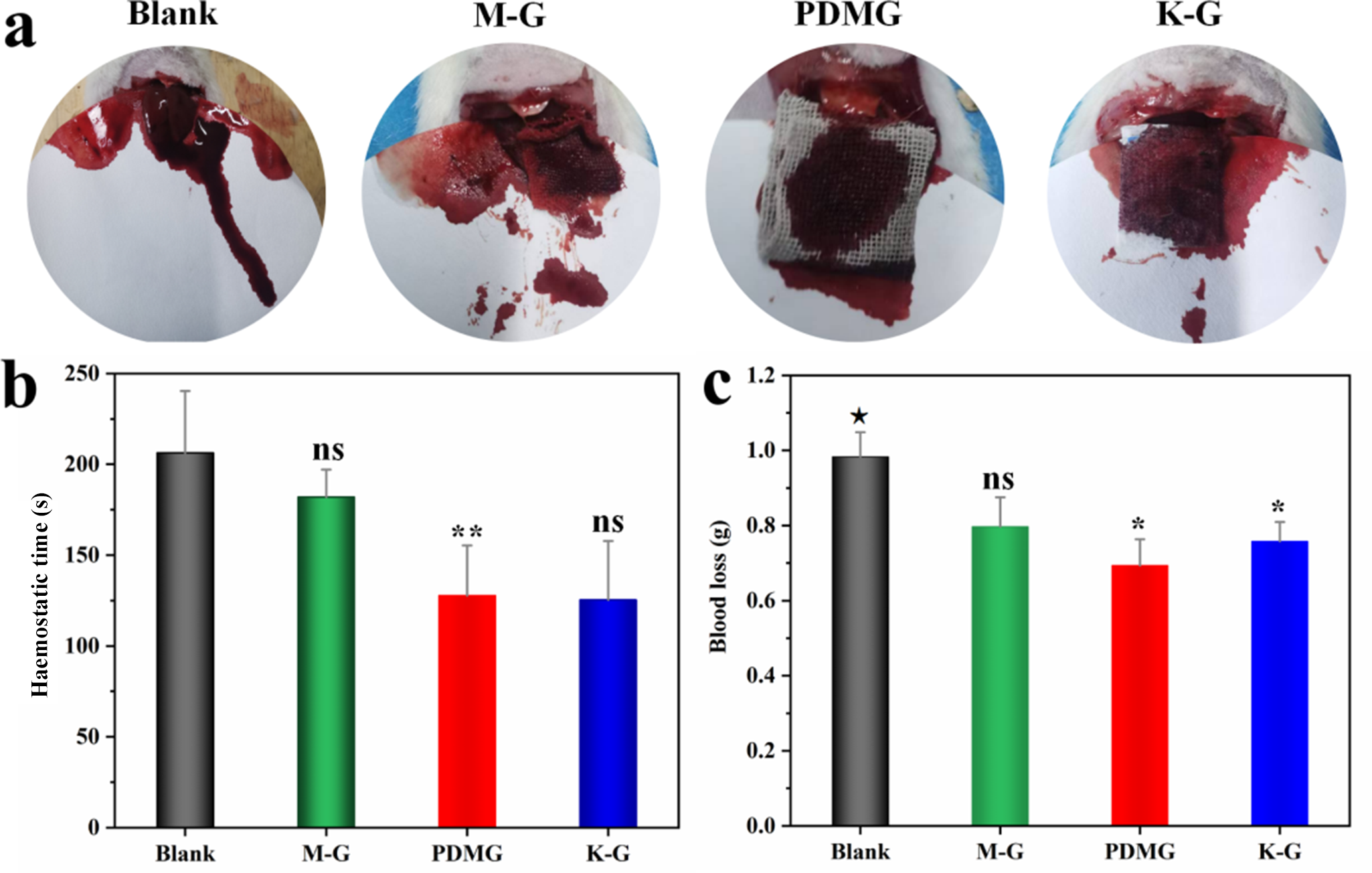
The gauze samples of G, M-G, PDMG and K-G were used to seal the wound sites of the rats. In the blank control group, there was more blood outflow onto the filter paper, and this phenomenon was significantly improved in the experimental groups, yet notable intergroup differences existed. Compared with the commercial gauze (K-G), significantly less blood was observed on the filter paper for the PDMG gauze, which also demonstrated advantages over the M-G group (Fig. 6a). Quantitative analysis of haemostatic time and blood loss was performed (Fig. 6b,c). The coagulation times of the M-G, PDMG and K-G gauze samples were 182.00 ± 15.12, 127.67 ± 27.76 and 125.33 ± 32.43 s, respectively, all of which were faster than that of the blank group (206.33 ± 33.98 s), but there were no significant differences compared with the K-G group. The bleeding volumes of the above groups were 0.7972 ± 0.0783, 0.6937 ± 0.070 and 0.7583 ± 0.0514 g, respectively, all of which were lower than that of the blank group (0.9833 ± 0.0655 g). These results demonstrated the potential procoagulant effect of PDMG.
(a) Photographs of bleeding and haemostatic processes of the liver haemorrhage model, (b) haemostatic time (s) and (c) blood loss (g). Compared with the K-G group: ★p < 0.05, ★★p <0.01, ★★★p < 0.001; compared with the G or blank group: *p < 0.05, **p < 0.01, ***p < 0.001. ns = non-significant.

Figure 6 Long description
The image A showing four circular photos arranged left to right, each with a label above it: Blank, M-G, PDMG, K-G. The image B showing a bar graph. The y-axis label is Haemostatic time (s). The y-axis range is 0 to 250. The x-axis categories are Blank, M-G, PDMG, K-G. Four vertical bars are shown: Blank: bar height at about 210. M-G: bar height at about 180, with text ns above the bar. PDMG: bar height at about 130, with text asterisk asterisk above the bar. K-G: bar height at about 125, with text ns above the bar. Each bar has a vertical error bar. The image C showing a bar graph. The y-axis label is Blood loss (g). The y-axis range is 0.0 to 1.2. The x-axis categories are Blank, M-G, PDMG, K-G. Four vertical bars are shown: Blank: bar height at about 1.0, with text asterisk above the bar. M-G: bar height at about 0.8, with text ns above the bar. PDMG: bar height at about 0.7, with text asterisk above the bar. K-G: bar height at about 0.75, with text asterisk above the bar. Each bar has a vertical error bar.
Discussion
The goal of this work was to develop a facile coating for reducing or avoiding the detaching of clay minerals from gauzes and to validate the great potential of such materials for practical application in wound haemostasis with in improving haemostatic properties. The modified gauze was prepared by coating it with PDA and loading MDPal via an impregnation method simultaneously. The composition and structural analysis indicated that MDPal was effectively loaded onto the PDA-modified gauze (Fig. 1). As the haemostatic performances of a material depends heavily on its structural characteristics, it was established that the polymerization time and dopamine concentration were critical factors to determining the physicochemical properties of PDA coatings. Our findings highlighted the role of these haemostatic gauzes in promoting coagulation, evaluated either with the BCI or via dynamic haemoglobin absorbance values. A reaction duration of 6.5 h and with 200 mg of dopamine were the optimal conditions for the PDA coating to load MDPal onto the gauzes, attributable to the synergistic effects of these conditions, which enabled improved coagulation activity. The stability of the optimized gauze was evaluated by peeling it off the wound, scratching the surface and twisting it and via thermal analysis, and the results suggested the good stability of the gauze composites (Fig. S2).
Moreover, it is widely recognized that blood (component)–material interactions are critical to revealing haemostatic mechanisms, especially for superior haemostatic property/contact-activation functionality, including plasma coagulation and enhanced erythrocyte-adhesion properties. Upon contact with plasma, the water and plasma proteins will quickly absorb onto the gauze to activate the coagulation factors, before then forming a preliminary blood clot. PDMG possessed fast moisture absorption capacity (Fig. 2b), which was helpful for the coagulation process, as well as suggesting the potential for the formation of protein crowns on nanomaterials, such as zeolite (Shang et al., Reference Shang, Chen, Castagnola, Liu, Boselli and Petseva2021), which would induce structural changes within the absorbed proteins, thereby mediating the haemostatic effects.
Combined with the CA results (Fig. 2c), it was found that the PDA-modified gauze had a highly hydrophobic surface, which could reduce unnecessary blood absorption and wound bacterial adhesion, and its haemostatic performance was significantly better than that of the original gauze. However, it was not the case that the higher the hydrophobicity, the better the haemostatic property of the modified gauze. Because the specific micro/nano-structure of the superhydrophobic surface enabled precise regulation of the exposed area and adhesive region available to blood components, excessive hydrophobicity would prevent the attachment of blood components. The results are in line with recent findings regarding superhydrophobic haemostatic gauzes/dressings (Chen et al., Reference Chen, Yang, Liu, Liu, Deng and Xu2024; Wu et al., Reference Wu, Cai, Xu, Wu, Li and Zhao2025).
The significantly improved haemostatic performance of the dopamine-modified gauze loaded with MDPal could be attributed to the synergistic effect of the hierarchical pore structure, high specific surface area and surface modification. Specifically, MDPal features a high specific surface area, a well-developed porous system (Table S2) and negative charge, providing numerous active adsorption sites for erythrocyte adhesion and platelet and coagulation factor activation in the blood, contributing to the acceleration of the formation of stable coagulation networks at the bleeding site. After dopamine modification, the dispersion and loading uniformity of MDPal on the gauze surface were greatly improved, which effectively alleviated particle agglomeration and limited the reduction of the specific surface area and the collapse of the porous structure. In addition, dopamine modification endowed the composite gauze with superior hydrophilicity, favourable bioadhesion and improved interfacial biocompatibility, further promoting blood adsorption, tissue adhesion and the firm construction of blood clots. Therefore, MDPal and dopamine modification synergistically accelerated the haemostatic process, which endowed the composite gauze with significantly better haemostatic properties than the samples of G and M-G.
During the later coagulation period, fibrinogen is converted into a fibrin network through the participation of thrombin, whereby erythrocytes can be aggregated and induced by haemostatic agents, especially those entrapped within the network, enabling the formation of firm blood clots (Long et al., Reference Long, Zhang, Huang, Chang, Hu and Yang2018). Erythrocytes are disc-shaped and biconcave in form on the surfaces of gauze samples. Among the gauze samples tested here, the PDMG group presented the highest number of the adherent erythrocytes, which were densely attached both to the surface and at the interstices of the gauze. The reason for this might be due to the physical absorption of erythrocytes onto MDPal, as well as the interaction between PDA and erythrocytes, resulting in a synergistic effect leading to the generation of optimal haemostatic performance. Moreover, a practical case applied in a liver haemorrhage model also demonstrated that PDMG has a potential procoagulant effect on wound haemorrhages, comparable to that of the commercially available K-G product, as well as it exhibiting good biocompatibility. These results provide a solid basis for extended studies, but the necessary experiments prior to its clinical use need to be conducted and evaluated in-depth.
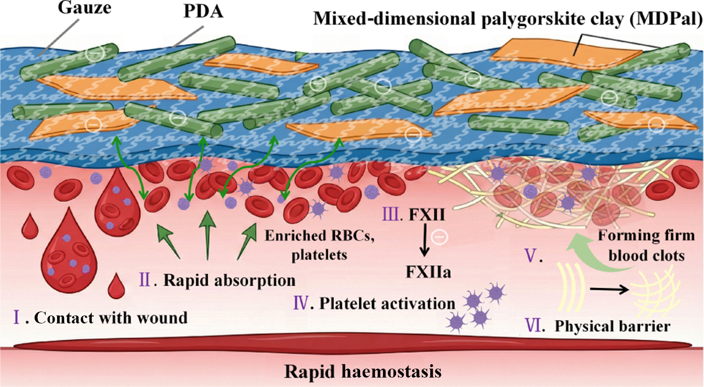
Therefore, the haemostatic mechanism of the as-prepared composite gauze is illustrated in Fig. 7. After being placed on a bleeding wound, the gauze quickly absorbs water molecules to concentrate the blood and promote erythrocyte and platelet activation and adhesion. It was reported that the negative charge could effectively promote the FXII self-activation pathway to trigger an intrinsic coagulation cascade (Jiang et al., Reference Jiang, Amantay, Tang, Osman, Jiang and Wang2026). Therefore, it is possible that the negative surface charge and other surface properties of MDPal would promote rapid haemostasis synergistically.
Haemostatic mechanism of PDMG (AI-assisted image generation).

Figure 7 Long description
The infographic illustrates the haemostatic mechanism of PDMG. It shows a layered structure with gauze, PDA and mixed-dimensional palygorskite clay (MDPal) at the top. The process begins with 'I. Contact with wound,' where blood droplets are shown. 'II. Rapid absorption' follows, with arrows indicating the movement of enriched RBCs and platelets. 'III. FXII' and 'FXIIa' are labeled, indicating activation. 'IV. Platelet activation' is depicted next. 'V. Forming firm blood clots' is shown with arrows leading to clot formation. Finally, 'VI. Physical barrier' is indicated, leading to 'Rapid haemostasis' at the bottom.
CD62P (P-selectin) is the most commonly used platelet activation marker in clinical practice. In this study, upon platelet activation, granular membrane proteins such as CD62P were detected on the platelet surface by flow cytometry. Our previous study demonstrated that the proportion of activated platelets in the MDPal group increased, providing direct evidence that strengthens the mechanistic conclusion that MDPal activated platelets (Wang et al., Reference Wang, Yang, Yang, Mu and Wang2024b). The exposed tissue factor triggers the extrinsic coagulation pathway and generates thrombin. Subsequently, the two pathways converge into a common pathway whereby prothrombinase complex assembly occurs on activated platelet surfaces, further promoting the production of thrombin for converting fibrinogen into fibrin (Stojanovski & Di Cera, Reference Stojanovski and Di Cera2022; Chen et al., Reference Chen, Yang, Liu, Liu, Deng and Xu2024). Then, fibrin polymerizes and assembles into a fibrous network to entrap blood cells, enabling the formation of firm blood clots (Kearney et al., Reference Kearney, Pechlivani, King, Tiede, Phoenix and Cheah2019; Miszta et al., Reference Miszta, Kopec, Pant, Holle, Byrnes and Lawrence2020). Moreover, the physical barrier effect of the gauze on facilitating rapid haemostasis is not negligible. Our haemostatic gauze, prepared using a facile approach, is efficient and biocompatible and has potential for rapid wound haemostasis applications. However, the long-term interfacial stability of the immobilized clay minerals and the effects of storage conditions on the performance of the composite gauze were not explored in this study. Relevant investigations into aging stability, particle shedding behaviour, efficacy in other models (e.g. arterial or venous injury, different animal species) and optimal storage environments will be conducted in our future work to support its practical application.
Conclusions
In summary, a highly safe and efficient MDPal-based haemostatic gauze was fabricated by simultaneous PDA coating and MDPal loading via an impregnation method. MDPal was tightly integrated into the gauze network with the assistance of PDA, which played a critical role in preventing the powder from shedding. The as-prepared gauze with suitable water vapour transmission, moisture absorption and stability exhibited efficient bleeding control. This could be attributed to the nano-heterogeneous structure of MDPal with its large surface area, which significantly improve prothrombin to thrombin conversion and accelerated clotting activity, and the synergistic effect of PDA and the network of gauze matrix improved the coagulation process. The in vivo experiments demonstrated that the loading of PDMG could effectively control bleeding so as to achieve rapid haemostasis, remarkably shortening bleeding times and reducing blood losses. This work provides a facile approach for the development of an efficient and biocompatible clay-based haemostatic gauze for improving wound haemostasis.
Supplementary material
The supplementary material for this article can be found at https://doi.org/10.1180/clm.2026.10039.
Author contributions
Xiaomei Wang and Yalong Li: Investigation, Methodology, Project administration, Writing – original draft; Fangfang Yang and Hongbin Zhao: Investigation, Resources, Data curation; Bin Mu: Writing – review & editing; Aiqin Wang: Conceptualization, Methodology, Writing – review & editing.
Financial support
This work was supported by the Major Project of the Joint Research Fund of Gansu Province (25JRRA1231), Young Talents of Gansu Province (2025QNGR65) and the AI software, supported by jieluoxuan, in drawing Fig. 7 of the haemostatic mechanisms.
Competing interests
The authors declare that they have no known competing financial interests or personal relationships that could have appeared to influence the work reported in this paper.
Data availability
No data were used for the research described in the article.