Recently, closure of ventricular septal defect has been performed safely and effectively with a low complication rate in infants or even in the neonatal period. Reference Meijboom, Szatmari and Utens1–Reference Aydemir, Harmandar and Karaci3 The complications of surgical closure could be residual defect, rhythm disturbance (complete atrioventricular block or right bundle branch block), or valvular dysfunction. Reference Lee, Cho and Kwak4,Reference Kouchoukos, Blackstone and Hanley5 In particular, rhythm disturbance is more likely to occur when we close a perimembranous type ventricular septal defect because of the conduction pathway along the postero-inferior margin of the ventricular septal defect, Reference McCarthy, Ho and Anderson6,Reference Titus, Daugherty and Edwards7 and tricuspid valve regurgitation also occasionally newly develops in association with a ventricular septal defect patch due to adhesion to valve structures or anchoring sutures passing through the septal leaflet. For suturing closing the perimembranous ventricular septal defect, closing sutures are traditionally placed 3 ∼ 4 mm or more away from the edge of the ventricular septal defect to avoid injury to the conduction pathway. Reference Jonas8
In our institution, since 2003, Dr KWH (one of the authors) introduced his alternative surgical technique for ventricular septal defect closure, placing the closing sutures just at the edge of the ventricular septal defect margin as shallowly as the suture needles were visible transparently through the endocardium to reduce the prevalence of complete atrioventricular block and right bundle branch block and tricuspid regurgitation. It was expected that not only injury of the His bundle but also block of the right bundle branch could be avoided. In this study, we sought to verify the hypothesis that this alternative method could reduce right bundle branch block and tricuspid valve regurgitation compared with the traditional method without increasing the risk for other complications in perimembranous ventricular septal defect.
Materials and methods
The study was approved by the Institutional Review Board of our institution. The individual informed consent was waived (approval number: 2111-102-1272).
Patients
We retrospectively reviewed the patients’ data who underwent closure of isolated perimembranous ventricular septal defect from January 2000 to December 2019. Of 813 patients who underwent this surgical procedure, 556 patients were included in this study. Cases which had insufficient operation record (n = 137), used continuous suturing method for ventricular septal defect patch closure (n = 116), and had already right bundle branch block preoperatively (n = 4) were excluded. We categorised these patients into two groups according to the method of ventricular septal defect closure: traditional (T) and alternative (A) methods. The traditional method was conducted by two surgeons from 2000 to 2016, and the alternative method has been applied since 2003 by the other two surgeons. We excluded cases with concomitant procedures that can affect the conduction pathway or tricuspid valve. Closure of the atrial septal defect or patent ductus arteriosus was included. Regarding the suturing, interrupted sutures were used in most patients (86.4%); therefore, we excluded the small number of patients in whom continuous sutures were used for ventricular septal defect closure to prevent a possible bias by the method of suturing methods. To evaluate long-term results, we enrolled patients who had been followed up at least 2 years after ventricular septal defect closure.
With these criteria, 556 patients were included in this study and we reviewed their operation records and analysed echocardiography or electrocardiogram of the perioperative period and last out-patient clinic visit. The median follow-up period of the cohort was 110.3 months (interquartile range: 49.2 ∼ 165.4 months).
Operative strategy and surgical techniques
All operations were performed under cardiopulmonary bypass and moderate hypothermia. After cardiopulmonary bypass support and aortic cross clamp, the ventricular septal defect was exposed through right atriotomy and closed via a tricuspid valve without detachment. All closures were made by an interrupted suture technique using polypropylene, polyester, or expanded polytetrafluoroethylene (GORE-TEX®, W. L. Gore & Associates Inc., Flagstaff, AZ, USA) sutures of 5–0, 6–0, 7–0, or 8–0 with a tiny spaghetti-shaped pledget (2 mm in length for 5–0 or 6–0 and 1.5 mm in length for 7–0 with 0.6 ∼ 0.7 mm of diameter hole). The patches used for closure were glutaraldehyde-fixed autologous pericardial patch or synthetic material, Dacron polyester patch (Bard® Sauvage® filamentous knitted polyester fabric, Bard Peripheral Vascular Inc., Tempe, AZ, USA). The material and size of the suture and patch were used depending on the surgeon’s preference, the size of the ventricular septal defect, and the size of the patients. After ventricular septal defect closure, the evaluation of tricuspid leaflet coaptation was routinely conducted by cold saline infusion into the right ventricle.
Traditional method (T) - The traditional method was that suture bites were made approximately 3 mm or more away from the edge of the ventricular septal defect (Supplementary Figure S1). In particular, the postero-inferior margin of ventricular septal defect is close at the conduction pathway. To avoid injury to the conduction system, the sutures were taken distant from this margin, outside of the conduction pathway.
Alternative method (A) - In the alternative method, the sutures were placed just at the edge of the ventricular septal defect margin, not away from the margin, as shallowly as the suture needles were visible transparently through the endocardium. The suture line along the edge of the ventricular septal defect excluded the conduction pathway (Fig. 1). A similar method was reported by Varghese et al in 2016. Reference Varghese, Saheed, Ravi, Sherrif, Agarwal and Kothandam9 When a fibrotic ridge was present in the sub-septal leaflet region, we attempted to expose this region by gentle retraction of the septal leaflet towards the atrial side and used this fibrotic tissue to anchor our ventricular septal defect sutures. If there was no sufficient tissue for anchoring sutures, we applied our interrupted ventricular septal defect sutures through the septal leaflet annulus. We no longer use continuous sutures near chordae or leaflets in order to avoid tricuspid valve deterioration by purse-string effect or entrapment by sutures. We also always try to position passing-septal leaflet sutures as close as possible to the annulus rather than the leaflet not to deteriorate tricuspid valve leaflet. To minimise tissue damage, we always use thin string and a short, small needle, and we handle all ventricular septal defect anchoring strings with care. We frequently use 8–0 polypropylene interrupted sutures for ventricular septal defect closure in neonates or infants with less than 3 kg of body weight with other CHDs requiring urgent corrective surgery, such as severely symptomatic tetralogy of Fallot, coarctation of aorta, or transposition of great arteries.
(A) An illustration of our alternative method (surgeon’s view), sutures were placed shallower, along the edge of a ventricular septal defect to exclude conduction pathway; (B), (C) operative pictures (surgeon’s view) that show passing needles are shown transparently through the endocardium (red circle) at postero-inferior margin of the ventricular septal defect of 28-day-old neonate.

Evaluation of clinical outcomes and statistical analysis
We primarily analysed the presence of right bundle branch block or tricuspid valve regurgitation according to operative technique. These features were evaluated not only in the immediate postoperative period but also at the out-patient clinic period, which was described as ‘late’ to observe changes. Risk factor analysis was performed to identify significant risk factors for the occurrence of right bundle branch block and the progression of tricuspid valve regurgitation using logistic regression analysis. Additionally, other complications related to heart rhythm, heart function, or performance status between the two methods were assessed.
Electrocardiogram - The evaluation of electrocardiogram included right bundle branch block, atrioventricular conduction block, and other conduction disturbances in patients. Right bundle branch block was defined based on AHA/ACCF/HRS Recommendations Reference Surawicz, Childers, Deal and Gettes10 by a QRS duration of ≥ 90 ms in children less than 4 years of age. R > S in V1; rSR’ in V1 or V2, and deep wide S-wave in I and V6. Incomplete right bundle branch block was defined by a QRS duration of 86 ∼ 90 ms with rSR’ in V1 or V2 in children less than 4 years of age. The changes in QRS duration throughout the follow-up period were measured. Postoperative right bundle branch block was defined only as complete right bundle branch block.
Echocardiography - Transthoracic echocardiography was performed during the perioperative period and follow-up period. Tricuspid regurgitation was evaluated according to the American Society of Echocardiography criteria: 0, none to trivial; 1, mild; 2, mild to moderate; 3, moderate; and 4, severe. Reference Zoghbi, Enriquez-Sarano and Foster11 The presence of residual shunt around ventricular septal defect patch, ventricular function, or other valvular problems were assessed.
Statistical analyses were performed using IBM SPSS statistical software (version 25.0; IBM Corp., Armonk, NY, USA). A p value of < 0.05 was considered statistically significant. Continuous variables are expressed as the mean or median value with standard deviation or inter-quartile ranges, and categorical variables are presented as frequencies with percentages. Differences in clinical factors between the groups were evaluated using Student’s t test for continuous variables and the chi-square test or Fisher’s exact test for categorical variables. A logistic regression analysis was used to evaluate risk factors associated with right bundle branch block or tricuspid regurgitation. The clinical or surgical variables were evaluated for their association with right bundle branch block using univariable logistic regression analysis. For multivariable analysis, the factors with a p value < 0.2 in univariable analysis were included.
Results
Patient characteristics
The baseline characteristics of the patients according to surgical technique are presented in Supplementary Table S1. The patients’ median age and body weight at the time of operation were 2.9 months (inter-quartile range: 1.9–5.3) and 5.1 kg (inter-quartile range: 4.3–6.5), respectively. The traditional method (Group T) was used in 370 cases (66.5%), and the alternative method (Group A) was used in 186 cases (33.5%). Autologous pericardial patch (n = 463, 83.3%) and polypropylene suture (n = 223, 40.1%) were the most frequently used materials for ventricular septal defect closure. Some patients (n = 102, 18.3%) had preoperative tricuspid valve regurgitation that was mild or more than mild.
The age and body weight of Group A patients were significantly lower than these of Group T. The cardiopulmonary bypass and aorta cross clamping time during surgery were longer in Group A than in Group T. The Dacron patch was used for closure of ventricular septal defect only in Group T (25.1%). In Group A, most of the surgeries used polypropylene suture material (98.4%) and 6-0 or 7-0 sutures, which was significantly smaller than that in Group T.
Clinical outcomes after ventricular septal defect closure
Overall, right bundle branch block was observed in 157 patients (28.2%) at immediate postoperative period, and in 192 patients (34.5%) at the last follow-up. Of all patients, 436 patients (78.4%) had follow-up data for at least 2 years after ventricular septal defect closure (Table 1). Postoperative right bundle branch block occurred more frequently in Group T (n = 109, 39.6%) than in Group A (n = 24, 14.9%, p < 0.001), and right bundle branch block still occurred more frequently in Group T (29.5%, p = 0.001) in patients with autologous pericardial patch. The status of right bundle branch block occurrence at the immediate postoperative period was maintained until the most recent follow-up (median follow-up duration: 110.3 months) (Fig 2); of 133 patients who had right bundle branch block at postoperative electrocardiogram, 126 patients (97.0%) still showed right bundle branch block at the most recent examination. Likewise, the patients without right bundle branch block had a similar rate of maintained electrocardiogram (84.3%). Divided as Group T (median follow-up duration: 139.0 months) and Group A (median follow-up duration: 71.3 months), a similar trend in right bundle branch block status was observed. Progression of tricuspid valve regurgitation was more common in Group T (16.9%) than in Group A (3.2%, p < 0.001) postoperatively.
Right bundle branch block difference between immediate postoperative and last follow-up according the group. (A; total patient, B; Group T, C; Group A).

ECG and echocardiography data according to suture lines (total N = 436)

All variables except EF showed significant difference with p-value < 0.05 between two groups.
ECG = electrocardiogram; EF = ejection fraction; QRSd = QRS duration; RBBB = complete right bundle branch block; TR = tricuspid valve regurgitation.
* Echocardiography data were found for only 256 patients (Group T: 198 and Group A: 158).
Postoperative electrocardiogram changes other than right bundle branch block occurred only in Group T. There were two patients with a 2nd degree atrioventricular block, one with bifascicular block and one with complete atrioventricular block requiring permanent pacemaker insertion during the follow-up period. Of all, the leakage at the closured ventricular septal defect was found in 7 patients, 5 (1.4%) in Group T and 2 (1.1%) in Group A. The all leakage of ventricular septal defect was very small sized and clinically not significant in terms of heart function and symptoms. Decreased functional status and limitation of ordinary activity were observed in 12 patients (2.2%) at out-patient clinic, mainly due to accompanying genetic syndrome. Most patients (99.4% of 343 patients who were checked with echocardiography at least 2 years after the operation) had normal cardiac function, > 56% ejection fraction or > 26% fractional shortening calculated on echocardiography at last examination.
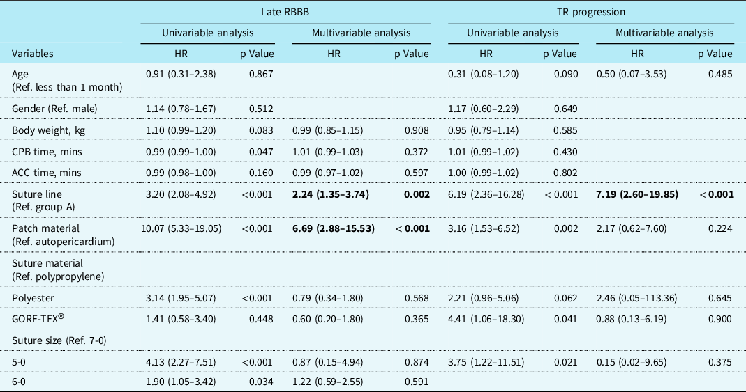
For late right bundle branch block occurrence, some clinical and surgical factors were analysed via logistic regression (Table 2). In univariable analysis, longer cardiopulmonary bypass time was related to lower right bundle branch block occurrence, and Group T, Dacron patch, polyester suture, and 5-0 sized suture were risk factors for late right bundle branch block. In multivariable logistic regression analysis, Group T and Dacron patch were significant risk factors for late right bundle branch block (hazard ratio = 2.24 (p = 0.002) and 6.69 (p < 0.001), respectively). For tricuspid valve regurgitation progression, only Group T was significant risk factor in multivariable logistic regression analysis (hazard ratio = 7.19, p < 0.001). In both Group T and A, progression of tricuspid valve regurgitation and late right bundle branch block did not have correlation to each other in Pearson chi-square test, however, it tended to show a marginal significant correlation in Group T (p value = 0.060 in Group T versus 0.425 in Group A).
Logistic regression analysis for late RBBB and TR progression

ACC = aortic cross clamp; CPB = cardiopulmonary bypass; HR = hazard ratio; RBBB = complete right bundle branch block; TR = tricuspid regurgitation.
Discussion
Postoperative right bundle branch block
Of the various types of ventricular septal defect, the perimembranous type has a risk of conduction injury due to the proximity of the conduction pathway along the postero-inferior margin of the ventricular septal defect during corrective surgery. Reference McCarthy, Ho and Anderson6,Reference Titus, Daugherty and Edwards7 The bundle branch of His is known to be located within 2 ∼ 3 mm from the posterior-inferior margin of the ventricular septal defect in the endomyocardium. Reference Yoshitake, Kaneko and Morita12 Direct injury to the conduction pathway or inflammation or oedema around the conduction pathway by sutures could cause conduction disturbances such as atrioventricular block or specific bundle branch block. In general, rhythm disturbance associated with ventricular septal defect closure occurs at relatively low incidence rate without critical problems except complete atrioventricular block. The incidence of complete atrioventricular block was reported to be approximately 1% Reference Tucker, Pyles, Bass and Moller13 and that of the right bundle branch block was reported to be almost 33%∼44% with traditional method. Reference Hobbins, Izukawa, Radford, Williams and Trusler14,Reference Fukuda, Suzuki, Kashima, Sato and Morikawa15
Closure of ventricular septal defect has been generally recommended for sutures to be placed 4–5 mm away from the ventricular septal defect margin to avoid injury to the His bundle and prevent the conduction disturbance. Reference Sabiston, Sellke, Del Nido and Sabiston16 In this study, we evaluated the safety of the alternative closing method of ventricular septal defect which involves placing the sutures shallowly just at the ventricular septal defect margin. With these shallow bites, we expected that our closing sutures could be still placed away from the conduction pathway that was presumed to go through the endocardium. Fukuda et al also reported a lower incidence of right bundle branch block (6.3%) with using shallow bites, which had a similar concept with ours. Reference Fukuda, Suzuki, Kashima, Sato and Morikawa15 Their methods showed significantly improved right bundle branch block rate compared with a conventional method (41.7%), in which the ventricular septal defect closing sutures were deeply placed at region more than 5 mm away from the ventricular septal defect margin. The similar alternative methods were also reported by Varghese et al, but above two studies had small number of perimembranous ventricular septal defect patients with older ages (about 5 ∼ 12 months) than our patients. Reference Varghese, Saheed, Ravi, Sherrif, Agarwal and Kothandam9,Reference Fukuda, Suzuki, Kashima, Sato and Morikawa15 Our study demonstrated the safety of our alternative method with younger patients (median 2.3 months, inter-quartile range:1.9 ∼ 5.3 months) in greater numbers and also evaluated the progression of tricuspid valve regurgitation and postoperative right bundle branch block with longer follow-up duration in isolated perimembranous ventricular septal defect.
In multivariable logistic regression analysis, the traditional method and Dacron patch were only significant factors for right bundle branch block occurrence. Although Dacron patch was known to induce inflammation and fibrosis, occasionally leading to conduction problems or outflow tract stenosis, Reference Desai, Hachana and Bukhari17,Reference Kalfa, Ghez, Kreitmann and Metras18 suture method was still significant factor for right bundle branch block occurrence. To exclude the effect of patch materials, we evaluated the right bundle branch block occurrence in patients with autologous pericardial patch. Of these patients, alternative suture method was still favourable factor for right bundle branch block occurrence (hazard ratio = 0.51, p value = 0.004). Regardless of suture materials, suturing method was significant risk factor for right bundle branch block after ventricular septal defect.
In our study, right bundle branch block occurred in 28.2% (n = 157) of patients after surgery for perimembranous ventricular septal defect closure which is similar to other studies. Reference Hobbins, Izukawa, Radford, Williams and Trusler14,Reference Fukuda, Suzuki, Kashima, Sato and Morikawa15 Although right bundle branch block is generally considered a benign feature in the normal population Reference Fleg, Das and Lakatta19 , several authors still reported some concerns, such as diastolic dysfunction, Reference Pedersen, Andersen, Knudsen, Christensen, Sørensen and Hjortdal20 or bradycardia by right bundle branch block resulting in abnormal functional capacity. Reference Heiberg, Eckerström and Rex21,Reference Heiberg, Redington and Hjortdal22 In addition, some studies have revealed that right bundle branch block in the general population could be a risk factor for reduced ventricular function and cardiovascular events, and a bifascicular block or complete atrioventricular block required for pacemaker implantation. Reference Bussink, Holst, Jespersen, Deckers, Jensen and Prescott23,Reference Fernández-Lozano and Brugada24 We had only one patient (0.2% of ours) who showed postoperative right bundle branch block that changed to complete atrioventricular block and eventually underwent permanent pacemaker implantation. We have another patient who developed bifascicular block from right bundle branch block at 18 years after surgery without any significant symptoms or signs. This patient should be closely monitored to ascertain the development of complete atrioventricular block.
Because the perimembranous ventricular septal defect is one of the most commonly accompanied CHDs with other cardiac anomalies (tetralogy of Fallot, pulmonary atresia, interrupted aortic arch, coarctation of aorta, etc.), right bundle branch block could be more critical in some patients with complex CHDs or in marginal ventricular function. We certainly know that right bundle branch block can occur due to many factors other than ventricular septal defect closure, right ventricular outflow tract reconstruction, right ventriculotomy, or clinical consequences induced by right ventricular dilatation or hypertrophy. However, we should try to reduce the incidence of right bundle branch block when we perform ventricular septal defect closure if we can.
Postoperative tricuspid valve regurgitation
Our alternative suture method was also thought to be related to a lower incidence rate of progression of tricuspid valve regurgitation at the last follow-up because the alternative method usually allows for a smaller patch than the traditional method. The ventricular septal defect patch could be adhesive to the tricuspid septal leaflet over time (Supplementary Figure S2) resulting in valvular incompetence. The smaller patch could be available by anchoring sutures being placed close to the ventricular septal defect margin with alternative method, and it might reduce a chance of adhesion to septal leaflet and of disturbance of leaflet motion.
We frequently observe chordae from the medial or inferior papillary muscle crossing the ventricular septal defect or running parallel to ventricular septal defect margin. We could prevent chordae entrapment by our sutures or ventricular septal defect patch if we have ventricular septal defect closure sutures placed just at the margin of the ventricular septal defect and use a smaller patch. We believe that our alternative method could reduce the incidence of newly developed tricuspid valve regurgitation in our patients with these reasons.
Study limitations
This study has several limitations. First, it was a single institutional and retrospective study. Selection bias is inevitable and the indication for surgery changed over time during study period, and little bit differed between the early period and late period of the study. Therefore, the patients’ characteristics were different in some baseline characteristics, such as age or body weight between the two groups. Second, the follow-up duration in the Group T (median: 123 months) was longer than that in Group A (median: 62 months), which might not allow us to review the long-term data of some of the patients in the Group A. However, as we described in Figure 2, the immediate postoperative findings tended to last until the most recent follow-up study; therefore, we do not expect the result to have changed even if we had followed up with the patients in Group A for a longer period of time.
The age and body weight of the patients in Group A were significantly younger and lower, which made the operation more difficult for surgeons (longer cardiopulmonary bypass and aortic cross clamp times in Supplementary Table S1). This factor could explain why a longer cardiopulmonary bypass time was statistically related to lower right bundle branch block occurrence in univariable analysis. We assume that the longer cardiopulmonary bypass and aortic cross clamp time were certainly affected not only by younger age and lower body weight of the patients but also by suturing methods; we think that shallow suturing at the edge of the ventricular septal defect should be performed very cautiously and that it requires more time to perform, especially for neonates or infants with much fragile endocardium. Considering all these disadvantages, we still had a lower incidence of postoperative right bundle branch block and progression of tricuspid valve regurgitation with the alternative suturing method, which must be beneficial compared with the traditional method.
Conclusion
Shallow sutures just at the edge of the perimembranous ventricular septal defect may reduce the rate of postoperative right bundle branch block occurrence and progression of tricuspid valve regurgitation without increased complications. Although the right bundle branch block and mild degree of tricuspid valve regurgitation were generally considered benign features, they could be critical for the patients with complex CHD, and we should try to reduce the incidence of right bundle branch block when we perform ventricular septal defect closure if we can.
Supplementary material
The supplementary material for this article can be found at https://doi.org/10.1017/S1047951123002470
Acknowledgements
We authors are grateful to Medizin Atelier’s illustrator, S.U. Kim for creating Figure 1(a).
Financial support
This research received no specific grant from any funding agency, commercial, or not-for-profit sectors.
Competing interests
None.