Thoracic aortovenous fistulas are extremely rare and the majority are secondary to trauma. It is rarely that these represent an isolated congenital anomaly and have never been reported to be associated with other structural cardiac diseases that also cause left-to-right shunt of blood. We describe a case of a 13-month-old girl who presented with symptomatic congenital aortocaval fistula with coexisting secundum atrial septal defect. She improved clinically following a successful percutaneous closure of the aortocaval fistula and is monitored regularly for her atrial septal defect.
Case history
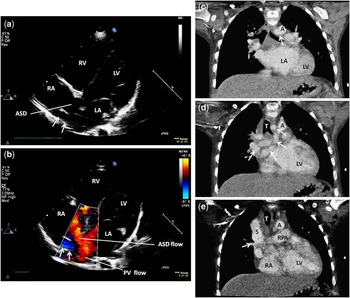
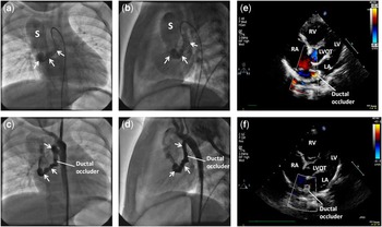
A 13-month-old girl with a secundum atrial septal defect was referred to our centre for further assessment and treatment because of an uncharacteristic significant symptom. Her growth was below the third percentile. At presentation, her weight was 8.1 kilograms and height was 75.5 centimetres. She was comfortable at rest but became sweaty and clammy on exertion or when crying. She had a grade I continuous murmur on the praecordium. Her electrocardiogram showed sinus rhythm with incomplete right bundle branch block and prominent P waves. Transthoracic echocardiogram showed a vascular structure above the dilated atria draining towards the right atrium and the presence of a secundum atrial septal defect (Fig 1a, b). Systolic function of both ventricles was normal but the right ventricle was mildly dilated. Thoracic computed tomography showed an aortocaval fistula originating from the junction between the descending and arch of aorta and draining into the posterolateral aspect of a dilated superior caval vein close to the right atrium (Figs 1c–e and 2a, b). She had cardiac catheterisation and the fistula was occluded using a 6/4 millimetre Amplatzer® Duct Occluder (AGA Medical Corporation, Minnesota, United States of America). There was residual shunting of blood through the device immediately after the procedure (Fig 2c and d), but complete occlusion was observed on transthoracic echocardiogram 2 months later (Fig 2e and f) with resolution of her symptoms. She continues to be under regular monitoring for her atrial septal defect.
(a) and (b) are the modified apical four-chamber transthoracic views showing the aortocaval fistula (white arrow →) and atrial septal defect (ASD) with colour flow mapping demonstrating that the direction of blood flow in the aortocaval fistula was opposite to that of the blood draining into left atrium (LA) via pulmonary vein (PV). On the coronal sections of the computed tomography angiogram (c–e), the fistula appears to originate from the junction between the descending and arch of aorta, ascending on the right of and parallel to the arch of aorta before descending anterior to the left main bronchus, and crosses the midline posterior to the right pulmonary artery (RPA) to drain into the posterolateral aspect of a dilated superior caval vein (S) close to the right atrium (RA). A, aorta; LV, left ventricle; RV, right ventricle.

The (a) anteroposterior and (b) lateral contrast fluoroscopic views of the aortocaval fistula (white arrow →) before an Amplatzer Ductal Occluder was deployed (c) and (d) at the proximal third of the fistula. Residual shunt was seen immediately after the procedure. Modified apical five-chamber transthoracic echocardiographic views showing (e) minor residual shunt a day after the procedure, which (f) resolved 2 months later. LVOT, left ventricular outflow tract.

Discussion
Congenital aortocaval fistulas are extremely rare, with only a few published case reports.Reference Soler, Mehta, Garcia, Kaiser and Tamer 1 , Reference Oomman, Mao, Krishnan and Girinath 2 To our knowledge, this is the first reported case of aortocaval fistula with an associated secundum atrial septal defect. Congenital aortovenous fistula is often used as a common term to describe a larger but rare group of congenital thoracic arteriovenous fistulas. The commonest forms include those that arise from the aortic root or ascending aorta and that are associated with rupture of the sinus of valsalva or a dissecting aneurysm and coronary fistula.Reference Antunes, Marhino, Rodrigues, Melo, Oliveira and Providencia 3 These arteriovenous fistulas commonly drain into the right cardiac chambers and pulmonary arteries.Reference Antunes, Marhino, Rodrigues, Melo, Oliveira and Providencia 3 However, these fistulas can originate from any segment of the thoracic aorta,Reference Soler, Mehta, Garcia, Kaiser and Tamer 1 – Reference Coto, Caffarena, Such and Marques 5 coronaries arteries,Reference Perry, Rome, Keane, Baim and Lock 6 and larger thoracic arteries including subclavian,Reference Awasthy, Tomar, Radhakrishnan and Kumar 7 – Reference Auge, Bosch, Crexells and Oriol 9 brachiocephalic,Reference Gutierrez, Monaco, Hartmann and McKnight 10 intercostal,Reference Awasthy, Tomar, Radhakrishnan and Kumar 7 and internal mammaryReference Awasthy, Tomar, Radhakrishnan and Kumar 7 arteries and drain into the innominate vein,Reference Antunes, Marhino, Rodrigues, Melo, Oliveira and Providencia 3 superior caval vein,Reference Soler, Mehta, Garcia, Kaiser and Tamer 1 , Reference Oomman, Mao, Krishnan and Girinath 2 azygote vein,Reference Soler, Mehta, Garcia, Kaiser and Tamer 1 right cardiac chambersReference Rosenberg, Williams and Trusler 4 , Reference Coto, Caffarena, Such and Marques 5 (cameral fistulas) coronary sinus, and pulmonary circulation.Reference Auge, Bosch, Crexells and Oriol 9 There may be multiple fistulasReference Antunes, Marhino, Rodrigues, Melo, Oliveira and Providencia 3 or a fistula that drains into more than one venous systems.Reference Soler, Mehta, Garcia, Kaiser and Tamer 1
The clinical presentation of congenital aortocaval fistula mimics that of conditions that cause left-to-right shunt of blood. Patients may be asymptomatic but can present with non-specific symptoms such as failure to thrive or suffer from respiratory distress depending on the severity of the shunt. A continuous murmur can commonly be heard on the praecordium and back. Imaging modalities such as echocardiography, computed tomography angiogram, magnetic resonance imaging, and invasive cardiac catheterisation help define the anatomy and assess the severity of the shunt. Percutaneous or surgical closures are the definitive treatment options. Antunes et alReference Antunes, Marhino, Rodrigues, Melo, Oliveira and Providencia 3 reported a case of an 8-year-old child who had presumably an incomplete closure of an aortocaval fistula with two Gianturco coils. Recanalisation of the fistula gradually increased and a repeat intervention with the insertion of a third coil was required 7 years later. Following complete embolisation of the fistula, there was no sign of recanalisation detected 6 months later.Reference Antunes, Marhino, Rodrigues, Melo, Oliveira and Providencia 3 Therefore, these patients should be reassessed following closure of the fistula, especially in the presence of a residual shunt as this may recanalise later.Reference Antunes, Marhino, Rodrigues, Melo, Oliveira and Providencia 3
Conclusion
Congenital aortocaval fistula is a very rare cause of left-to-right shunt of blood and percutaneous closure is often feasible. Patients should be thoroughly assessed for the presence of multiple fistulas or other structural cardiac disease and reassessed following the closure. Close monitoring is mandatory in the presence of residual shunt as recanalisation or increase in the severity of shunt may occur.