Introduction
The Aspidogastrea Faust & Tang, 1936 is a basal group of parasitic trematodes characterized by having a ventral holdfast organ with rows of alveoli or suckerlets, or just presenting a row of rugae or suckers (Rohde, Reference Rohde and Rohde2005). Aspidogastridae Poche, 1907 are known to parasitize molluscs (obligate host) and vertebrates such as teleosts and turtles (facultative or obligate final hosts) (Rohde, 2002). Aspidogastrinae Poche, 1907 currently comprises the genera Aspidogaster Baer, 1827, Lophotaspis Loss, 1902, Lobatostoma Eckmann, Reference Eckmann1932, Multicotyle Dawes, 1941, Sychnocotyle Ferguson, Cribb & Smales, 1999 and Neosychnocotyle Snyder & Tkach, 2007.
The genus Lobatostoma was reported to parasitize molluscs and teleosts, and out of the nine valid species, six have been described from fish of the genus Trachinotus Lacépède, 1801 (Perciformes: Carangidae). The remaining species were described from fish of the families Haemulidae, Scianidae and Cichlidae (the only freshwater host).
During a survey of fish parasites off the Brazilian coast, adult specimens of L. kemostoma (MacCallum & MacCallum, Reference MacCallum and MacCallum1913) were recovered from the intestine of Trachinotus carolinus (Linnaeus, 1766). This carangid fish is distributed in the western Atlantic from Massachusetts, USA to Argentina, feeding on invertebrates and small fish (Froese & Pauly, Reference Froese and Pauly2013).
The description of L. kemostoma was carried out in the early 20th century and some taxonomically important morphological features, e.g. detail of the ventral disc with the arrangement and number of alveoli, were incompletely described, indicating the need for a morphological review. Therefore, L. kemostoma is redescribed on the basis of examination of type series and voucher specimens deposited in the US National Parasite Collection (USNPC) and specimens from the Helminthological Collection of the Instituto Oswaldo Cruz (CHIOC), Rio de Janeiro, Brazil, along with freshly collected specimens from T. carolinus.
The present paper also provides new data on surface ultrastructure of the aspidogastrid observed using scanning electron microscopy (SEM). Further, we discuss a lectotype designation to L. kemostoma.
Materials and methods
Collection and examination of fish
Thirty-eight specimens of T. carolinus were caught by commercial trawlers in Angra dos Reis (23°00′S 44°10′W), coastal zone of Rio de Janeiro State, Brazil. Sampling was carried out from June 2012 to March 2013. Fish were kept in thermal boxes filled with ice prior to being transported to the laboratory and then immediately dissected. Recovered trematodes were fixed and stored in 70% cold alcohol or 5% formaldehyde solutions. The mean intensity of infection is given, followed by the standard deviation with the range in parentheses. Fish nomenclature and authorities follow Froese & Pauly (Reference Froese and Pauly2013).
Morphological analysis
For light microscopy, specimens were stained with Mayer's carmalum or Delafield's haematoxylin, cleared in eugenol and mounted in Canada balsam. Illustrations were made with the aid of a drawing tube attached to a Leica™ DM LS2 microscope (Leica Microsystems, Wetzlar, Hesse, Germany). Measurements are given in micrometres (μm) unless otherwise stated, with the means in parentheses.
For scanning electron microscopy, specimens were fixed for 1 h at room temperature with 2.5% glutaraldehyde in 0.1 m sodium cacodylate buffer. After washing in the same buffer, specimens were post-fixed for 3 h at room temperature in 1% osmium tetroxide in 0.1 m sodium cacodylate buffer. Then the material was dehydrated in an ascending acetone series, dried by the critical point method with CO2, mounted with silver cellotape on aluminium stubs and sputter-coated with a 20 nm thick layer of gold. Samples were examined using a Jeol JSM-6791F microscope (Jeol, Tokyo, Japan) at an accelerating voltage of 15 kV.
Molecular analysis
For genetic analysis one specimen of L. kemostoma was prepared for total genomic DNA extraction using the phenol/chloroform method as described by Billings et al. (Reference Billings, Yu, Teel and Walker1998). To amplify gene fragments of 28S rDNA, a set of primers was used: LSU5/Dig 12/1500R (Tkach et al., Reference Tkach, Littlewood, Olson, Kinsella and Swiderski2003). Polymerase chain reactions (PCR) were performed in a volume of 50 μl with 20 mm Tris–HCl at pH 8.4; 50 mm KCl; 250 μm each deoxynucleoside triphosphate (dNTPs); 1.5 mm MgCl2; 0.5 μm each oligonucleotide primer; 1.25 U of Platinum Taq DNA polymerase (Invitrogen, Carlsbad, California, USA) and 2 μl of genomic DNA. PCR was carried out using cycling parameters as described previously (Tkach et al., Reference Tkach, Littlewood, Olson, Kinsella and Swiderski2003). PCR products were visualized with Sybergreen (Invitrogen, Eugene, Oregon, USA) staining before electrophoresis on 1.5% agarose gels. All amplified PCR products generated were purified with Wizard SV gel and PCR clean-up system kit (Promega, Madison, Wisconsin, USA) following the manufacturer's instructions, and sequenced in both directions using the same primer sets as in the respective PCR assay. DNA cycle-sequencing reactions were performed using BigDye v.3.1 chemistry (Applied Biosystems, Foster City, California, USA). Sequencing reactions were performed in the ABI Prism 3100 sequence analyser (PE Biosystems, Foster City, California, USA). Sequences were assembled and edited in DNASTAR SeqMan (DNASTAR, Inc., Madison, Wisconsin, USA). The edited sequences were compared for similarities with sequences from GenBank using BLAST 2.0 (Basic Local Alignment Search Tool) (Altschul et al., Reference Altschul, Gish, Miller, Myers and Lipman1990). To examine the phylogenetic relationships, nucleotide sequences were analysed by the CLUSTAL W algorithm of the Bioedit Package (Thompson et al., Reference Thompson, Higgins and Gibson1994; Hall, Reference Hall1999). The phylogenetic tree was inferred by using the software MEGA 5.0 (Tamura et al., Reference Tamura, Peterson, Peterson, Stecher and Nei2011) utilizing the Kimura 2 parameters model (K2P) and gamma distribution selected by MEGA 5.0. The maximum likelihood method was used to construct the tree (Felsenstein, Reference Felsenstein1981) and resampling with 5000 bootstrap replicates was used to evaluate the reliability of the groups. New sequences of L. kemostoma were deposited in GenBank with accession numbers KF561238 and KF561239.
Parasite identification and museum collections
The aspidogastreans were identified at generic and specific level according to Rohde (2002) and Oliva & Carvajal (Reference Oliva and Carvajal1984). For comparative purposes, four syntypes (USNPC 035565.02; 036393.02 with three specimens) of L. kemostoma collected by G.A. MacCallum in the intestine of T. carolinus were studied, together with four slides (USNPC 083016) deposited by Bunkley-Williams et al. (1996). In addition, six voucher specimens of L. kemostoma from T. carolinus off the Brazilian coast (CHIOC 31.450a–c; 31.451a–b; 32.129) were examined. Voucher specimens are deposited in the Helminthological Collection of Oswaldo Cruz Institute (CHIOC) at Fundação Oswaldo Cruz (FIOCRUZ), Rio de Janeiro, Brazil, and in the United States National Parasite Collection (USNPC), Beltsville, Maryland, USA.
Results
A total of 49 specimens of L. kemostoma were collected in T. carolinus from the coastal zone of Rio de Janeiro. Of the 38 fish examined, 16 were infected (prevalence 42.1%) and the mean intensity was 3.06 ± 3.99 (range 1–16).
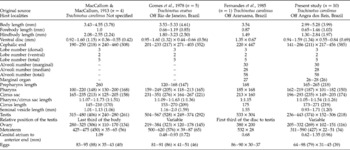
A redescription of L. kemostoma based on a comparative investigation of freshly collected material with a critical analysis of syntypes and voucher specimens is presented, with a lectotype designation. New measurements taken from the specimens available at USNPC and CHIOC, previously described by MacCallum & MacCallum (Reference MacCallum and MacCallum1913), Gomes et al. (Reference Gomes, Fábio and Rolas1978) and Fernandes et al. (Reference Fernandes, Kohn and Magalhães1985), were compared with specimens collected herein (table 1).
Comparative morphological and morphometric characteristics (measurements in μm unless stated) of Lobatostoma kemostoma (MacCallum & MacCallum, Reference MacCallum and MacCallum1913); n= number of specimens examined; accession numbers are given in the text.

Redescription
Description of Lobatostoma kemostoma (MacCallum & MacCallum, Reference MacCallum and MacCallum1913)
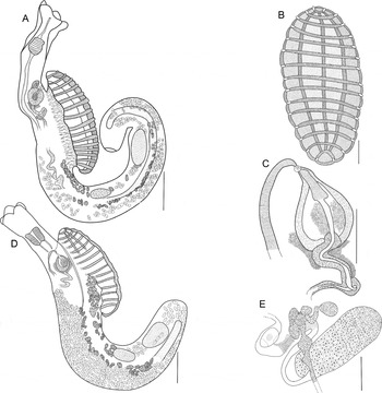
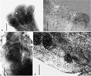
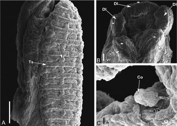
Body elongate 2.99–5.28 (3.99) mm long, wider at level of ventral disc (fig. 1A). Tegument aspinose. Ventral disc oval, 0.94 – 1.59 (1.34) mm long, projects from body by bands of muscles and covers c. one-third of the body length (fig. 1A, B); disc width 554–841 (689). Internal septum separating the ventral disc from the body proper observed in just some specimens. Forebody, from anterior end to proximal base of the ventral disc, 0.65–1.46 (1.03) mm long; hindbody (tail), from distal base of the ventral disc to posterior end, 1.30–2.84 (1.97) mm long (fig. 1A). Ventral disc with single alveolus on both poles followed by four longitudinal rows of alveoli divided by three longitudinal septa and 15 transverse septa, forming a total of 58 alveoli (counted before mounting) (fig. 1B, see also fig. 3A). Each transverse septum possesses marginal organs 26–28 (26) in diameter with ampulla on both sides (figs 1B, 2B). Anterior end lobed 141–286 (211) long × 217–456 (385) wide; two ventral and three dorsal petal-like lobes, highly muscular and similar in size and shape (figs 1A, 2A, 3B); longitudinal grooves in each lobe end (fig. 3B). Oral opening in the midst of cephalic lobes (figs 2A, 3B). Prepharynx 165–265 (218) long; pharynx well developed 162–219 (187) long × 101–182 (150) wide. Oesophagus not observed. Caecum thick-walled, blind, reaching the posterior third of body (fig. 1A).
The morphology of Lobatostoma kemostoma from Trachinotus carolinus from the Atlantic Ocean: (A) lateral view; (B) ventral view of the disc; (C) terminal genitalia of lectotype (USNPC 035565.02); (D) ventro-lateral view of lectotype (USNPC 035565.02); (E) ovarian complex of paralectotype (USNPC 036393.02). Scale bars: 500 μm (A, D); 200 μm (B); 100 μm (C, E).

The morphology of Lobatostoma kemostoma from Trachinotus carolinus off the Brazilian coast: (A) ventral view of oral opening (arrow) surrounded by five cephalic lobes; (B) marginal alveoli with two marginal organs (arrows) on transverse septa; (C) lateral view of terminal genitalia; (D) embryonated eggs with opercular opening (arrow). Abbreviations: c, cirrus; cs, cirrus sac; ed, ejaculatory duct; e, embryo; ga, genital atrium; m, metraterm; pg, prostatic glands. Scale bars: 100 μm (A); 50 μm (B, C, D).

The morphology of Lobatostoma kemostoma from Trachinotus carolinus off the Brazilian coast: (A) ventro-lateral view of ventral disc with grooves (arrow) at transverse septa; (B) cephalic lobes surrounding the oral opening with grooves in each lobe end (asterisks); (C) knob-shaped cirrus through the genital pore (arrowheads). Abbreviations: Co, cirral opening; Dl, dorsal lobes; Ls, longitudinal septum; Ts, transverse septum; Vl, ventral lobes. Scale bars: 100 μm (A); 20 μm (B); 10 μm (C).

Testis oval to round, 236–443 (374) long × 152–306 (218) wide, may be located in the posterior part of the disc but generally in the third part of the hindbody (fig. 1A). Terminal part of vas deferens forms a coiled, thick-walled seminal vesicle 0.93–1.71 (1.20) mm long; distal part surrounded by a cluster of gland cells (fig. 1A, C). Cirrus sac pyriform to globular, strongly muscular, 196– 93 (235) long × 149–203 (174) wide, encloses an ejaculatory duct encircled by numerous prostatic glands, reaching the cirrus (figs 1A, C and 2C). Pharynx/cirrus sac length ratio 1:1.05–1:1.54 (1:1.26). Cirrus muscular, pyriform to elongate, 173–271 (219) long (fig. 1C), knob-shaped end (fig. 3C). Genital atrium muscular at 0.62–1.35 (0.96) mm from anterior end, located immediately anterior to proximal base of ventral disc (figs 1A and 3C).
Ovary elongate 205–332 (269) long × 102–151 (116) wide; pretesticular, contiguous with testis, occasionally separated (fig. 1A). Post-ovarian oviduct loops forming ootype with Mehlis' gland and vitelloducts (fig. 1E); septa not seen. Laurer's canal long, narrow, opens dorsally at level of middle of testis, only observed in fresh material. Uterine seminal receptacle present (fig. 1E). Uterus long, pre- and post-ovarian; metraterm pre-ovarian, strongly muscular, 311–590 (427) long × 22–51 (34) wide (figs 1A, C and 2C). Eggs large, numerous, tanned, operculated, 64–98 (79) long × 31–45 (39) wide (figs 1A and 2D). Vitellarium follicular, in lateral fields; usually from last third of the ventral disc to posterior end of the testis (fig. 1A). Excretory vesicle not observed; excretory pore terminal.
Taxonomic summary
-
Host: Trachinotus carolinus (Linnaeus, 1776).
-
Locality: Off the Angra dos Reis, RJ, Brazil (23°00′S 44°10′W).
-
Sites of infection: Intestine and pyloric caecum.
-
Specimens deposited: Voucher specimens CHIOC 37907a–b and USNPC 107210.
Syntype specimen designed as a lectotype
Body elongate 4.55 mm long (fig. 1D). Tegument aspinose. Ventral disc small, oval, 1.60 mm long × 550 wide, approximally one-third of body length. Internal septum separates the ventral disc from the body proper (fig. 1D). Forebody 1.0 mm long; hindbody 2.55 mm long. Ventral disc in lateral position (majority of alveoli not visualized). Anterior end lobed 250 long × 460 wide; two ventral and three dorsal petal-like lobes, highly muscular and similar in size and shape. Oral opening in the middle of cephalic lobes. Prepharynx 260 long; pharynx 220 long × 190 wide. Oesophagus not observed. Caecum blind, reaching the posterior third of body. Testis oval 480 long × 280 wide in third part of hindbody. Seminal vesicle 1.12 mm long; distal part with gland cells. Cirrus sac globular, 235 long × 205 wide; ejaculatory duct circled by prostatic glands. Pharynx/cirrus sac length ratio 1:1.07. Cirrus elongate, 210 long. Genital atrium muscular at 1.09 mm from anterior end, near the base of the ventral disc. Ovary elongate 325 long × 170 wide; pretesticular, in the same area of testis. Oviduct forms ooype with Mehlis' gland and vitelloducts; septa not observed. Laurer's canal not seen. Uterine seminal receptacle present. Uterus long, pre- and post-ovarian; metraterm pre-ovarian, strongly muscular, 425 long × 35 wide. Eggs large, numerous, 85 long × 40 wide. Vitellarium follicular, in lateral fields; extending from middle disc to anterior end of the testis. Excretory pore terminal.
DNA characterization
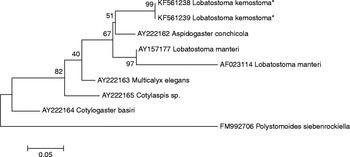
Molecular analysis of partial 28S rDNA resulted in an 1117 bp long sequence. Newly generated sequences were aligned and compared with the aspidogastrids sequences from GenBank to assess the phylogenetic relationship of L. kemostoma. The species compared were Lobatostoma manteri Rohde, Reference Rohde1973 (AY157177, AF023114), Aspidogaster conchicola Baer, 1827 (AY222162); Cotylogaster basiri Siddiqi & Cable, 1960 (AY222164), Multicalyx elegans (Olsson, 1868) (AY222163), Cotylaspis sp. (AY222165) and Polystomoides siebenrockiella Rohde, 1965 (FM992706), the latter being used as an outgroup. The highest BLAST score between the sequences was shown by L. manteri (90% similarity). The phylogenetic tree from the maximum likelihood (ML) analysis (fig. 4) shows the present sequences of L. kemostoma as a sister-group of A. conchicola, while the two sequences of the congeneric species L. manteri clustered together and were closely related to L. kemostoma and A. conchicola. Multicalyx elegans, which belongs to Multicalycidae, presented an intermediate position between members of Aspidogastridae.
The relationship between aspidogastrean species obtained in the present study (asterisks) and sequences from GenBank, based on maximum likelihood analysis of the partial 28S rDNA dataset. Polystomoides siebenrockiella was used as the outgroup, and bootstrap values are shown at the nodes.

Remarks
The freshly collected specimens belong to the Aspidogastrinae, as defined by Rohde (2002), because of the ventral disc, which has four longitudinal rows of alveoli. It is placed in Lobatostoma by the presence a single testis, cirrus sac and cephalic lobes (Eckmann, Reference Eckmann1932; Rohde, 2002). At present, the genus contains nine valid species distributed worldwide, namely L. ringens (Linton, Reference Linton1905); L. kemostoma (MacCallum & MacCallum, Reference MacCallum and MacCallum1913); L. pacificum Manter, Reference Manter1940; L. manteri Rohde, Reference Rohde1973; L. jungwirthi Kritscher, Reference Kritscher1974; L. platense Mañé-Garzon & Holcman-Spector, Reference Mañé-Garzon and Holcman-Spector1976; L. hanumanthai Narasimhulu & Madhavi, Reference Narasimhulu and Madhavi1980; L. anisotremum Oliva & Carvajal, Reference Oliva and Carvajal1984; and L. veranoi Oliva & Luque, Reference Oliva and Luque1989. Lobatostoma kemostoma can be differentiated from all congeners by the following combinations of characters in adults full of eggs: (1) the hindbody (tail) is longer than the ventral disc (although L. pacificum has been described as sometimes presenting the same condition); and (2) cephalic lobes have the same shape and size. In addition, this species differs from all others but one by having just 30 marginal alveoli on the disc. The similar L. jungwirthi, which has between 30 and 40 marginal alveoli (Lunaschi, Reference Lunaschi1984), can also be separated by lacking the hindbody, besides being the only species recovered from a freshwater fish.
The present specimens were morphologically consistent with those described by MacCallum & MacCallum (Reference MacCallum and MacCallum1913), as well as the specimens from collections examined, except for the four voucher specimens deposited by Bunkley-Williams et al. (1996) (USNPC 083016.00) which we preferred to assign just as Lobatostoma sp., since their high degree of contraction made specific identification impossible.
In the original description of L. kemostoma, the arrangement and number of alveoli on the ventral disc were incompletely described. MacCallum & MacCallum (Reference MacCallum and MacCallum1913) pointed out that there were 14 or 15 ‘marginal depressions’ in four longitudinal rows (see MacCallum & MacCallum, Reference MacCallum and MacCallum1913, p. 252), even though, in this way, the alveoli on poles could be overcounted. Actually, there are 14 alveoli in four longitudinal rows, in addition to two alveoli on both extremities. For comparative purposes, we can describe the arrangement of alveoli as 30 distributed marginally and 28 distributed centrally, resulting in a total of 58 alveoli.
Overall, the morphometric data were similar for all the analysed material (see table 1). On the other hand, the measurements taken from the present specimens represented a larger range of values, thus enlarging the morphometric variability for most features.
In the original description of L. kemostoma, MacCallum & MacCallum (Reference MacCallum and MacCallum1913) did not designate a holotype, but the original figure is similar to the specimen mounted on the slide USNPC 035565.02 deposited in 1912 and registered as ‘type’. In addition, another five slides designated as ‘types’ were deposited between 1911 and 1912. According to the International Code of Zoological Nomenclature (ICZN, Reference ICZN1999), ‘A lectotype may be designated from syntypes to become the unique bearer of the name of a nominal species-group taxon and the standard for its application’ (article 74.1). Therefore, considering that the figure in the original publication conforms to the syntype USNPC 035565.02, we propose it to be considered as the lectotype of L. kemostoma, and all other syntypes (USNPC 035564.02, 035565.01, 035563.01, 035563.02, 035564.01) become automatically paralectotypes, as stated by ICZN (article 74.1.3).
Discussion
The genus Lobatostoma was erected by Eckmann (Reference Eckmann1932) to accommodate Aspidogaster ringens Linton, Reference Linton1905 and A. kemostoma MacCallum & MacCallum, Reference MacCallum and MacCallum1913 based on features described above. Lobatostoma kemostoma was described from T. carolinus, probably collected from the Atlantic coastal zone of USA; the type locality is not mentioned in the original description, but the material deposited at USNPC was obtained from the New York Aquarium. Since then, L. kemostoma has been reported parasitizing fish of genus Trachinotus, mainly from the Atlantic Ocean (Gomes et al., Reference Gomes, Fábio and Rolas1978; Fernandes et al., Reference Fernandes, Kohn and Magalhães1985; Bunkley-Williams et al., Reference Bunckley-Williams, Dyer and Williams1996; Lamothe-Argumedo et al., Reference Lamothe-Argumedo, García-Pietro, Osorio-Sarabia and Pérez-Ponce de León1997; Sanchez-Ramirez & Vidal-Martinez, Reference Sanchez-Ramirez and Vidal-Martinez2002; Pereira et al., Reference Pereira, Velloso, Chaves, Moraes and Oliveira2004). Luque & Oliva (Reference Luque and Oliva1993) reported L. kemostoma in the Pacific Ocean from T. paitensis. On the other hand, L. ringens, which was also described from T. carolinus and from Micropogon undulatus (Linnaeus, 1766), has a wide host range, being reported from ten families of fish and from one molluscan host (Hendrix & Overstreet, Reference Hendrix and Overstreet1977; Suriano & Martorelli, Reference Suriano and Martorelli1983; Cordeiro & Luque, Reference Cordeiro and Luque2005). In general, the aspidogastreans are known by their low host specificity (Rohde, Reference Rohde1972, Reference Rohde1994), e.g. Aspidogaster conchicola von Baer, 1827 was recovered from 23 of 37 species of mussels in Ohio, USA (Stromberg, Reference Stromberg1970). The presence of L. kemostoma in Trachinotus spp. only, can be due to the presence of toothed pharyngeal plates in this fish (Grubich, Reference Grubich2003) which can crush the snail shells into small pieces. These molluscs are considered as the first intermediate host to immature specimens of Lobatostoma, at least for the two known life cycles of the genus, L. manteri and L. jungwirthi (Rohde, Reference Rohde1973; Zylber & Ostrowski de Núñez, Reference Zylber and Ostrowski de Núñez1999).
The arrangement and number of alveoli on the ventral disc have been broadly used to identify the species of Lobatostoma (Rohde, Reference Rohde1973; Narasimhulu & Madhavi, Reference Narasimhulu and Madhavi1980; Lunaschi, Reference Lunaschi1984; Oliva & Carvajal, Reference Oliva and Carvajal1984; Oliva & Luque, Reference Oliva and Luque1989), but apparently confusion has occurred since the first description of Lobatostoma. Linton (Reference Linton1905) described L. ringens as having 36 marginal alveoli and 17 median alveoli divided by a longitudinal septum, which would mean 34 median alveoli. After that, all species, except two, were also described as having the median alveoli divided by a longitudinal septum but without adding them in the total number of alveoli (MacCallum & MacCallum, Reference MacCallum and MacCallum1913; Rohde, Reference Rohde1973; Mañé-Garzon & Holcman-Spector, Reference Mañé-Garzon and Holcman-Spector1976; Narasimhulu & Madhavi, Reference Narasimhulu and Madhavi1980; Oliva & Carvajal, Reference Oliva and Carvajal1984; Oliva & Luque, Reference Oliva and Luque1989). Lobatostoma pacificum and L. jungwirthi were the only species in which the number of median alveoli was correctly described, showing 30 and 28 alveoli, respectively (Manter, Reference Manter1940; Kritscher, Reference Kritscher1974). In order to standardize the total number of alveoli in agreement with subfamily diagnosis (see Remarks), all species of Lobatostoma should be treated as having two rows of median alveoli divided by a longitudinal septum, plus the marginal alveoli, forming two rows in the border of the disc. For a better visualization of the disc, it should be studied in a concave slide under a light microscope before mounting. Additionally, only adults full of eggs should be analysed, avoiding some variations that can occur during ontogenetic development (Rohde & Sandland, Reference Rohde and Sandland1973).
Narasimhulu & Madhavi (Reference Narasimhulu and Madhavi1980) pointed out that the only reliable feature to differentiate the species of Lobatostoma is the number of alveoli on the disc, although L. hanumanthai, described by them, somewhat overlapped the number of total alveoli of L. platense (59–66 versus 63–69) described 4 years earlier off the Uruguayan coast in South America. Apparently they were unaware of the similar species, and, therefore, the occurrence of L. hanumanthai, originally described from T. blochii (Lacepède, 1801) off the Indian coast, reported by Pereira et al. (Reference Pereira, Velloso, Chaves, Moraes and Oliveira2004) from the intestine of T. marginatus Cuvier, 1832 off the Rio Grande do Sul coast, Brazil, should be re-evaluated. Unfortunately, the authors did not deposit any specimens in an institutional collection.
For Oliva & Luque (Reference Oliva and Luque1989), besides the number of alveoli, other reliable features that should be taken into consideration are: the hindbody longer or smaller than the ventral disc, relative position of the testis, and the ratio between the pharynx and the cirrus sac. However, the position of the testis seems to be variable (Narasimhulu & Madhavi, 1980; Oliva & Carvajal, Reference Oliva and Carvajal1984; present study) leading us to agree with Narasimhulu & Madhavi (Reference Narasimhulu and Madhavi1980) that this feature should not be considered of taxonomic value. Moreover, the relationship between the hindbody and the ventral disc in terms of proportion, should also not be used as a diagnostic characteristic, since its variation relies heavily on the degree of contraction of the specimens when fixed (Narasimhulu & Madhavi, Reference Narasimhulu and Madhavi1980; Oliva & Luque, Reference Oliva and Luque1989).
SEM studies are currently available for just L. manteri, L. jungwirthi and L. anisotremum (Rohde, Reference Rohde1973; Oliva & Carvajal, Reference Oliva and Carvajal1984; Paola & Damborenea, Reference Paola and Damborenea2001). The present work increases the number of SEM studies in Lobatostoma to four, and is the first SEM study of L. kemostoma. The molecular data showed a divergence of higher-level classification from the previous phylogenetic analyses based on morphology (Gibson, Reference Gibson1987; Brooks et al., Reference Brooks, Bandoni and O'Grady1989; Zamparo & Brooks, Reference Zamparo and Brooks2003), whereas it was similar to those based on molecular sequences (Olson et al., Reference Olson, Cribb, Tkach, Bray and Littlewood2003; Chen et al., Reference Chen, Gao and Nie2007). These molecular analyses support the Aspidogastridae as an artificial lineage with the Multicalycidae nested within them, as stated by Littlewood (Reference Littlewood, Maule and Marks2006). The present results also supported the Aspidogastridae as a paraphyletic group, with Multicalyx elegans (Multicalycidae) in an intermediate position between the former, but further analysis, based on a wider range of genes (ITS1, ITS2, COX), should be performed to confirm this assessment, together with new morphological data. The closest relatedness, between L. kemostoma and A. conchicola, could be due to the highly conserved sequences of 28S rDNA.
In the current study, we provided new morphological and molecular information on L. kemostoma, such as a partial 28S rDNA sequence, a complete description of number of alveoli on the ventral disc, line drawings, photomicrographs and scanning electron micrographs detailing morphological features, as well as morphometric data. Moreover, we assigned a lectotype to L. kemostoma from the type series.
Acknowledgements
The authors are indebted to Patricia Pillit for the loan of specimens of Lobatostoma kemostoma available at the US National Parasite Collection. Thanks are also due to Marcelo Knoff, Curator of the CHIOC, for the loan of voucher specimens of L. kemostoma from off the Brazilian coast.
Financial support
This study was supported financially by Fundação Carlos Chagas Filho de Amparo à Pesquisa no Rio de Janeiro (grant FAPERJ E-26/100.571/2013), Conselho Nacional de Desenvolvimento Científico e Tecnológico (CNPq), Coordenação de Aperfeiçoamento de Pessoal de Ensino Superior (CAPES) and Fundação Oswaldo Cruz-PROEP.
Conflict of interest
None.