Infundibular pulmonary stenosis secondary to isolated ventricular septal defect may interfere with the diagnosis of tetralogy of Fallot.Reference Kouchoukos, Blackstone, Doty, Hanley and Karp 1 Total correction of tetralogy of Fallot through transannular approach is associated with increased rates of mortality and morbidity.Reference Kouchoukos, Blackstone, Doty, Hanley and Karp 2 , Reference Kouchoukos, Blackstone, Doty, Hanley and Karp 3 In tetralogy of Fallot, dextroposition of the aorta means overriding of the aorta to the right ventricle with an extent of 30–90%.Reference Kouchoukos, Blackstone, Doty, Hanley and Karp 4 In case more than 90% of the aorta originates from the right ventricle, it is termed as double-outlet right ventricle with tetralogy of Fallot.Reference Mavroudis and Backer 5
In some instances, despite being actually not dextroposed, the aorta may be misinterpreted as dextroposed in the surgeon's point of view. Therefore, accurate determination of the dextroposition status of the aorta is of immense importance, as it will affect the prospective treatment strategy. Tips and pearls for deciding the actual dextroposition status of the aorta are discussed in this paper. In view of the uncertainty regarding an important anatomic feature of tetralogy of Fallot, we aimed to clarify this point using the anatomical relationship between various structures.
Methods
Dextroposition of the aorta occurs when the aorta overrides the interventricular septum to the right ventricular side. The aorta may seem to be dextroposed during operation, in echocardiogram, and angiogram, although it is not so actually. We state that some clues derived from the illustrations and figures of the literature can be useful to decide whether or not the aorta is really dextroposed.
Results
Dextroposition of the aorta can be assessed in three principal subgroups:
-
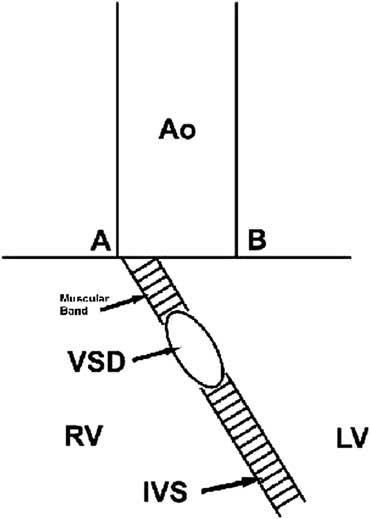
• Although it seems to be dextroposed during operation, in echocardiogram, and angiogram, the aorta could not be assumed as dextroposed if a muscular band exists between the aorta and the ventricular septal defect that are not adjacent (Fig 1).
Figure 1The aorta is “not truly dextroposed” in case the aorta and ventricular septal defect are non-adjacent. AO = aorta; IVS = interventricular septum; LV = left ventricle; RV = right ventricle; VSD = ventricular septal defect.

-
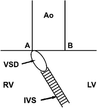
• The aorta is not truly dextroposed in case the aorta and ventricular septal defect are adjacent. The aorta may seem to be dextroposed during operation, in echocardiogram, and angiogram because interventricular septum shifts to the left because of the characteristics of the flow. The point where the ventricular septal defect plane meets the aortic annulus is not to the left ventricular side of point A (Fig 2).
Figure 2The aorta is “not truly dextroposed” in case the aorta and ventricular septal defect are adjacent. AO = aorta; IVS = interventricular septum; LV = left ventricle; RV = right ventricle; VSD = ventricular septal defect.
-
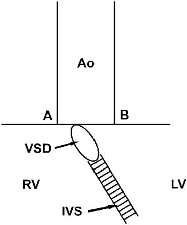
• The aorta and the ventricular septal defect are adjacent. The aorta lies to the right of the ventricular septal defect plane and is truly dextroposed (Fig 3).
Figure 3The aorta is “truly dextroposed” in case the aorta and ventricular septal defect are adjacent. AO = aorta; IVS = interventricular septum; LV = left ventricle; RV = right ventricle; VSD = ventricular septal defect.


Figures and illustrations
The figures and illustrations in the following references have been examined in order to outline the tips and pearls in the diagnosis of “true” dextroposition of the aorta in tetralogy of Fallot:
-
• Figures 4–8Reference Castenada and Mayer 6 and Figure 9Reference De Leval 7 do not demonstrate a true dextroposition of the aorta.
-
• Figure 10Reference Van Doorn and De Leval 8 does not demonstrate a true dextroposition of the aorta.
-
• Figures 11–16 do not demonstrate a true dextroposition of the aorta, whereas Figures 17–20 demonstrate a true dextroposition of the aorta.Reference Anderson, Macartney, Shinebourne and Tynan 9
-
• Figures 21–36 do not demonstrate a true dextroposition of the aorta.Reference Kirklin and Barrat-Boyes 10
-
• Figures 37–51 do not demonstrate a true dextroposition of the aorta.Reference Kouchoukos, Blackstone, Doty, Hanley and Karp 11
-
• Figures 52–55 do not demonstrate a true dextroposition of the aorta.Reference Castaneda, Jones, Mayer and Hanley 12
-
• Figures 56–58 do not demonstrate a true dextroposition of the aorta.Reference Sabiston 13
-
• Figures 59–65 do not demonstrate a true dextroposition of the aorta.Reference Laks and Bread 14
-
• Figures 66–70 do not demonstrate a true dextroposition of the aorta.Reference Laks 15
Discussion
Tetralogy of Fallot occurs in approximately 1 in 3600 live births and constitutes 3.5% of all congenital heart defects.Reference Hoffmann and Kaplan 16 The first and the principal condition for the aorta to be termed as “dextroposed” is that the ventricular septal defect should be adjacent to the aorta. In other words, the superior wall of the ventricular septal defect should be formed by the aorta, and sutures to repair this defect should bite aorta. In addition to this, the plane of the aortic annulus should be on the right-hand side of the plane of the ventricular septal defect.
In the past, the terms “dextroposition” and “overriding” have been used interchangeably. Overriding of the aorta over the right ventricle, which occurs in the normal heart, should not be confused with dextroposition. Overriding of the aorta can be considered a normal anatomic feature that occurs because of the sigmoid interventricular septum’, as well as in hearts with isolated infundibular perimembranous ventricular septal defect. The term dextroposition indicates a pathologic condition with a rightward displacement of the aortic root. There has been considerable debate concerning whether a true dextroposition of the aorta exists in tetralogy of Fallot. Some investigators have suggested that in tetralogy there is only an exaggeration of the normal overriding aorta caused by dilatation of this vessel secondary to increased aortic flow and simulating a dextroposition. Other authors have suggested that the condition is typified by a true dextroposition of the aorta with a rightward displacement and a clockwise rotation of the aortic root, particularly in cases with severe infundibular obstruction.Reference Isaaz, Cloez, Marçon, Worms and Pernot 17
Tetralogy of Fallot is characterised by the lack of conal rotation and conal malseptation. The conal malseptation, involved in the abnormal overriding of the aorta, is anatomically demonstrated by the anterior deviation of the conal septum. In this circumstance, the septal insertion is in front of the trabecula septomarginalis. The latter anomaly has been previously studied by two-dimensional echocardiography, allowing the diagnosis and quantification of the deviation of the infundibular septum. However, the concept of conotruncal malseptation as the only morphogenetic mechanism in tetralogy of Fallot can neither explain all the features of the condition, nor is it sufficient to produce the rightward shift of the aorta with the clockwise rotation. The lack of the normal conal rotation may bring the aorta to a dextroposterior position during the normal embryogenesis.Reference Isaaz, Cloez, Marçon, Worms and Pernot 17
The aortic root can be displaced towards the right – dextroposed – as a consequence of three presumable mechanisms:Reference Kouchoukos, Blackstone, Doty, Hanley and Karp 1 rightward translation, possibly secondary to the lack of left migration of the conus from an embryologic point of view;Reference Kouchoukos, Blackstone, Doty, Hanley and Karp 2 clockwise rotation – looking downstream – about an eccentric axis, supposed to be secondary to the lack of conal rotation; orReference Kouchoukos, Blackstone, Doty, Hanley and Karp 3 a combination of these two mechanisms.
The various measurements performed in patients with tetralogy of Fallot have shown that the aorta is abnormally displaced towards the right because of a clockwise rotation around an eccentric pivot, the aortic anterior commissure. All these abnormalities of the spatial position of the aortic root can only be the result of a true anomaly developed during embryogenesis. All in all, the aortic dilatation above a perimembranous ventricular septal defect as a result of abnormal haemodynamics and an exaggerated normal overriding would not alter the spatial position of the aortic root.
The existence and extent of dextroposition of the aorta in tetralogy of Fallot is important as it affects the further treatment strategy. Therefore, the definitive diagnosis should be made pre-operatively.
As a result, we suggest that:
-
1. The aorta and the ventricular septal defect should be adjacent to mention dextroposition of the aorta; however, this condition is insufficient for diagnosis.
-
2. The plane from which the aorta leaves the ventricle should superimpose on the plane of ventricular septal defect into the right ventricle. Consideration of the planes of aortic annulus and ventricular septal defect together may help understanding easier.
-
3. If the aorta and ventricular septal defect join at one edge, the aorta may seem to be dextroposed.
-
4. New diagnostic and therapeutic methods are necessary for accurate pre-operative evaluation of dextroposition of the aorta.