Tetralogy of Fallot is the most common form of cyanotic congenital heart disease, with a prevalence of 0.26–0.8 per 1000 live births.Reference Khositseth, Manop and Khowsathit 1 Total repair for tetralogy of Fallot has been available for 55 years with a favourable outcome in most patients. Late survival has also improved, with reports showing a 20-year survival rate nearing 90%.Reference Nollert, Fischlein, Bouterwek, Bohmer, Klinner and Reichart 2 , Reference Murphy, Gersh and Mair 3 Today, we are faced with an increasing number of adult patients who require regular follow-up for complications after initial correction of tetralogy of Fallot. These complications mostly consist of pulmonary regurgitation, residual or recurrent pulmonary stenosis, ventricular septal defect, or right ventricular outflow tract aneurysm.
Echocardiography provides information on the presence of right ventricular dilatation and/or hypertrophy, the presence of pulmonary regurgitation, and by Doppler estimation of right ventricle systolic pressure. However, assessing the function of the morphological right ventricle is challenging because of its complex anatomy. Difficulties are compounded by irregularities in the ventricular cavities and abnormalities in wall motion in patients with congenital heart lesions. None of the geometric assumptions used to assess left ventricular function hold true for the systemic right ventricle. The complex shape of the right ventricle makes quantification difficult.Reference Jiang, Levine and Weyman 4 Thus, in clinical scenarios, many centres rely on visual estimation of right ventricle systolic function, which is then subject to variability because of incomplete visualisation of the entire right ventricle and the experience of the observer. Therefore, cardiac magnetic resonance has evolved to be a better quantitative standard, especially for serial comparisons.Reference Hornung, Derrick, Deanfield and Redington 5 However, this technique, which is contraindicated in some patients, is expensive, has limited availability, and requires significant resources and expertise to acquire and interpret the images.
The tissue Doppler imaging-derived systolic myocardial velocities are considered more useful compared with conventional echocardiography in the assessment of the right ventricle contractile function.Reference Kukulski, Hubbert, Arnold, Wranne, Hatle and Sutherland 6 In the literature, a limited number of studies have shown the potential usefulness of the tissue Doppler imaging-derived myocardial performance index (Tei index) and isovolumic acceleration in the evaluation of right ventricular function in post-operative tetralogy of Fallot patients.Reference Cetin, Tokel, Varan, Orün and Aşlamaci 7 However, no research study has been performed yet to compare cardiac magnetic resonance and tissue Doppler imaging echocardiography in post-operative tetralogy of Fallot patients.
Thus, the aim of this study was to compare tissue Doppler imaging assessment of right ventricular function measuring the myocardial performance index and isovolumic acceleration values with cardiac magnetic resonance findings.
Methods
Study design and patients
This cross-sectional study was conducted at the Unit of Pediatric Cardiology of İstanbul University Cerrahpaşa Medical Faculty, Istanbul, Turkey. Before recruitment of patients and controls, the study protocol was reviewed and approved by the local ethics committee, in accordance with the ethical principles for human investigations, as outlined by the Second Declaration of Helsinki, and written informed consents were obtained from all the patients. Between November, 2010 and January, 2011, we recruited 31 consecutive age- and gender-matched patients operated for tetralogy of Fallot and 36 healthy controls. No evidence of structural cardiovascular disease was detected by two-dimensional or Doppler echocardiography in the control group.
The study comprised two groups: group 1 (n = 31) consisted of patients operated for tetralogy of Fallot and group 2 (n = 36) consisted of healthy controls. Patients (mean ± standard deviations, 14.25 ± 3.61 years) who had undergone surgery for tetralogy of Fallot were selected according to the following exclusion criteria: resting significant arrhythmias by electrocardiographic Holter monitoring; echocardiogram of inadequate quality; any anamnestic and clinical evidence of heart failure; and treatment with digitalis, β-blockers, and antiarrhythmics. Patients were included in the study if (1) the echocardiographic assessment was made within 2 months of the cardiac magnetic resonance, and (2) they were clinically stable, in sinus rhythm. The symptoms of patients have been classified by using New York Heart Association Functional Classification. All patients were in New York Heart Association I–II and had regular sinus rhythm with complete right bundle branch block.
Cardiac magnetic resonance and tissue Doppler imaging echocardiography was performed in group 1. Only echocardiography was performed in group 2.
Baseline definitions and measurements
Height and weight were measured according to standardised protocols. Body surface area was calculated as the body surface area = (weight0.425 × height0.725) × 0.007184 (Dubois formula) (m2). Blood pressure was measured using a mechanical sphygmomanometer in the medical office setting. In each individual, after 15 min of comfortably sitting, the average of three blood pressure measurements was calculated.
Echocardiography and Doppler measurements
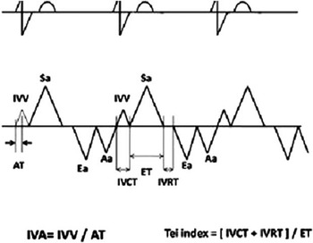
A detailed echocardiographic evaluation, which included an M-mode, two-dimensional, and colour Doppler – continuous and pulsed wave – examination, was performed. Images were obtained on a Siemens Acuson CV-70 with a 4–2 MH transducer (East Flanders, Belgium). Tissue Doppler imaging echocardiography was performed by activating the tissue Doppler imaging functions in the same unit. The tissue Doppler imaging programme was set to the pulsed wave Doppler mode reported previously.Reference Sengupta, Mohan and Pandian 8 , Reference Garcia-Fernandez, Zamorano and Azevedo 9 Filters were set to exclude high-frequency signals. Gains were minimised to allow a clear tissue signal with minimal background noise. Using the four-chamber view, a 2-mm sample volume was placed at the lateral corner of the tricuspid valve annulus, and peak systolic, early diastolic, and late diastolic myocardial velocities were obtained at a sweep speed of 100 mm/s and analysis was carried out as has been described before.Reference Sengupta, Mohan and Pandian 8 Isovolumic contraction time was defined as the time period between the end of the late diastolic myocardial wave and the beginning of the peak systolic wave. Isovolumic relaxation time was defined as the time period between the end of the peak systolic wave and the beginning of the early diastolic myocardial wave. Ejection time was measured as the duration of ventricular outflow velocity pattern. Myocardial performance index was calculated according to the following equation: myocardial performance index = (isovolumic contracton time + isovolumic relaxation time)/ejection time (Fig 1). The myocardial acceleration during isovolumic contraction was measured by dividing the myocardial velocities during isovolumic contraction by the time interval from onset of the myocardial velocity during isovolumic contraction to the time at peak velocity of this wave.Reference Tayyareci, Tayyareci, Nişanci, Umman and Buğra 10 The mean values were recorded by averaging the results of three consecutive measurements.
Tissue Doppler-derived systolic velocities obtained at the lateral corner of tricuspid annulus. IVV begins before the R-wave on electrocardiogram and followed by peak systolic myocardial velocity (Sa). IVA was calculated by dividing the peak IVV by time interval from the onset of the wave during isovolumic contraction to the time at peak velocity of this wave (acceleration time). AT = acceleration time; ET = ejection time; IVA = isovolumic myocardial acceleration; IVCT = isovolumetric contraction time; IVRT = isovolumetric relaxation time; IVV = isovolumic velocity.

Pulsed wave Doppler studies of right ventricular outflow tract were performed to assess the systolic pressure gradient across the right ventricle and the presence and degree of pulmonary regurgitation. Mild pulmonary regurgitation was considered to be present if diastolic retrograde flow could be detected under the pulmonary valve. Moderate pulmonary regurgitation was diagnosed if the retrograde flow could be seen in the right ventricle further apically from the pulmonary valve and in the pulmonary trunk. Severe pulmonary regurgitation was diagnosed if abnormal retrograde diastolic flow could also be detected in the branch pulmonary arteries.Reference Meijboom, Szatmari and Deckers 11
Cardiovascular magnetic resonance imaging protocol
Cardiac magnetic resonance were performed using a 1.5-T Philips Achieva magnetic resonance system (Rhineland-Palatinate, Germany) using SENSE XL Torso coil 16 elements. The timing of a typical protocol is approximately 35–40 min. After obtaining scout images and a reference scan, an axial stack of black blood turbo spin echo images is acquired to outline cardiac and non-cardiac anatomy. For analysis of valvular and ventricular function, we follow the axis of the heart rather than the axis of the body. For visualisation of valvular insufficiency, two- and four-chamber cines – steady-state free precession sequences – are acquired. To quantify right ventricular function, multiple cines are obtained in the short-axis orientation. An additional cine is aligned along the right ventricular outlet tract to visualise pulmonary insufficiency and right ventricular outlet tract enlargement. From the additional right ventricular outlet tract cine and a transversal black blood image, a velocity map across the pulmonary artery is acquired for calculation of pulmonary regurgitant volume. Quantification of regurgitation entails a post-processing technique in which through-plane velocity and area are measured by tracing the vessel border on sequential phase-contrast images obtained over one cardiac cycle.
For exact measurement of right ventricular volumes and ejection fraction, cardiac magnetic resonance is the most accurate imaging modality. Furthermore, cardiac magnetic resonance may give an unrestricted view of the right ventricular outlet tract, and an aneurysmatic enlargement can be observed. The right ventricular end-diastolic volume and right ventricular end-systolic volume are calculated by manually drawing endocardial contours at end diastole and end systole, respectively, on cine loops, oriented axial, or along the short axis of the right ventricle. A contrast-enhanced magnetic resonance angiography is used to visualise the pulmonary tree. As mentioned in the literature, pulmonary regurgitation fraction >40% was considered severe pulmonary regurgitation.Reference Cetin, Tokel, Varan, Orün and Aşlamaci 7
Cardiovascular magnetic resonance scans protocol:
-
• scout images single-phase steady-state free precession;
-
• parallel imaging reference scan;
-
• axial black blood turbo spin echocardiogram;
-
• 2-chamber multiphase steady-state free precession;
-
• 4-chamber multiphase steady-state free precession;
-
• right ventricular outlet tract cine multiphase steady-state free precession;
-
• multislice, multiphase short-axis steady-state free precession;
-
• magnetic resonance angiography pulmonary artery; and
-
• flow mapping pulmonary trunk (phase-contrast images).
Statistical analysis
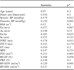
All statistical analyses were performed using statistical package for the social sciences for Windows version 17.0 (SPSS, Chicago, Illinois, USA). Kolmogorov–Smirnov tests were used to test the normality of data distribution (Table 1). The data were expressed as arithmetic means and standard deviations. The χ 2-test was used to compare the categorical variables between groups. Independent sample t-test was used for comparison of continuous variables among the two homogeneous study groups. Mann–Whitney U-test was used for comparison of continuous variables among the two non-homogeneous study groups. Pearson's and Spearman's correlation analyses were used, respectively, to analyse continuous and categorical variables. Two-sided p-value <0.05 was considered statistically significant.
The distribution of the groups of variables.

Aa = late diastole; BP = blood pressure; BSA = body surface area; Ea = early diastole; ET = ejection time; IVA = isovolumetric acceleration; IVCT = isovolumetric contraction time; IVRT = isovolumetric relaxation time; MPI = myocardial performance index; PRF = pulmonary regurgitant fraction; RV-EDV = right ventricular end-diastolic volume; RV-EF = right ventricle ejection fraction; RV-ESV = right ventricular end-systolic volume; Sa = systole
*Kolmogorov–Smirnov
Results
Clinical characteristics of the study population
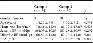
Clinical and demographic characteristics of controls and operated tetralogy of Fallot patients are presented in Table 2. There were no statistical differences with regard to gender, age, body surface area, heart rate, and both systolic and diastolic blood pressure between the controls and operated tetralogy of Fallot patients (p > 0.05 for all). By electrocardiographic analysis, all the patients were in sinus rhythm, and all patients with tetralogy of Fallot had a right bundle block. A total of 31 patients with a mean age of 11.98 ± 3.03 years at follow-up who underwent repair of tetralogy of Fallot at a mean age of 2.56 ± 1.61 years and 36 age- and sex-matched healthy children with a mean age of 13.72 ± 3.21 years were enrolled in this study. The 31 patients had transannular patch enlargement of the right ventricular outflow tract. In all, seven patients underwent Blalock–Taussig shunt before corrective surgery. Of the 31 patients, 24 were in New York Heart Association functional class I, whereas the remaining seven were in class II.
Comparison of demographic and clinical characteristics of the study subjects.

BP = blood pressure; BSA = body surface area; ns = non-significant
All measurable values were given with mean ± standard deviation
Standard echocardiographic examination
By Doppler criteria, severe pulmonary regurgitation was present in eight (25%) patients, moderate pulmonary regurgitation was present in 15 (50%) patients, and mild pulmonary regurgitation was present in eight (25%) patients.
Tissue Doppler imaging velocities and isovolumic time intervals in patients with tetralogy of Fallot and healthy children
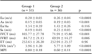
Compared with those of normal children, tissue Doppler imaging velocities of tetralogy of Fallot patients showed decreases in early diastolic myocardial velocity (0.20 ± 0.03 versus 0.26 ± 0.04 cm/s, p < 0.0001, respectively), late diastolic myocardial velocity (0.15 ± 0.03 versus 0.19 ± 0.03 cm/s, p < 0.001, respectively), and peak systolic velocity (0.19 ± 0.03 versus 0.23 ± 0.03 cm/s, p < 0.0001, respectively). Both isovolumic contraction time and isovolumic relaxation time in tetralogy of Fallot patients were significantly longer than those in normal children (103.77 ± 27.78 versus 71.91 ± 15.86 and 84.72 ± 21.13 versus 69.91 ± 14.27 ms, p < 0.001 and 0.006, respectively). Isovolumic acceleration value from the right ventricular tricuspid lateral annulus was significantly lower in patients than in controls (3.96 ± 1.20 versus 5.77 ± 1.09 m/s2, p < 0.0001, respectively). However, myocardial performance index value from the right ventricular tricuspid lateral annulus was significantly longer in patients than in controls (0.80 ± 0.18 versus 0.60 ± 0.11, p < 0.0001, respectively) (Table 3).
Tissue Doppler findings.

Aa = late diastole; Ea = early diastole; ET = ejection time; IVA = isovolumetric acceleration; IVCT = isovolumetric contraction time; IVRT = isovolumetric relaxation time; MPI = myocardial performance index; Sa = systole
All measurable values were given with mean ± standard deviation
Cardiovascular magnetic resonance findings
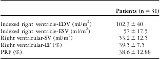
The time interval between cardiac magnetic resonance scans and echocardiography was 2 months. Table 4 lists cardiac magnetic resonance parameters.
Cardiovascular magnetic resonance findings.

EDV = end-diastolic volume; EF = ejection fraction; ESV = end-systolic volume; PRF = pulmonary regurgitant fraction; SV = stroke volume
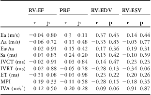
Derived from the right ventricle lateral tricuspid annulus myocardial performance index and isovolumic acceleration was not correlated with the right ventricle ejection fraction and pulmonary regurgitation fraction source from the cardiac magnetic resonance (Table 5).
Comparison of right ventricular tissue Doppler imaging parameters with cardiac magnetic resonance-derived parameters.

Aa = late diastole; Ea = early diastole; EDV = end-diastolic volume; EF = ejection fraction; ESV = end-systolic volume; ET = ejection time; IVA = isovolumetric acceleration; IVCT = isovolumetric contraction time; IVRT = isovolumetric relaxation time; MPI = myocardial performance index; PRF = pulmonary regurgitant fraction; RV = right ventricle; Sa = systole; SV = stroke volume
Discussion
Most of the problems reported during the late follow-up of patients after repair of tetralogy of Fallot have been related to abnormal right ventricle physiology. Residual pulmonary incompetence, residual or recurrent pulmonary stenosis, exercise intolerance, residual ventricular septal defect, and malignant ventricular arrhythmias have been identified as predictors of unfavourable long-term outcome.Reference Murphy, Gersh and Mair 12 Long-term volume overload, in patients with moderate or severe pulmonary regurgitation, progressive right ventricle dysfunction takes place in time. For this reason, in patients with right ventricle dilatation, pulmonary valvular replacement should be done before irreversible changes take place.Reference Jang, Kim, Choi, Lim, Kim and Lee 13 This finding signifies the importance of post-operative follow-up of the patients and the methods that should be used in the follow-up.
To the best of our knowledge, this is the first report to evaluate the relationship between right ventricle tissue Doppler imaging parameters and findings of cardiac magnetic resonance in patients with operated tetralogy of Fallot. In this study, right ventricular function was assessed by tissue Doppler imaging and cardiac magnetic resonance. Cardiac magnetic resonance findings by comparing with myocardial performance index, isovolumic acceleration and tried to determine the prognostic values. However, in this study there was no correlation between myocardial performance index, isovolumic acceleration and right ventricular ejection fraction, pulmonary regurgitation fraction, and other results of cardiac magnetic resonance.
Although echocardiography, which has good correlation with radionuclide angiography,Reference Amico, Lichtenberg, Reisner, Stone, Schwartz and Meltzer 14 is used every day in clinical practice to quantitatively assess left ventricular function, it was previously considered an inaccurate tool to quantitatively assess right ventricular function because of a lack of an ideal geometric model for evaluation of ventricular volumes.Reference Aebischer and Czegledy 15 In children, three-dimensional echocardiography has been found to have an excellent correlation with cardiac magnetic resonance in the assessment of right ventricular volumes and function.Reference Papavassiliou, Parks, Hopkins and Fyfe 16 However, questions remain about the accuracy in adults because of inadequate windows and larger right ventricular volumes.
The myocardial performance index is a useful clinical index of global ventricular function for evaluating both systolic and diastolic function.Reference Cheung, Smallhorn, Redington and Vogel 17 , Reference Harada, Tamura, Toyono and Yasuoka 18 The tissue Doppler imaging method of calculation may have advantages over the pulsed wave Doppler echocardiography calculation. The pulsed wave Doppler myocardial performance index has gained acceptance as a clinical examination for assessing cardiac function and is particularly useful as a predictor of clinical outcome in patients with cardiac disease.Reference Arnlov, Ingelsson, Riserus, Andren and Lind 19 – Reference Eto, Ishii, Tei, Tsutsumi, Akagi and Kato 21 The main advantage of this index is that it appears to be independent of ventricular geometry and heart rate.Reference Bruch, Schmermund and Marin 20 However, one of the main limitations of the pulsed wave Doppler myocardial performance index is that it cannot be calculated over a single cardiac cycle because the interval between the end and onset of tricuspid inflow and the ejection time is measured sequentially. By contrast, the tissue Doppler imaging can provide the timing elements necessary to calculate the myocardial performance index on a beat-to-beat basis.Reference Cannesson, Jacques, Pinsky and Gorcsan 22 In addition, the tissue Doppler imaging derived from the tricuspid annulus velocities allows the determination of the isovolumic contraction and relaxation times and the ejection time over a single cardiac cycle in normal conditions.Reference Cannesson, Jacques, Pinsky and Gorcsan 22 These results suggest that the pulsed wave Doppler myocardial performance index may be misinterpreted because it cannot be calculated over a single cardiac cycle, whereas the tissue Doppler imaging myocardial performance index should indicate right ventricular global function. In our study, myocardial performance index value from the right ventricular tricuspid lateral annulus was significantly longer in patients than in controls.
Therefore, evaluation of right ventricular systolic function is important in this group of patients. Assessment of right ventricular function is difficult because of its asymmetrical shape and narrow acoustic window. In our study, we used tissue Doppler imaging-derived right ventricular isovolumic acceleration. It is a new parameter and has been validated to be a reliable and relatively load-independent measure of right ventricular systolic function.Reference Pauliks, Chan and Chang 23 , Reference Vogel, Derrick and White 24 The main finding of our study is the evidence for its clinical use in assessing right ventricular systolic function to determine the severity of right ventricular dysfunction. In many studies, peak systolic velocity has also been shown to reflect right ventricular systolic function. This parameter was found to have a very good correlation with right ventricular fractional area and right ventricular ejection fraction assessed by radionuclide ventriculography.Reference Nageh, Kopelen, Zoghbi, Quinones and Nagueh 25 Peak systolic velocity is significantly afterload dependent,Reference Kukulski, Hubbert, Arnold, Wranne, Hatle and Sutherland 6 whereas isovolumic acceleration reflects right ventricular systolic function during isovolumic contraction. In contrast to peak systolic velocity, isovolumic acceleration has the advantage of being relatively preload and afterload independent. This parameter has been successfully validated by both experimental and clinical studies. Vogel et al.Reference Vogel, Schmidt and Kristiansen 26 demonstrated that isovolumic acceleration was an accurate parameter to assess right ventricular systolic dysfunction and was able to measure the force-frequency relation. Harada et al.Reference Vogel, Derrick and White 24 showed that peak systolic velocity was lower in patients after repair of tetralogy of Fallot compared with the control group. Frigiola et al.Reference Frigiola, Redington, Cullen and Vogel 27 proposed that isovolumic acceleration was not related directly to right ventricular dilatation but could give information about global right ventricular function in a study composed of 124 patients. In another study, Toyono et al.Reference Nageh, Kopelen, Zoghbi, Quinones and Nagueh 25 reported decreased right ventricular myocardial velocities and isovolumic acceleration after repair of tetralogy of Fallot.
Schwerzmann et al.Reference Schwerzmann, Samman and Salehian 28 (mean operated age: 6.6 years, range: 2.1–19.9 years) in a series of 36 patients with tetralogy of Fallot found that inverse linear correlation between myocardial performance index and cardiac magnetic resonance right ventricular ejection fraction (r = 0.73, p < 0.001). In this study, myocardial performance index was measured using pulsed wave Doppler echocardiography. The follow-up period is longer than that in our study (median 18.2, range 3–37.1 years). Cheung et al.Reference Cheung, Lam, Cheung and Cheung 29 (mean operated age 4 ± 1.8 years) in their study of 30 patients with tetralogy of Fallot found a correlation between myocardial performance index and right ventricular ejection fraction, pulmonary regurgitation fraction (respectively, r = −0.4, p = 0.028 and r = −0.4, p = 0.031). In this study, myocardial performance index was measured using pulsed wave Doppler echocardiography. Again, in our patients the follow-up period was shorter than in the previous study.
Conclusion
In our study, a significant relationship has not been detected between the findings obtained from right ventricular cardiac magnetic resonance and myocardial performance index and isovolumic acceleration. Our results do not support the use of myocardial performance index and isovolumic acceleration as a sensitive or a specific tool in patients with operated tetralogy of Fallot.
We did not compare our results with findings of other modalities such as cardiac catheterisation and three-dimensional echocardiography. Cardiac catheterisation was not available for asymptomatic patients. The management of asymptomatic patients with an abnormal myocardial performance index and isovolumic acceleration is not clear because the prognostic importance of subclinical myocardial dysfunction as detected by the myocardial performance index and isovolumic acceleration is still uncertain. Further studies that would present comparative results with new diagnostic modalities are needed to evaluate the diagnostic value of right ventricular myocardial performance index and isovolumic acceleration in patients with operated tetralogy of Fallot. Therefore, in the future large prospective cohort studies are needed to address this issue.
Limitations of the study
When evaluating our findings, it should be considered that the post-operative follow-up period is not very long and that there were no patients older than the second decade and the extrapolation of these findings to long term may not be applicable.
The relatively small sample size and the cross-sectional design are limitations of the present study. The cross-sectional design of this study did not allow us to investigate whether tissue Doppler imaging measurements are predictive of clinical outcomes.
In this study, respiratory cycles were not recorded. Respiratory variations of preload and their effects on echocardiographic measurements were not assessed and may influence the relation of myocardial performance index and isovolumic acceleration to cardiac magnetic resonance-measured right ventricular function.
In addition, the non-simultaneous acquisition of the tissue Doppler imaging and cardiac magnetic resonance data might have introduced an error. However, the echocardiographic evaluation was made within 2 months of the cardiac magnetic resonance and truly simultaneous image acquisition was not feasible.
Acknowledgement
The Institute's ethical approval was obtained from the local research ethics committee. The authors thank children and parents who participated in this study.
Financial Support
None.
Conflicts of Interest
None.