Introduction
Iatrogenic facial nerve injury is one of the most devastating complications of ear surgery and can be a significant cause of physical disability and emotional distress to the patient.
Despite technological advancements, such as the modern operating microscope, improved drills and better imaging techniques, the risk of iatrogenic trauma to the facial nerve remains unchanged, particularly during revision surgery.
Intra-operative facial nerve monitoring is frequently used during ear surgery, especially in revision cases where the anatomy has been disturbed by previous surgery. However, one cannot rely exclusively upon this method. Knowledge of core surgical anatomy is the key to avoiding damage to the nerve.
Methods
In the non-infected ear, the horizontal facial nerve can be easily located by identifying the oval window niche, with the stapes suprastructure and incudostapedial joint. The horizontal facial nerve lies immediately superior to the oval window. However, in an ear with a large amount of chronic granulation tissue and/or cholesteatoma, not only are the normal landmarks on the medial wall obscured, but the oval window niche is often full of infected tissue. Removing infected tissue in this area can damage the stapes suprastructure or cause inadvertent removal of the stapes footplate resulting in total hearing loss.
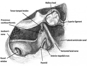
In the infected ear, the safest landmark to use to locate the horizontal facial nerve is the processus cochleariformis. This is a hard bony protrusion which has the tensor tympani tendon projecting from its apex. A blood vessel accompanies the tendon; this area therefore has a good blood supply and as such infection-related resorption is rare. The horizontal facial nerve can be identified immediately superior to the processus cochleariformis (Figure 1).Reference Sullivan and Smith1, Reference Haynes2 Although the horizontal facial nerve superior to the oval window niche is often dehiscent, the portion of the facial nerve immediately superior to the processus cochleariformis is rarely exposedReference Baxter3 and therefore less likely to be damaged.
Anatomical diagram showing the relations of the facial nerve in the middle ear after a canal wall down mastoidectomy with removal of the tympanic nerve and lateral two ossicles. PSCC = posterior semicircular canal; LSCC = lateral semicircular canal; GSPN = greater superficial petrosal nerve; Horiz = horizontal; Vert = vertical

There are three methods of safely identifying the processus cochleariformis, and thus the horizontal facial nerve, in an ear filled with granulation tissue and/or cholesteatoma. These methods are described below.
The first method involves removing granulation tissue from the posterior-inferior quadrant of the middle ear in the area of the round window. The round window is protected by its bony overhang and is not liable to be damaged in this procedure. The bulge of the promontory that is anterior to the round window niche is then identified and followed anteriorly in order to locate Jacobson's nerve. This nerve lies in a bony groove on the promontory, just anterior to the round window niche, and runs superiorly, leading directly to the processus cochleariformis (Figure 1).
The second method entails removing any infected tissue from the anterior-superior quadrant of the middle ear and identifying the eustachian tube orifice. Removing the tissue from this area should allow the surgeon to identify the smooth plate of bone in the roof of the orifice that covers the tensor tympani muscle. This smooth plate is then followed posteriorly until the tensor tympani tendon is identified protruding from the processus cochleariformis (Figure 2).
Anatomical diagram of a section through the middle ear, with the tympanic membrane and incus removed, showing the anatomical relations of the facial nerve.

The third method is to identify the lateral process of the handle of malleus in the superior portion of the medial external canal. As this receives its blood supply from the artery running alongside the tensor tympani tendon, the lateral process is usually present even when all the other parts of the ossicular chain have been eroded. Once the lateral process has been identified, the neck of the malleus is located. The tensor tympani tendon, which is attached to the medial surface of the neck of the malleus, is identified protruding from the processus cochleariformis (Figure 2).
Once the processus cochleariformis has been found using one of these methods, the horizontal facial nerve can then be located. The nerve runs 1 mm superior to the processus cochleariformis,Reference Măru, Cheiţă, Mogoantă and Prejoianu4 at an angle of 10 degrees, immediately inferior to the bony protrusion of the lateral semicircular canal. The anterior genu, and therefore the anterior limit of the horizontal facial nerve, is 2–3 mm anterior to the processus cochleariformis.Reference Goin5 Here the facial nerve has an acute turn medially and posteriorly. However, the greater superficial petrosal nerve is a similar size to the facial nerve and runs anteriorly in a straight line as a continuation of the facial nerve. It is therefore difficult to identify the point where the supra-labyrinthine portion turns medially. This is best achieved by carefully drilling in the small triangular area between the middle fossa dura superiorly, the horizontal facial nerve inferiorly and the anterior end of the horizontal semicircular canal posteriorly.
Conclusion
In an infected ear that is full of granulation tissue and/or cholesteatoma, the horizontal facial nerve can be reliably identified by locating the processus cochleariformis using the three methods described. This avoids damage to the nerve and important structures around it.