Introduction
Lichen-forming fungi produce a striking variety of vegetative tissues to enclose, protect, support and display their photosynthetic symbionts. Such tissues are frequently compared structurally and functionally to those of plants, which often share convergent features. In many fruticose lichens, fungal tissue composed of elongated cells with highly thickened walls (prosoplectenchyma) provides structural support to the thallus, inviting comparison to the collenchyma and sclerenchyma fibres of plants. However, the supportive tissues of lichens appear to differ from those of plants in at least one fundamental property. Whereas the secondary walls that reinforce plant tissue are deposited after cell expansion has ceased and are usually incompatible with further growth, the fungal cells within lichen prosoplectenchyma appear capable of continued elongation and proliferation. Although the distribution of growth in lichen thalli is still poorly known, diffuse elongation has been demonstrated in the branched axes of Usnea longissima (Rolstad & Rolstad Reference Rolstad and Rolstad2008) and Ramalina usnea (Sanders & Tokamov Reference Sanders and Tokamov2015), in the thallus nets of Ramalina menziesii (Sanders Reference Sanders1989, Reference Sanders1992), and to some extent in the podetia of Cladonia (Kärenlampi Reference Kärenlampi1970). All of the lichens in question are supported by longitudinally oriented prosoplectenchyma (“chondroid tissue”) with highly thickened cell walls. Evidently, growth is not necessarily limited to meristem-like apical zones where tissue layers are first organized, but may instead occur throughout the mature, differentiated regions of the thallus. How cells and cell walls behave in such tissues remains a significant question. In the reticulate thallus of Ramalina menziesii, supportive prosoplectenchyma forms a thick cortex surrounding the algal layer; in Usnea longissima, such tissue makes up the cartilaginous central cord that supports the main thallus axes. TEM studies of the cortex in R. menziesii revealed massive intercellular accumulations, consisting of electron-dense cell wall materials in concentric layers interspersed with amorphous, electron-transparent substances (Sanders & Ascaso Reference Sanders and Ascaso1995). The more peripheral rings of wall material appeared increasingly fragmented within a matrix of electron-transparent substances, suggesting that tissue elongation was continuously disrupting cell walls while new walls (and intervening layers of electron-transparent material) replaced them to the interior (Sanders & Ascaso Reference Sanders and Ascaso1995). TEM examination of the medullary cord in Usnea longissima showed similar characteristics, although the intervening electron-transparent material was less abundant (Sanders & de los Ríos Reference Sanders and de los Ríos2012). These observations suggested a mechanism of wall disruption and replacement that is substantially different from the cellular growth patterns usually described in fungi or plants. However, the two lichens in question, Ramalina menziesii and Usnea longissima, might be viewed as morphological outliers, the former with a uniquely reticulate thallus and the latter with unusually long main axes largely devoid of an algal layer or cortex. In the present work, we use TEM to assess cortical cell behaviour in a more typical beard lichen, Ramalina usnea (Fig. 1), for which diffuse growth of its branching thallus axes was recently demonstrated (Sanders & Tokamov Reference Sanders and Tokamov2015). We summarize the mechanism of diffuse expansion suggested by our observations, contrast it with growth patterns known in other kinds of fungal cells, and discuss features that remain to be elucidated by further study.
Ramalina usnea, a common beard lichen in coastal Florida, on branches of Taxodium.

Materials and Methods
Thalli of Ramalina usnea were collected air-dry from the campus of Florida Gulf Coast University (Lee County, FL, USA). Samples were wetted with distilled water using a spray mister and maintained in covered Petri dishes for 24 h to allow rehydration. Moist thalli were then cut into 1–2 mm segments that were placed immediately in fixative solution (2·5% glutaraldehyde in phosphate buffer, pH 7·1), followed by post-fixation in 1% OsO4 solution, dehydration in an alcohol series, infiltration with Spurr's low-viscosity resin and subsequent polymerization, according to de los Ríos & Ascaso (Reference de los Ríos, Ascaso, Kranner, Beckett and Varma2002). Ultrathin sections were cut c. 79 nm thick using a Leica EM UC-6 ultramicrotome, post-stained with uranyl acetate followed by lead citrate, then examined in a JEOL JEM-1011 transmission electron microscope. Semi-thin sections were also obtained using a Leica EM UC-6 ultramicrotome and later stained with toluidine blue for light microscope observation. Hand-cut sections of freshly collected material were examined with an Olympus BX-51 microscope using polarizing filters, or UV illumination after treatment with the fluorescent stain Uvitex 2B (Polysciences, Inc.).
The extensive intercellular materials often impeded good penetration of fixatives and embedding resin, resulting in suboptimal preservation and difficulty in obtaining intact ultrathin sections, especially in longitudinal orientation. However, selected areas of many sections examined provided an informative picture of the arrangement and development of the intercellular materials.
Results
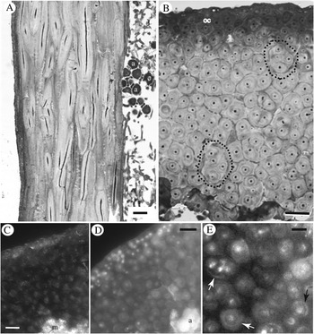
The cortex was comprised of long, filamentous elements with a chiefly longitudinal orientation. Cell protoplasts were no more than c. 1·5 μm in diameter and were typically separated from those of adjacent cells by accumulated wall and extracellular substances several micrometres thick (Fig. 2A). The outermost portion of the cortex was often distinguished by somewhat more irregularly oriented cells with relatively reduced but more densely staining intercellular material. In transverse section, cortical cells often appeared to be associated with one or several of their neighbours in fascicles or bundles (dotted lines in Fig. 2B). However, optical sectioning of thick and semi-thin sections revealed that in the course of their longitudinal trajectory, such filaments often did not remain within the same groupings (compare Fig. 2A). Concentric layering of wall material was evident in the embedded sections (Fig. 2B), and sometimes distinguishable in the UV-illuminated hand sections treated with a chitin-binding fluorophore (Fig. 2D & E).
Ramalina usnea. Light microscope images showing prosoplectenchymatous cortex. A & B, semi-thin sections of resin embedded lichen material; cell lumina staining darkly with toluidine blue; A, longitudinal section, showing longitudinal orientation of cortical filaments. Periphery of algal zone (a) visible at right; B, transverse section. A more darkly stained outer cortical layer (oc) is visible at the upper surface. Two groupings of seemingly bundled cells in the main cortex are indicated by dashed lines. C–E, hand-cut transverse sections of fresh material; C, with polarized light, crystalline materials mainly evident in medulla (m) and outer cortex; D & E, UV-illuminated after staining with Uvitex; D, cortex with irregular algal layer (a) beneath; E, detail of main cortex. Arrows indicate outer wall layers. Scales: A–D = 10 μm; E = 5 μm.

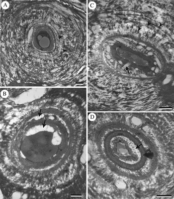
Transmission electron microscopy revealed that the accumulations between cell lumina consisted of electron-dense and electron-transparent materials distributed concentrically around individual cells in transverse section (Fig. 3A). The electron-dense material had a distinct, micro-layered structure whereas electron-transparent areas appeared to contain amorphous materials in globular or irregular deposits (Fig. 3B, arrows). In some cases, the shapes of electron-transparent areas suggested the former presence of lichen secondary substance crystals possibly removed in the preparatory processes (Fig. 3C, arrow). However, hand-cut sections of fresh material examined with polarized light showed most crystalline materials localized in the medulla and outermost cortex (Fig. 2C).
TEM images of Ramalina usnea cortex, transverse section, showing concentric arrangement of wall materials. Current cell wall proper (w) adjacent to protoplast, with older cell walls (ow) and interspersed electron-transparent materials (arrows) visible peripherally. Note fragments of disrupted cell walls (arrowheads) in concentric layers with greater distance from cell. Scales: A & C = 500 nm; B & D = 250 nm.

The electron-dense material comprised a discrete cell wall immediately adjacent to the protoplast but similar structures were also visible peripherally, separated from each other by intervening electron-transparent material (Fig. 3B–D). With distance from the protoplast, the concentric wall layers were increasingly disrupted into electron-dense fragments within the intervening electron-transparent material (Fig. 3B–D).
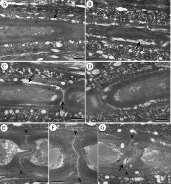
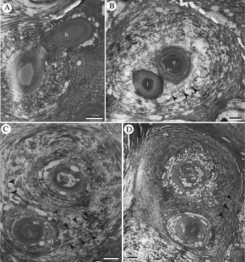
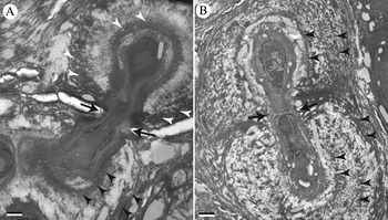
In longitudinal section, layers of electron-dense and electron-transparent materials were likewise visible (Fig. 4). In this orientation, layers appeared as lines rather than circles, making their relationship to the cell that produced them much less obvious. Septa possessed an electron-transparent median layer thickened at junctions with the lateral wall (Fig. 4E–G). Fungal filaments within the cortex produced branch filaments, which grew intrusively through the surrounding matrix of wall materials and intervening substances (Fig. 5A). In transverse section, the concentric layers associated with one cell were often clearly interrupted by the later intrusion of another cell, which itself had fewer concentric layers associated with it (Fig. 5B–D). Occasionally, short anastomoses or connecting branches between adjacent filaments were seen in longitudinal orientation within the transverse section (Fig. 6). Notably, these bridging cells lacked the accumulated wall layers present around the filaments they interconnected (Fig. 6A & B).
TEM images of Ramalina usnea cortex, longitudinal section. A–D, current cell wall proper (w) adjacent to protoplast, with older cell walls (ow) and interspersed electron-transparent materials (arrows) visible peripherally. E–G, septa between adjacent filament compartments, with median layer (arrowheads) and accumulated wall materials deposited to either side. Scales: A–D = 500 nm; E–G = 250 nm.

TEM images of Ramalina usnea cortex, transverse section. Intrusive growth of branch filaments through the wall layers of neighbouring cells. A, branch filament (b) emerging in longitudinal view from cell in transverse orientation; B, younger filament (i) growing intrusively through wall materials produced by older cell (o). Note interruption of concentric layer of older cell's wall fragments (arrowheads) by younger intruding cell (i); C & D, walls of older cell (w) distinguished from those of younger intruding cell (wi) by their concentric orientation. Interruption of older neighbour's wall layers (arrowheads) by intruding filament. Scales: A–D = 500 nm.

TEM images of Ramalina usnea cortex, transverse section. Short lateral connections between neighbouring filaments. Lateral connecting portion has a single cell wall (arrows), while the connected filaments have multiple walls (arrowheads). Scales: A = 250 nm; B = 500 nm.

Discussion
The continued accumulation of wall layers and their relationship to those of neighbouring cells permit several inferences about cell behaviour in the diffusely expanding tissue. The increasingly disrupted appearance of the electron-dense wall layers with distance from the cell implies that the newest layers are those deposited adjacent to the protoplast (Fig. 3). This contrasts with the pattern of secondary wall deposition considered more usual in fungal hyphae, where additional layers may be secreted to the exterior of the primary wall (Read Reference Read2011). Yet for cortical filaments that grow intrusively through the wall materials produced by their neighbours, as occurs in R. usnea, it would be difficult to imagine how an embedded cell could add multiple layers at the exterior surface of its own wall. Interior deposition of secondary wall layers was observed previously in lichen prosoplectenchyma (Sanders & Ascaso Reference Sanders and Ascaso1995; Sanders & de los Ríos Reference Sanders and de los Ríos2012) as well as other fungal cells, particularly spores (Marchant Reference Marchant, Burnett and Trinci1979; Hafellner & Bellemère Reference Hafellner and Bellemère1981). Since older wall layers are increasingly cut off from the protoplast by the deposition of newer layers to their interior, it seems unlikely that the cell could continue to integrate new structural components into them as the tissue expands. Cell walls not actively maintained by insertion of new structural components would be disrupted by tissue expansion. We therefore infer that the disrupted appearance of exterior wall layers is a direct consequence of tissue expansion, which is mainly longitudinal. Transversely oriented segments that interconnect the longitudinal filaments showed little disruption or replacement of wall layers (Fig. 6).
As filament branches grow longitudinally alongside their neighbours, the relative age of the adjacent cells is often apparent in transverse section from the pattern and extent of their concentric wall layers. A filament growing through the concentric wall materials produced by an older neighbour visibly interrupts those layers while producing its own concentric wall layers that will be less extensive than those of its older neighbour (Fig. 5B–D). This comparison provides further corroboration that wall layers are accumulating over time in the course of growth. The course of wall layer deposition and the intrusion of new filaments is summarized schematically in Fig. 7.
Schematic illustration of the sequence of events in the development of wall layers and the intrusion of younger filaments through them. Cell protoplast shown in grey; electron-dense cell wall layers in black; intervening, amorphous, electron-transparent materials in white. A–H, deposition of electron-transparent materials to interior of cell wall, often irregularly (broken white line) at first, followed by formation of new wall layer in alternation. Older cell walls toward periphery are increasingly disrupted (broken black lines). I–L, intrusion of newer branch filament through wall layers of neighbouring older cell, interrupting older cell's concentric wall layers. Both cells continue to produce new wall layers to their interior.

The continued deposition of wall layers and their penetration by neighbouring filament branches also explains why cells appear to be arranged in bundles when observed in transverse section (Fig. 2B; see also Brandt Reference Brandt1906; Kashiwadani Reference Kashiwadani1990; Sanders & Ascaso Reference Sanders and Ascaso1995), whereas no such association is apparent from longitudinal sections (Fig. 2A). The outermost wall layers surrounding an older cell will encircle not only the cell that produced them, but also any younger cells growing through its wall layers, giving the impression that these cells are bundled together. Although their orientation is strongly longitudinal, filaments are not strictly parallel. As revealed by optical sectioning of thick transverse sections, embedded filaments do not necessarily remain within the wall layers of a single neighbour but may instead meander from one to the next. Thus, the bundled appearance of filaments in a transverse section does not reflect an association strictly maintained along their length.
Studies of fungal cell growth in filamentous fungi have long emphasized a hyphal model in which growth is restricted, with few exceptions, to the filament apex (Gooday Reference Gooday1995). Most cited exceptions have been limited to specialized structures such as mushroom stipe tissue (Craig et al. Reference Craig, Gull and Wood1977), but diffuse (intercalary) expansion of vegetative hyphae is likely to be more common than previously thought (Read Reference Read2011). A recent study of the grass endophyte Epichloe, for example, has shown that fungal hyphae interpenetrating the cells of plant meristems must necessarily elongate along their entire length as the host tissue expands (Christensen et al. Reference Christensen, Bennett, Ansari, Koga, Johnson, Bryan, Simpson, Koolaard, Nickless and Voisey2008). Such cells, like the mushroom stipe filaments, do not show the massive accumulation of distinct walls or wall layers as documented here and in previous studies of diffusely expanding lichen prosoplectenchyma (Sanders & Ascaso Reference Sanders and Ascaso1995; Sanders & de los Ríos Reference Sanders and de los Ríos2012; see also Pérez-Ortega et al. Reference Pérez-Ortega, Fernández-Mendoza, Raggio, Vivas, Ascaso, Sancho, Printzen and de los Ríos2012). In the studied examples of diffuse expansion in non-lichen-forming fungi, the filament maintains a single, discrete cell wall, along the length of which new structural components would have to be inserted during growth. Autoradiographic studies using labelled N-acetyl glucosamine, the monomer from which chitin is polymerized, confirm that this material is incorporated throughout the growing filament's wall (Craig et al. Reference Craig, Gull and Wood1977). Although similar experiments have not been carried out in lichen tissues, the structural observations presented here suggest a mechanism of diffuse growth in the prosoplectenchyma of Ramalina usnea that is quite different from the one evident in the non-lichen examples. Rather than maintaining their original wall, the elongating cells of the lichen tissue instead appear to produce new walls continually as their older walls are disrupted in the course of diffuse tissue expansion. These contrasting mechanisms are summarized schematically in Fig. 8. Reports of diffuse cell growth in other kinds of lichen tissue (Schwendener Reference Schwendener1860), including the expansion of reticulate spongiostratum (Henssen & Dobelmann Reference Henssen and Dobelmann1987), might fall into either of these categories and deserve further investigation.
Schematic comparison of three modes of growth reported in fungal filaments. A, apical growth of typical fungal hypha; new cell wall material (black) inserted into wall at tip. B, diffuse (“intercalary”) elongation of fungal filaments, such as the cells of a mushroom stipe or endophyte hypha penetrating meristematic plant tissue. New wall material (black) inserted into cell wall throughout its length; the cell wall maintains its integrity as a single, recognizable structure over time. C, diffuse elongation of cells in prosoplectenchymatous lichen tissue of R. usnea. Repeated disruption of cell wall and formation of new walls (black) to the interior as cell expansion continues; older cell walls, shown in grey, are increasingly disrupted as cell expands.

The cortical prosoplectenchyma, with its massive accumulation of wall materials, has several important functions in Ramalina usnea. It must provide supportive strength to the thallus, bind moisture to sustain sufficient photosynthetic activity, and transmit light effectively through its considerable thickness to the algal layer below. A clearer understanding of how these diverse objectives are achieved may require identification of the component wall materials in this multipurpose tissue. Cell wall composition in lichen-forming fungi has not been extensively investigated, particularly with respect to the diversity of tissue types, but several significant works have been published. Studies of axenically cultured mycobionts have shown that chitin is indeed present, along with significant quantities of beta-glucose polymers with 1,3 and 1,6 linkages (Galun et al. Reference Galun, Braun, Frensdorf and Galun1976; Honegger & Bartnicki-García Reference Honegger and Bartnicki-García1991). Chitin microfibrils are localized at the inner surface of the wall, as has been reported for fungal cell walls in general (Bowman & Free Reference Bowman and Free2006). However, since isolated mycobionts do not develop the range of cell types expressed in the symbiotic state, one might reasonably question to what extent their wall structure coincides with that found in lichen tissue. Analysis of fungal walls extracted from lichenized materials has demonstrated that chitin and other beta-linked glucans are indeed present, although chitin often comprises a relatively small percentage of wall composition, in some cases detectable only in septa (Boissière Reference Boissière1987; Schlarmann et al. Reference Schlarmann, Peveling and Tenberge1990). Interestingly, the proportion of chitin, a nitrogenous compound, tends to be much lower in lichens that do not have cyanobionts or nitrophilic habitat preferences (Schlarmann et al. Reference Schlarmann, Peveling and Tenberge1990). In addition to the microfibrillar components, the cell wall appears to include large amounts of non-linear glucans of various linkage patterns that are known to be hydrophilic and are likely to contribute to the tissue's moisture-binding properties. Using labelled antibodies, Honegger & Haisch (Reference Honegger and Haisch2001) localized the presence of lichenin, a hydrophilic beta-glucan with 1,3 and 1,4 linkages, to various places within the thallus of Cetraria islandica, including electron-transparent bands surrounding the cells of its thick-walled cortex. We hypothesize that the electron-transparent material surrounding the cortical cells in R. usnea likely represents some similarly hydrophilic glucans that contribute to the swelling of the hydrated cortex. The electron-dense wall layers likely include structural components such as chitin and/or other microfibrillar glucans, as suggested by UV-fluorescent wall layers after Uvitex 2B staining (Fig. 2D & E), comparable to previous observations in the similarly structured prosoplectechyma of U. longissima and R. menziesii (Sanders & de los Ríos Reference Sanders and de los Ríos2012). Approaches such as those applied by Boissière (Reference Boissière1987) and Honegger & Haisch (Reference Honegger and Haisch2001) will be needed to identify specific components of the prosoplectenchymatous wall complex and relate them to the structural arrangements observed with TEM.
In addition to providing water and translucence to photobiont cells, the swelling of moisture-binding carbohydrates associated with cell walls may also play a role in driving diffuse growth. In typical fungal hyphae, turgor pressure has long been considered a driving force for growth, although some experiments suggest this might not always be the case (Wessels Reference Wessels1994). Tension applied to the exterior of cells, or at the whole-tissue level, might also be a significant driver of growth. In accounting for the expansion of endophyte hyphae, Voisey (Reference Voisey2010) proposed that their attachment to the surfaces of expanding host cells may provide the tension that supports diffuse growth. Without discounting a role for turgor pressure within the protoplasts of lichen prosoplectenchyma, the swelling of moisture-binding carbohydrates in the surrounding apoplast might supply at least as much of the force driving diffuse expansion of the tissue. Where such hydrophilic materials are deposited as a layer to the interior of structural walls, their expansion will contribute directly to the expansion and eventual disruption of walls to their exterior. If the behaviour of these fungal filaments appears to contrast so markedly with that of the typical fungal hypha, it is again useful to bear in mind how sharply the functions of the lichen cortex diverge from those of the typical fungal hypha.
Author ORCIDs
William B. Sanders: 0000-0001-9572-4244. Asunción de los Ríos: 0000-0002-0266-3516
Funding was provided by the Spanish Ministry of Economy and Competitiveness (Grant CTM2015-64728-C2-2-R, to A. de los Ríos.). We thank Cristina Patiño (Centro National de Biotecnología, CSIC, Madrid) for accommodating the TEM work, and Javier Bueno and Beatriz Martín for their skilful preparation of the ultrathin sections. The manuscript benefited from critical review by two anonymous referees.