Case report
A 15-day-old patient, born prematurely at 36 weeks of gestation and weighing 2200 g, presented with ventricular septal defect, critical coarctation/interrupted aortic arch, and suspicion of DiGeorge syndrome. A CT scan (total contrast dose – 5 ml, dose length product – 33.9 mGy×cm), performed to explore aortic anatomy, confirmed interrupted aortic arch type B. In the context of low body weight and suspected genetic syndrome, the patient underwent initial bilateral pulmonary artery banding. Control echocardiography showed effective bands with gradients of 54 and 47 mmHg on the right and left pulmonary arteries, respectively.
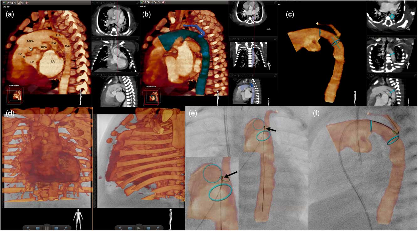
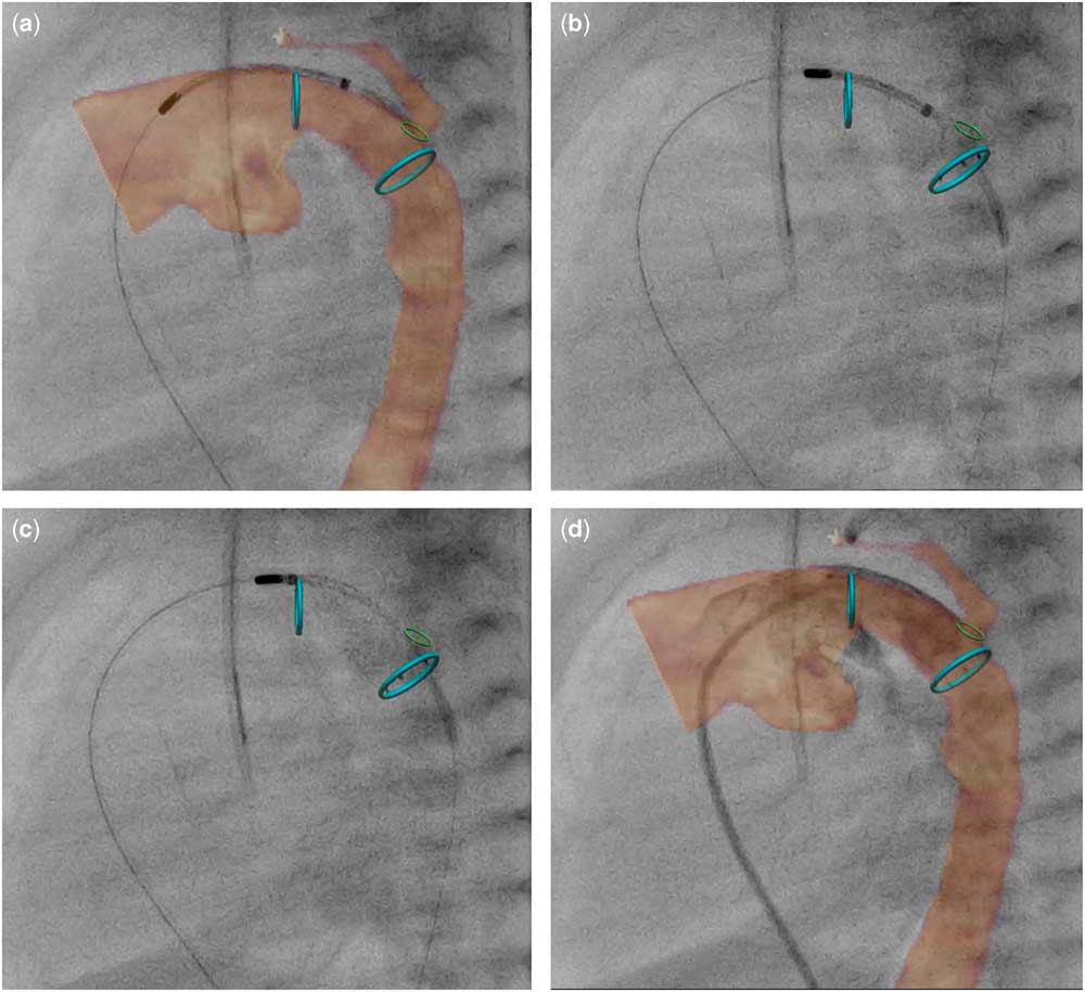
A day after pulmonary artery banding, the patient underwent stenting of the ductus arteriosus. During induction of anaesthesia and introduction of a short 5-F endovascular sheath in the femoral vein, the pre-registered CT scan was manually segmented with a new three-dimensional image fusion software (VesselNavigator; Philips Healthcare, Best, The Netherlands) to create a roadmap for stent delivery (Fig 1a–c). The desired stent position was marked with two, blue, rings, and an additional ring marker was placed at the origin of the left subclavian artery (Fig 1c). For registration and fusion of the roadmap, fluoroscopy in posterior–anterior and lateral projections was used with bony structures serving as reference points (Fig 1d). Movement of the soft coronary wire within the borders of the duct and the subclavian artery together with a small (1 ml) contrast injection confirmed the proper alignment (Fig 1e). An 8×20-mm Sinus-SuperFlex-DS stent (OptiMed, Ettlingen, Germany) was positioned and successfully deployed under sole three-dimensional guidance (Fig 2, Supplementary video 1). Final contrast injection (2 ml) showed perfect stent position with unobstructed ductal flow and easy filling of the left subclavian artery. The total contrast dose administered was 3 ml, and total fluoroscopy and procedural times were 4 and 15 min, respectively. The radiation exposure, expressed as dose area product, was 86.2 µGy×m2. The patient’s further course was uneventful.
VesselNavigator-assisted segmentation and registration of the roadmap with live fluoroscopy. A CT scan was automatically three-dimensional volume-rendered to identify the heart and big vessels. A “cut plane” tool in the sagittal plane was used to expose the desired structures ( a ). Subsequently, the ductus arteriosus and nearby vessels were manually segmented with a “single click” option ( b ). The selected volume is highlighted on the reconstruction (large, left panel) and on three perpendicular planes (right panels). To mark the stent’s landing zone (blue), ring markers were placed at the pulmonary end of the duct and just distally to the subclavian artery ( c ). After short fluoroscopy in posterior–anterior and lateral projections was acquired, the reconstruction was manually aligned with the spine, vertebrae, and sternum, serving as reference points ( d ). In the coronal plane, a soft coronary wire outside the proximal blue marking ring reflects minor misalignment of the roadmap ( e ). Movement of the wire within the reconstructed left subclavian artery shows accurate alignment in the sagittal plane ( f ). *Ventricular septal defect; DA=ductus arteriosus; DAo=descending aorta; LA=left atrium; LPA=left pulmonary artery; LV=left ventricle; MPA=main pulmonary artery; RPA=right pulmonary artery; RV=right ventricle.

VesselNavigator-assisted ductus arteriosus stent implantation. A self-expanding Sinus-SuperFlex-DS stent (Optimed, Ettlingen, Germany) was introduced from the femoral vein and positioned solely under three-dimensional roadmap guidance ( a ). To enhance visualisation of the fine nitinol structure of the stent, only ring markers were left during the implantation. At the beginning of the deployment, golden stent markers are matched with the distal marking ring, indicating the end of the landing zone. As the stent is being further deployed, it conforms to the curvature of the ductus ( c ) and a final contrast injection ( d ) confirms a perfect result with the entire length of the ductus covered by the stent.

Discussion
Bilateral pulmonary artery banding coupled with concomitant or delayed ductus arteriosus stent implantation has become an alternative initial treatment of various forms of obstructive left-heart lesions.Reference Goreczny, Qureshi and Rosenthal 1 , Reference Moszura, Dryzek and Goreczny 2 In premature patients with interrupted aortic arch, this approach enables prostaglandin infusion cessation and postponement of final arch reconstruction to a later stage with increased body weight.
Our previous experiences with three-dimensional rotational angiography for ductal interventions in neonates have shown reliable guidance with no additional contrast injections.Reference Goreczny, Morgan and Dryzek 3 Despite the benefits, this method requires specific set-up of the C-arm for rotational angiography, and more importantly the three-dimensional reconstruction may only be prepared after the angiography, adding to the total procedural time. With the availability of pre-intervention imaging, however, segmentation and planning of the procedure may be performed before the patient’s arrival to the catheterisation laboratory.
During the intervention, additional time is spared with quick and easy two-dimensional-to-three-dimensional registration, which excludes the need for obtaining three-dimensional volume for fusion of the roadmap with live fluoroscopy.Reference Goreczny, Dryzek and Moszura 4 , Reference Stangenberg, Shuja and Carelsen 5
To increase accuracy of the overlay, several reference points may be used including bony structures, movement of wires and catheters, and low-volume contrast injection. Similar to the three-dimensional rotational angiography, the alignment of the overlay must be critically evaluated for each target structure, and appropriate corrections have to be made to accommodate for distortion produced by pulsatility, wires, delivery systems, or devices.
Reliable three-dimensional guidance enables resignation of serial angiographies, consequently eliminating the need to introduce a long, upsized vascular sheath and expose the low-weight patient to a higher contrast volume. The colour ring markers, an additional feature when compared with the previous method, clearly highlight the stent’s landing zone. When used without the three-dimensional volume, the ring features permit stent deployment with continuous visualisation of fine nitinol stent wires.
Acknowledgements
The authors acknowledge Dr Maciej Lukaszewski (Radiology Department, Polish Mother’s Memorial Hospital) for the CT scan and Alexandra Hul (Medical University of Lodz) for editing.
Financial Support
This research received no specific grant from any funding agency or from commercial or not-for-profit sectors.
Conflicts of Interest
None.
Supplementary material
To view supplementary material for this article, please visit https://doi.org/10.1017/S1047951117000154