




No CrossRef data available.
Article contents
Differences in the neurochemical characteristics of the cortex and striatum of mice with cerebral malaria
Published online by Cambridge University Press: 13 December 2004
Abstract
Fatal murine cerebral malaria is an encephalitis and not simply a local manifestation in the brain of a systemic process. Histopathologically, murine cerebral malaria has been characterized by monocyte adherence to the endothelium of the microvasculature, activation of microglial cells, swelling of endothelial cell nuclei, microvasculature damage, and breakdown of the blood-brain barrier with cerebral oedema. Brain parenchymal cells have been proposed to be actively involved in the pathogenesis of murine cerebral malaria. We, therefore, compared the neurochemical characteristics of Plasmodium berghei ANKA-infected mice with controls to determine whether cerebral malarial infection significantly impairs specific neuronal populations. Between 6 and 7 days after infection, we found a significant loss of neurones containing substance P, with preservation of cells containing somatostatin, neuropeptide Y and calbindin in the striatum of infected mice compared with controls. In the cortex of infected mice, we found a significant reduction in the number of cells containing substance P, somatostatin and neuropeptide Y. The number of calbindin-containing neurones was unchanged. This study found significant changes in the neurochemical characteristics of the cortex and striatum of mice infected with P. berghei ANKA, which may contribute to their cerebral symptoms.
Information
- Type
- Research Article
- Information
- Copyright
- © 2004 Cambridge University Press
INTRODUCTION
Cerebral malaria is one of the major pathological complications of Plasmodium falciparum infection in humans (Hunt & Grau, 2003). Among the prominent histopathological features in post-mortem brains of cerebral malaria patients are sequestration of parasitized erythrocytes in microvessels, ring haemorrhages and oedema (Oo et al. 1987). The causes underlying this pathology in human cerebral malaria are not clear. It has been proposed that erythrocytes infected with P. falciparum express surface antigens that promote their adherence to the vascular endothelium, with a resulting reduction in blood flow and oxygen supply (Warrell et al. 1988). However, the absence of hypoxic neuronal damage and the occurrence of endothelial adherence without signs of cerebral malaria make this an unlikely major or sole cause.
A second hypothesis is that the production of pro-inflammatory cytokines is responsible for neural dysfunction. The pattern of cytokine production in cerebral malaria resembles that seen in other inflammatory states of the central nervous system. The mRNA levels for the pro-inflammatory cytokines tumor necrosis factor-α, interferon-γ and interleukin-1β increase in parallel with the amount of parasite mRNA in the brains of mice with cerebral malaria (Jennings et al. 1997). The levels of mRNA for other cytokines (interleukin-2, 4, 5, 6, 10 and 12), including those with anti-inflammatory activity, are unchanged (Jennings et al. 1997), although the findings of Kossodo et al. (1997) suggest that interleukin-10 may play a protective role against experimental cerebral malaria in mice.
As it is not possible to examine the progressive development of histopathological changes in the human central nervous system or to carry out experimental manipulations, a number of animal models of cerebral malaria have been developed. A useful model is the fatal murine cerebral malaria model, namely, P. berghei ANKA (PbA) infection in CBA or C57BL/6 mice (Rest, 1982; Thumwood et al. 1988; Neill & Hunt, 1992). These mice develop behavioural changes at approximately day 5 post-infection (p.i.) and progress to coma and death at approximately days 6–8 p.i. (Grau et al. 1986; Thumwood et al. 1988; Neill & Hunt, 1992). At the terminal stage of the disease, the central nervous system tissue of these mice exhibits petechial haemorrhages, oedema and vascular occlusion (Grau et al. 1986; Thumwood et al. 1988; Chang-Ling, Neill & Hunt, 1992; Neill & Hunt, 1992; Neill, Chang-Ling & Hunt, 1993), all of which are features of post-mortem human cerebral malaria tissue (Oo et al. 1987). Modifications of blood-brain barrier function in these mice provoke changes in the distribution and properties of astrocytes and microglia, which are key non-neuronal elements in the brain parenchyma (Hunt & Grau, 2003). In particular, retinal microglia, representative of central nervous system microglia, become activated within 48–72 h post-inoculation with PbA (Hunt & Grau, 2003). Later in the disease process, microglia take on an amoeboid appearance, typical of an immunologically activated state, and express tumor necrosis factor (Hunt & Grau, 2003). Astrocytes show reactive changes in the retina and optic nerve early in the course of the disease, linked to permeabilization of the blood-brain barrier (Hunt & Grau, 2003). Later, when mice are exhibiting neurological symptoms, astrocyte damage is observed and this requires immune system activation (Hunt & Grau, 2003). Given the essential roles played by astrocytes in maintaining brain homeostasis and blood-brain barrier properties, such damage is likely to affect neuronal function adversely (Hunt & Grau, 2003). Therefore, we used this model of fatal murine cerebral malaria to compare the neurochemical characteristics of infected mice with controls to examine whether PbA infection significantly impairs specific neuronal populations, which may contribute to the attendant cerebral dysfunction.
MATERIALS AND METHODS
Infection procedure
Four-month-old female C57BL/6J mice were supplied by Harlan Olac Ltd, Oxon, UK. The malaria parasites used were Plasmodium berghei ANKA (from Professor D. Walliker, Institute of Cell, Animal and Population Biology, University of Edinburgh, Edinburgh, Scotland). Mice were infected by intravenous injection of 5×10⁴ parasitized erythrocytes suspended in 250 μl of phosphate-buffered saline (PBS), pH 7·4, via the lateral tail vein. Mice infected in this manner normally die between days 6 and 8 after infection, after exhibiting signs of cerebral involvement such as reduced locomotion, convulsions, ataxia and coma. Parasitaemias were recorded during the course of the disease from thin smears of tail blood stained with Giemsa's stain, being 6·0%, 19·6% and 23·0% on days 4, 6 and 7 p.i., respectively. Controls were uninfected C57BL/6J mice of the same age and sex. Eight mice were studied in each group from 3 separate infection experiments.
Tissue preparation
On day 6 or 7 p.i., mice were asphyxiated with CO2 and perfused through the left ventricle with ice-cold PBS followed by 4% paraformaldehyde in PBS, pH 7·4. Brains were removed and post-fixed in 4% paraformaldehyde for 24 h at 4 °C. The brains were cryopreserved in 30% sucrose in phosphate buffer for 48 h at 4 °C, frozen in isopentane maintained at approximately −50 °C and then cut into 50 μm-thick coronal sections on a cryostat (Jung Frigocut 2800E, Leica Microsystems, Milton Keynes, UK).
Immunohistochemistry
Floating sections were immunostained by the avidin-biotin-peroxidase method. Throughout all washes and incubations, sections were shaken. Sections were washed extensively in PBS and treated with 3% H2O2 in PBS for 20 min to destroy endogenous peroxidase activity. Non-specific sites were blocked for 1 h in PBS containing 0·3% Triton X-100 and either 1% normal goat serum (neuropeptide Y) or 1% normal rabbit serum (substance P and somatostatin) or 3·6% mouse IgG blocking reagent (calbindin; M.O.M. immunodetection kit, Vector Laboratories Ltd, Peterborough, UK). Sections were then incubated overnight at 4 °C in the primary antibody. After a series of preliminary experiments to establish optimal immunostaining conditions, the primary antisera, their sources and the dilutions used were: neuropeptide Y polyclonal antibody raised in rabbit, Calbiochem-Novabiochem Ltd, Nottingham, UK, 1[ratio ]10000; monoclonal rat anti-substance P, Oxford Biotechnology Ltd, Kidlington, UK, 1[ratio ]25; rat anti-somatostatin monoclonal antibody, Chemicon International Ltd, Harrow, UK, 1[ratio ]100 and monoclonal anti-calbindin-D-28K, Sigma–Aldrich Company Ltd, Dorset, UK, 1[ratio ]3000. Immunohistochemical characterization of these antibodies, including pre-absorption controls, has previously been reported (Cuello, Galfre & Milstein, 1979; Kawaguchi & Shindou, 1998).
After incubation in the primary antibody, sections were washed in PBS and incubated for 1 h at 4 °C in either 7·5 μg/ml biotinylated goat anti-rabbit IgG (neuropeptide Y) or 7·5 μg/ml biotinylated rabbit anti-rat IgG (substance P and somatostatin) or 18·75 μg/ml biotinylated anti-mouse IgG (calbindin; all secondary antibodies were from Vector Laboratories Ltd). After washes in PBS, sections were reacted with the avidin-biotin Vectastain Elite kit (Vector Laboratories Ltd) according to the manufacturer's instructions, with 3,3′-diaminobenzidine as chromogen in the presence of H2O2 (DAB substrate kit for peroxidase, Vector Laboratories Ltd). Nickel chloride was added to the substrate solution, resulting in a grey-black stain. Sections were dried for at least 48 h at room temperature on poly-L-lysine-coated slides, mounted in Entellan (VWR International Ltd, Poole, UK) and examined microscopically. Negative controls for each animal and each primary antibody were included in every assay, by omitting the primary antibody from the procedure. Staining was not detected in neurones under this condition.
Quantification of cell number and diameter
A Zeiss KS400 (version 3.0) quantitative 2D image analysis system, incorporating a Matrox Meteor frame grabber and a high resolution Sony XC-770 CCD monochrome camera, was used to count cell number and measure cell diameter. The Zeiss KS400 is a Microsoft Windows application with all the advantages of a standardized graphical user interface. The system is based on complete object oriented programming, providing fast and reproducible ways of measuring structures in monochrome images. Due to the complexity of the immunostaining images, the macro-programme used was semi-automated. Stained cells were counted in a 0·45 mm2 visual field (0·11 mm2 for calbindin-positive cells in the striatum). A cell was counted if it had an intact cell body and soma membrane. Microscopic fields were selected by one observer and another observer blind to the experimental condition performed the cell counts. Cell counts were converted from the number of cells in the visual field to the number of cells per mm2 by dividing by 0·45 (0·11 for calbindin-positive cells in the striatum). Cell counts (cells/mm2) and striatal cell diameters (μm) are presented as mean±standard error of the mean obtained from 8 animals per group. Statistical comparisons were made using the unpaired t-test (two tail) with Welch correction where required. P values<0·05 were considered statistically significant.
RESULTS
Somatostatin, neuropeptide Y, substance P and calbindin immunoreactivities were compared in mice infected with PbA (day 6–7 p.i.) and controls. The patterns of neurotransmitter immunoreactivities were consistent among the 8 animals in each group. Sections between interaural 4·39 mm, bregma 0·74 mm and interaural 4·18 mm, bregma 0·38 mm (approximate stereotaxic coordinates) were immunostained.
The number of somatostatin- and neuropeptide Y-containing neurones was significantly reduced in the cortex of day 6–7 p.i. mice compared with controls (Fig. 1). The number of these cells was unchanged in the striatum (Fig. 2), although, their morphology was markedly different (Fig. 3). Whereas the somatostatin- and neuropeptide Y-containing cells of control striatum were associated with an extensive network of dendrites (Fig. 3A), this was absent from the infected striatum (Fig. 3B). In addition, the diameter of the cell body was significantly smaller (Fig. 4).

Fig. 1. Quantification of immunohistochemically labelled cells in the cortex of PbA-infected and control mice. Values are mean±standard error of the mean of 8 mice in each group. Statistical comparisons were made using unpaired t-test (two tail) with Welch correction where required; *P<0·01, **P<0·005, ***P<0·001 versus control.

Fig. 2. Quantification of immunohistochemically labelled cells in the striatum of PbA-infected and control mice. Values are mean±standard error of the mean of 8 mice in each group. The calbindin values have been reduced by a factor of 10 to aid graphical representation. Statistical comparisons were made using unpaired t-test (two tail); ****P<0·0001 versus control.

Fig. 3. Neuropeptide Y immunoreactivity in coronal sections. These micrographs illustrate neuropeptide Y immunoreactivity in the striatum of control (A) and PbA-infected (B) mice. Neuropeptide Y-containing neurones in the striatum of PbA-infected mice have smaller cell bodies (Fig. 4) and rudimentary dendritic trees compared with controls.

Fig. 4. Quantification of cell diameter of somatostatin- and neuropeptide Y-containing cells in the striatum of PbA-infected and control mice. Values are mean±standard error of the mean of 8 mice in each group. Statistical comparisons were made using unpaired t-test (two tail); **P<0·005, ****P<0·0001 versus control.
We observed a highly significant reduction in the number of substance P-containing neurones in the cortex and striatum of day 6–7 p.i. mice compared with uninfected controls (Figs 1 and 2). Very few substance P-containing neurones survived in these regions of the brains of infected mice.
The number of calbindin-containing neurones was similar in the cortex and striatum of both groups of mice (Figs 1 and 2), but there appeared to be an increase in neuropil immunostaining in infected mice, although this was not quantified (Fig. 5). In control mice (Fig. 5A), moderately stained neuropil was detected with calbindin immunoreactivity seen primarily in the proximal dendrites of neurones. In PbA-infected mice (Fig. 5B), the neuropil appeared to be stained to a greater extent with calbindin labelling and increased throughout the second and third order dendritic branches of neurones.

Fig. 5. Calbindin immunoreactivity in coronal sections. These micrographs illustrate calbindin immunoreactivity in the cortex of control (A) and PbA-infected (B) mice. In control mice, proximal dendrites are primarily labelled while in PbA-infected mice, labelling appears to be increased throughout the second and third order dendrites.
DISCUSSION
This study found significant changes in the neurochemical characteristics of the cortex and striatum of mice infected with PbA compared with controls. In particular, we found a highly significant loss of neurones containing substance P in the striatum of PbA-infected mice. The striatum is a major subcortical area for the control of voluntary and postural movements (Stone, 1995). Interestingly, a loss of γ-aminobutyric acid/substance P striatal neurones has been postulated to result in hypokinetic disorders, best exemplified by Parkinson's disease (Tohgi et al. 1997). Additionally, a significant loss of substance P immunopositive cells has been reported in the striatum of a murine model of ataxia telangiectasia (Eilam et al. 2003). These mice display neurological deficits, as assessed by poor performance on three separate tests of motor function, indicative of ataxia (Barlow et al. 1996). Therefore, it is possible that the highly significant reduction of striatal substance P-containing neurones in PbA-infected mice may contribute to their reduced locomotion and marked ataxia.
A significant increase in the concentration of quinolinic acid has been reported in the cerebrospinal fluid of humans with cerebral malaria (Dobbie et al. 2000; Medana et al. 2002a, 2003) and in the brains of PbA-infected mice (Sanni et al. 1998). This compound activates receptors sensitive to N-methyl-D-aspartate (NMDA) (Stone & Perkins, 1981; Schwarcz, Whetsell & Mangano, 1983; Stone, 1993, 2001). As these receptors are associated with calcium influx into neurones, they can produce cell damage when over-activated (Kim & Choi, 1987). Therefore, a high local concentration of quinolinic acid in the brain could lead to neuronal damage, and so we compared the neurochemical characteristics of PbA-infected mice with the neurochemical effects reported to be mediated by quinolinic acid to determine if this compound could play a role in causing the neuronal damage presented here. Beal et al. (1989, 1991) examined the effects of intrastriatal injections of quinolinic acid in rats and found that somatostatin- and neuropeptide Y-containing neurones were less sensitive to quinolinic acid than projection neurones. On the other hand, Figueredo-Cardenas et al. (1994) and Figueredo-Cardenas, Chen & Reiner (1997) claimed that striatal somatostatin- and neuropeptide Y-containing cells were more sensitive to quinolinic acid than projection neurones, and that this vulnerability was dependent on the age of the rats. They found that somatostatin- and neuropeptide Y-containing cells typically survived better than projection neurones in young rats and more poorly in mature rats. It would then be expected that adult striatal somatostatin- and neuropeptide Y-containing cells would be lost after exposure to quinolinic acid, whereas we have found a complete preservation despite a dramatic loss of substance P-containing cells in PbA-infected mice. Several other groups (Boegman, Smith & Parent, 1987; Davies & Roberts, 1987; Boegman & Parent, 1988; Forloni et al. 1992) also failed to find an absolute preservation of somatostatin- and neuropeptide Y-containing neurones in the rat striatum after quinolinic acid exposure. Previous work in the rat has shown that excitotoxic injury induced by NMDA receptor agonists, such as quinolinic acid, causes a rise in intracellular Ca2+ (Mattson et al. 1991). The level of calbindin (a calcium-binding protein) immunoreactivity has been shown to be elevated in rat striatal neurones following exposure to quinolinic acid. Following an intrastriatal injection of quinolinic acid, the necrotic core of the lesion was devoid of calbindin-positive cells while in the transition zone (the peripheral portion of a lesioned area), surviving spiny cells exhibited increased calbindin-immunoreactive product in somata, proximal and distal dendritic branches and spines (Huang et al. 1995). Unlike this scenario in the rat where calbindin-containing neurones are vulnerable to quinolinic acid (Huang et al. 1995), we found that the number of calbindin-containing neurones was similar in the striatum of both groups of mice. However, there appeared to be an increase in neuropil immunostaining in PbA-infected mice. Therefore, an increased availability of calbindin protein at dendrites may reflect a greater demand for Ca2+ buffering, precipitated by the abnormal rise in intracellular Ca2+ induced by quinolinic acid. Although previous work has been conducted almost exclusively on rats receiving intrastriatal injections of quinolinic acid, the pattern of substantial loss of substance P-containing neurones with complete preservation of those with somatostatin, neuropeptide Y and calbindin in the striatum of PbA-infected mice is quite different from the profile reported in these studies, suggesting that quinolinic acid is an unlikely major or sole mediator of the neuronal damage reported here.
Neuronal damage has previously been reported in humans and mice with cerebral malaria. Medana et al. (2002b) proposed that impairment of transport within nerve fibres could induce neurological dysfunction, and quantified β-amyloid precursor protein (β-APP) to detect defects in axonal transport in brains from patients with P. falciparum cerebral malaria. Axonal injury disrupts neural integrity, the distribution of neurosecretory granules, and the transport of enzymes and chemicals involved in the formation of neurotransmitters and substances associated with trophic activity. β-APP is a protein that is normally transported along the axon, and accumulates at the sites of axonal injury. It was found that the frequency and extent of β-APP immunostaining were more severe in patients with cerebral malaria than in those with no clinical cerebral involvement. β-APP immunostaining was often associated with haemorrhages and areas of demyelination, suggesting that multiple processes may be involved in neuronal injury. Ma et al. (1997) also demonstrated loss of axonal viability in central nervous system tissue of PbA-infected mice. They reported that the optic nerves in these mice displayed a significantly increased incidence of patchy axonal demyelination and degeneration, and suggested that increased vascular permeability, vascular obstruction with monocytes and/or the activation of monocytes and microglia to produce cytokines may be responsible.
In summary, previous studies of the brains of PbA-infected mice have examined the histopathology (Rest, 1982; Jennings, Lal & Hunter, 1998) or the expression and distribution of putative pathological factors e.g. tumor necrosis factor-α (Medana, Hunt & Chaudhri, 1997) and indoleamine 2,3-dioxygenase (Hansen et al. 2000). However, to date, there has been no such information relating to neurotransmitters. Therefore, the present study describes, for the first time, significant changes in the neurochemical characteristics of the cortex and striatum of mice with cerebral malaria, which may contribute to their cerebral symptoms. Alternative causes or contributors (e.g. cytokines), other than a high level of quinolinic acid, to the cell dysfunction in this disease will be explored in future work.
This study was supported by The Wellcome Trust. The authors thank Dr J. M. Campbell, Mr D. Russell, Mr A. Lockhart, Dr R. C. Spike and Mr M. Neilson, Institute of Biomedical and Life Sciences, University of Glasgow for excellent advice and help.
References
REFERENCES
BARLOW, C.,
HIROTSUNE, S.,
PAYLOR, R.,
LIYANAGE, M.,
ECKHAUS, M.,
COLLINS, F.,
SHILOH, Y.,
CRAWLEY, J. N.,
RIED, T.,
TAGLE, D. &
WYNSHAW-BORIS, A. (
1996).
Atm-deficient mice: a paradigm of ataxia telangiectasia.
Cell
86,
159–171.CrossRefGoogle Scholar
BEAL, M. F.,
KOWALL, N. W.,
SWARTZ, K. J.,
FERRANTE, R. J. &
MARTIN, J. B. (
1989).
Differential sparing of somatostatin-neuropeptide Y and cholinergic neurons following striatal excitotoxin lesions.
Synapse
3,
38–47.CrossRefGoogle Scholar
BEAL, M. F.,
FERRANTE, R. J.,
SWARTZ, K. J. &
KOWALL, N. W. (
1991).
Chronic quinolinic acid lesions in rats closely resemble Huntington's disease.
Journal of Neuroscience
11,
1649–1659.CrossRefGoogle Scholar
BOEGMAN, R. J.,
SMITH, Y. &
PARENT, A. (
1987).
Quinolinic acid does not spare striatal neuropeptide Y-immunoreactive neurons.
Brain Research
415,
178–182.CrossRefGoogle Scholar
BOEGMAN, R. J. &
PARENT, A. (
1988).
Differential sensitivity of neuropeptide Y, somatostatin and NADPH-diaphorase containing neurons in rat cortex and striatum to quinolinic acid.
Brain Research
445,
358–362.CrossRefGoogle Scholar
CHANG-LING, T.,
NEILL, A. L. &
HUNT, N. H. (
1992).
Early microvascular changes in murine cerebral malaria detected in retinal wholemounts.
American Journal of Pathology
140,
1121–1130.Google Scholar
CUELLO, A. C.,
GALFRE, G. &
MILSTEIN, C. (
1979).
Detection of substance P in the central nervous system by a monoclonal antibody.
Proceedings of the National Academy of Sciences, USA
76,
3532–3536.CrossRefGoogle Scholar
DAVIES, S. W. &
ROBERTS, P. J. (
1987).
No evidence for preservation of somatostatin-containing neurons after intrastriatal injections of quinolinic acid.
Nature, London
327,
326–329.CrossRefGoogle Scholar
DOBBIE, M.,
CRAWLEY, J.,
WARUIRU, C.,
MARSH, K. &
SURTEES, R. (
2000).
Cerebrospinal fluid studies in children with cerebral malaria: an excitotoxic mechanism?
American Journal of Tropical Medicine and Hygiene
62,
284–290.Google Scholar
EILAM, R.,
PETER, Y.,
GRONER, Y. &
SEGAL, M. (
2003).
Late degeneration of nigro-striatal neurons in ATM−/−mice.
Neuroscience
121,
83–98.CrossRefGoogle Scholar
FIGUEREDO-CARDENAS, G.,
ANDERSON, K. D.,
CHEN, Q.,
VEENMAN, C. L. &
REINER, A. (
1994).
Relative survival of striatal projection neurons and interneurons after intrastriatal injection of quinolinic acid in rats.
Experimental Neurology
129,
37–56.CrossRefGoogle Scholar
FIGUEREDO-CARDENAS, G.,
CHEN, Q. &
REINER, A. (
1997).
Age-dependent differences in survival of striatal somatostatin-NPY-NADPH-diaphorase-containing interneurons versus striatal projection neurons after intrastriatal injection of quinolinic acid in rats.
Experimental Neurology
146,
444–457.CrossRefGoogle Scholar
FORLONI, G. L.,
ANGERETTI, N.,
RIZZI, M. &
VEZZANI, A. (
1992).
Chronic infusion of quinolinic acid in rat striatum: effects on discrete neuronal populations.
Journal of Neurological Science
108,
129–136.CrossRefGoogle Scholar
GRAU, G. E.,
PIGUET, P. F.,
ENGERS, H. D.,
LOUIS, J. A.,
VASSALLI, P. &
LAMBERT, P. H. (
1986).
L3T4+ T lymphocytes play a major role in the pathogenesis of murine cerebral malaria.
Journal of Immunology
137,
2348–2354.Google Scholar
HANSEN, A. M.,
DRIUSSI, C.,
TURNER, V.,
TAKIKAWA, O. &
HUNT, N. H. (
2000).
Tissue distribution of indoleamine 2,3-dioxygenase in normal and malaria-infected tissue.
Redox Report
5,
112–115.CrossRefGoogle Scholar
HUANG, Q.,
ZHOU, D.,
SAPP, E.,
AIZAWA, H.,
GE, P.,
BIRD, E. D.,
VONSATTEL, J. P. &
DIFIGLIA, M. (
1995).
Quinolinic acid-induced increases in calbindin D28k immunoreactivity in rat striatal neurons in vivo and in vitro mimic the pattern seen in Huntington's disease.
Neuroscience
65,
397–407.CrossRefGoogle Scholar
HUNT, N. H. &
GRAU, G. E. (
2003).
Cytokines: accelerators and brakes in the pathogenesis of cerebral malaria.
Trends in Immunology
24,
491–499.CrossRefGoogle Scholar
JENNINGS, V. M.,
ACTOR, J. K.,
LAL, A. A. &
HUNTER, R. L. (
1997).
Cytokine profile suggesting that murine cerebral malaria is an encephalitis.
Infection and Immunity
65,
4883–4887.Google Scholar
JENNINGS, V. M.,
LAL, A. A. &
HUNTER, R. L. (
1998).
Evidence for multiple pathologic and protective mechanisms of murine cerebral malaria.
Infection and Immunity
66,
5972–5979.Google Scholar
KAWAGUCHI, Y. &
SHINDOU, T. (
1998).
Noradrenergic excitation and inhibition of GABAergic cell types in rat frontal cortex.
The Journal of Neuroscience
18,
6963–6976.CrossRefGoogle Scholar
KIM, J. P. &
CHOI, D. W. (
1987).
Quinolinate neurotoxicity in cortical cell culture.
Neuroscience
23,
423–432.CrossRefGoogle Scholar
KOSSODO, S.,
MONSO, C.,
JUILLARD, P.,
VELU, T.,
GOLDMAN, M. &
GRAU, G. E. (
1997).
Interleukin-10 modulates susceptibility in experimental cerebral malaria.
Immunology
91,
536–540.CrossRefGoogle Scholar
MA, N.,
MADIGAN, M. C.,
CHAN-LING, T. &
HUNT, N. H. (
1997).
Compromised blood-nerve barrier, astrogliosis, and myelin disruption in optic nerves during fatal murine cerebral malaria.
Glia
19,
135–151.3.0.CO;2-#>CrossRefGoogle Scholar
MATTSON, M. P.,
RYCHLIK, B.,
CHU, C. &
CHRISTAKOS, S. (
1991).
Evidence for calcium-reducing and excito-protective roles for the calcium-binding protein calbindin-D28k in cultured hippocampal neurons.
Neuron
6,
41–51.CrossRefGoogle Scholar
MEDANA, I. M.,
HUNT, N. H. &
CHAUDHRI, G. (
1997).
Tumor necrosis factor-alpha expression in the brain during fatal murine cerebral malaria: evidence for production by microglia and astrocytes.
American Journal of Pathology
150,
1473–1486.Google Scholar
MEDANA, I. M.,
HIEN, T. T.,
DAY, N. P.,
PHU, N. H.,
MAI, N. T. H.,
VAN CHU'ONG, L.,
CHAU, T. T. H.,
TAYLOR, A.,
SALAHIFAR, H.,
STOCKER, R.,
SMYTHE, G.,
TURNER, G. D. H.,
FARRAR, J.,
WHITE, N. J. &
HUNT, N. H. (
2002 a).
The clinical significance of cerebrospinal fluid levels of kynurenine pathway metabolites and lactate in severe malaria.
Journal of Infectious Diseases
185,
650–656.Google Scholar
MEDANA, I. M.,
DAY, N. P.,
HIEN, T. T.,
MAI, N. T. H.,
BETHELL, D.,
PHU, N. H.,
FARRAR, J.,
ESIRI, M. M.,
WHITE, N. J. &
TURNER, G. D. (
2002 b).
Axonal injury in cerebral malaria.
American Journal of Pathology
160,
655–666.Google Scholar
MEDANA, I. M.,
DAY, N. P. J.,
SALAHIFAR-SABET, H.,
STOCKER, R.,
SMYTHE, G.,
BWANAISA, L.,
NJOBVU, A.,
KAYIRA, K.,
TURNER, G. D. H.,
TAYLOR, T. E. &
HUNT, N. H. (
2003).
Metabolites of the kynurenine pathway of tryptophan metabolism in the cerebrospinal fluid of Malawian children with malaria.
Journal of Infectious Diseases
188,
844–849.CrossRefGoogle Scholar
NEILL, A. L. &
HUNT, N. H. (
1992).
Pathology of fatal and resolving Plasmodium berghei cerebral malaria in mice.
Parasitology
105,
165–175.CrossRefGoogle Scholar
NEILL, A. L.,
CHANG-LING, T. &
HUNT, N. H. (
1993).
Comparisons between microvascular changes in cerebral and non-cerebral malaria in mice, using the retinal whole-mount technique.
Parasitology
107,
477–487.CrossRefGoogle Scholar
OO, M. M.,
AIKAWA, M.,
THAN, T.,
AYE, T. M.,
MYINT, P. T.,
IGARASHI, I. &
SCHOENE, W. C. (
1987).
Human cerebral malaria: a pathological study.
Journal of Neuropathology and Experimental Neurology
46,
223–231.CrossRefGoogle Scholar
REST, J. R. (
1982).
Cerebral malaria in inbred mice. I. A new model and its pathology.
Transactions of the Royal Society of Tropical Medicine and Hygiene
76,
410–415.CrossRefGoogle Scholar
SANNI, L. A.,
THOMAS, S. R.,
TATTAM, B. N.,
MOORE, D. E.,
CHAUDHRI, G.,
STOCKER, R. &
HUNT, N. H. (
1998).
Dramatic changes in oxidative tryptophan metabolism along the kynurenine pathway in experimental cerebral and noncerebral malaria.
American Journal of Pathology
152,
611–619.Google Scholar
SCHWARCZ, R.,
WHETSELL, W. O. &
MANGANO, R. M. (
1983).
Quinolinic acid: an endogenous metabolite that produces axon-sparing lesions in rat brain.
Science
219,
316–318.CrossRefGoogle Scholar
STONE, T. W. &
PERKINS, M. N. (
1981).
Quinolinic acid: a potent endogenous excitant at amino acid receptors in the CNS.
European Journal of Pharmacology
72,
411–412.CrossRefGoogle Scholar
STONE, T. W. (
1993).
Neuropharmacology of quinolinic and kynurenic acids.
Pharmacological Reviews
45,
309–379.Google Scholar
STONE, T. W. (
1995).
Neuropharmacology, 1st Edn.
W. H. Freeman and Company Limited,
Oxford.
STONE, T. W. (
2001).
Kynurenines in the CNS: from endogenous obscurity to therapeutic importance.
Progress in Neurobiology
64,
185–218.CrossRefGoogle Scholar
THUMWOOD, C. M.,
HUNT, N. H.,
CLARK, I. A. &
COWDEN, W. B. (
1988).
Breakdown of the blood-brain barrier in murine cerebral malaria.
Parasitology
96,
579–589.CrossRefGoogle Scholar
TOHGI, H.,
UTSUGISAWA, K.,
YOSHIMURA, M.,
YAMAGATA, M.,
NAGANE, Y. &
SAITOH, K. (
1997).
Reduction in the ratio of β-preprotachykinin to preproenkephalin messenger RNA expression in postmortem human putamen during aging and in patients with status lacunaris. Implications for the susceptibility to parkinsonism.
Brain Research
768,
86–90.Google Scholar
WARRELL, D. A.,
WHITE, N. J.,
VEALL, N.,
LOOAREESUWAN, S.,
CHANTHAVANICH, P.,
PHILLIPS, R. E.,
KARBWANG, J.,
PONGPAEW, P. &
KRISHNA, S. (
1988).
Cerebral anaerobic glycolysis and reduced cerebral oxygen transport in human cerebral malaria.
Lancet
2,
534–538.CrossRefGoogle Scholar
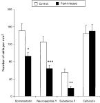
Fig. 1. Quantification of immunohistochemically labelled cells in the cortex of PbA-infected and control mice. Values are mean±standard error of the mean of 8 mice in each group. Statistical comparisons were made using unpaired t-test (two tail) with Welch correction where required; *P<0·01, **P<0·005, ***P<0·001 versus control.
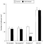
Fig. 2. Quantification of immunohistochemically labelled cells in the striatum of PbA-infected and control mice. Values are mean±standard error of the mean of 8 mice in each group. The calbindin values have been reduced by a factor of 10 to aid graphical representation. Statistical comparisons were made using unpaired t-test (two tail); ****P<0·0001 versus control.
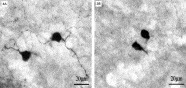
Fig. 3. Neuropeptide Y immunoreactivity in coronal sections. These micrographs illustrate neuropeptide Y immunoreactivity in the striatum of control (A) and PbA-infected (B) mice. Neuropeptide Y-containing neurones in the striatum of PbA-infected mice have smaller cell bodies (Fig. 4) and rudimentary dendritic trees compared with controls.
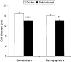
Fig. 4. Quantification of cell diameter of somatostatin- and neuropeptide Y-containing cells in the striatum of PbA-infected and control mice. Values are mean±standard error of the mean of 8 mice in each group. Statistical comparisons were made using unpaired t-test (two tail); **P<0·005, ****P<0·0001 versus control.
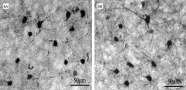
Fig. 5. Calbindin immunoreactivity in coronal sections. These micrographs illustrate calbindin immunoreactivity in the cortex of control (A) and PbA-infected (B) mice. In control mice, proximal dendrites are primarily labelled while in PbA-infected mice, labelling appears to be increased throughout the second and third order dendrites.
 You have
Access
- 2
- Cited by