



Schizophrenia is a severe psychiatric disorder characterised by positive symptoms such as hallucinations and delusions, long linked to dysfunction of the striatum, a subcortical structure central to motor control, cognition and emotional regulation. Reference Starzer, Hansen, Hjorthøj, Albert, Nordentoft and Madsen1–Reference Howes, Montgomery, Asselin, Murray, Valli and Tabraham3 Disruptions in striatal dopamine activity, particularly hyperactivity, have been widely implicated in psychotic experiences. Reference Grace4,Reference Howes and Nour5 Our prior work identified a neuroimaging biomarker of striatal dysfunction – the functional striatal abnormality (FSA) score – integrating resting-state functional magnetic resonance imaging (fMRI) measures of striatal activity and connectivity using machine learning, which differentiated patients from controls and predicted antipsychotic treatment response. Reference Li, Zalesky, Yue, Howes, Yan and Liu6 The striatum comprises associative, limbic and sensorimotor subregions with distinct functions; Reference Levitt, Nestor, Levin, Pelavin, Lin and Kubicki7 while traditional models emphasised dopamine dysregulation in the limbic striatum, Reference McCutcheon, Abi-Dargham and Howes8–Reference Fornito, Harrison, Goodby, Dean, Ooi and Nathan10 more recent evidence highlights the role of associative striatal circuits, particularly connections with the dorsolateral prefrontal cortex (dlPFC), dorsal anterior cingulate cortex (dACC) and insula, in the misattribution of salience and the persistence of delusions and hallucinations. Reference Levitt, Nestor, Levin, Pelavin, Lin and Kubicki7,Reference Fornito, Harrison, Goodby, Dean, Ooi and Nathan10–Reference Menon, Palaniyappan and Supekar13 Beyond functional connectivity studies, Reference Voineskos, Hawco, Neufeld, Turner, Ameis and Anticevic14 white matter tractography provides critical insight into the structural basis of these circuits. For example, our previous work demonstrated the predictive value of cortico-striatal tractography in addiction research Reference Yuan, Zhao, Yu, Manza and Volkow15 and, more recently, we reported dynamic connectivity changes following antipsychotic treatment in first-episode schizophrenia (FE-SCZ), offering clues to the structural evolution of the disorder. Reference Barth, Kelly, Nerland, Jahanshad, Alloza and Ambrogi16,Reference Xi, Wu, Cui, Bai, Gan and Jia17 In the present study, we used diffusion tensor imaging (DTI)-based probabilistic tractography to examine striatal-cortical connectivity in FE-SCZ, both at baseline and after antipsychotic treatment, and tested their relationship with positive symptoms. We hypothesised that FE-SCZ would show reduced connectivity, particularly in associative and limbic circuits, and that these abnormalities would relate to positive symptom severity and change with treatment.
Method
Participants
A total of 80 right-handed FE-SCZ patients and 80 gender-, age- and handedness-matched healthy controls were recruited for this study. Patients were out-patients recruited from the Department of Psychiatry at Xijing Hospital, and healthy controls were recruited via advertisements from the local community. All participants were aged between 18 and 45 years. The FE-SCZ group comprised patients in their first episode of schizophrenia, all within 2 weeks of initial exposure to antipsychotic treatment, indicating the acute stage of their condition. Diagnoses were confirmed by experienced psychiatrists according to the DSM-IV 18 criteria. In patients where the illness duration was less than 6 months, a conclusive FE-SCZ diagnosis was confirmed after a 6-month follow-up. The antipsychotic medication dosages were converted to olanzapine equivalents based on the defined daily dose (DDD) system.
Inclusion criteria for the FE-SCZ group were as follows: (a) a diagnosis of schizophrenia based on the DSM-IV criteria and confirmed through the Chinese version of the Structured Clinical Interview for DSM-IV (SCID-I); (b) minimal prior antipsychotic exposure (no more than 2 weeks before baseline assessment); and (c) a negative urine toxicology screen. Inclusion criteria for the healthy control group were (a) no lifetime history of psychotic, mood or substance use disorders, confirmed by the Structured Clinical Interview for DSM-IV Non-Patient version (SCID-NP), and (b) no family history of psychiatric disorders. Exclusion criteria for all participants included significant medical or neurological illnesses, current misuse of substances other than nicotine, pregnancy and nursing. Demographic and clinical characteristics were based on 73 patients and 77 controls who completed baseline assessments. Of these, 2 patients and 7 controls were excluded from imaging analyses because of excessive head motion or poor image quality, resulting in 71 patients and 70 controls included in the final imaging analyses. The authors assert that all procedures contributing to this work comply with the ethical standards of the relevant national and institutional committees on human experimentation and with the Helsinki Declaration of 1975, as revised in 2013. All procedures involving human participants/patients were approved by the Ethics Committee of Xijing Hospital, Fourth Military Medical University (Approval No. XJYYLL-2021065). Written informed consent was obtained from all participants. Patient participants were independently assessed by a psychiatrist to ensure their capacity to provide consent.
Clinical and neurocognitive assessments
At baseline, schizophrenia patients underwent clinical evaluation using the Positive and Negative Syndrome Scale (PANSS) and a battery of cognitive tests. Follow-up assessments occurred 4.79 months (s.d. = 1.46 months) after baseline and included repeat PANSS evaluation and neuroimaging (DTI) acquisition. Cognitive function was assessed through a series of tasks designed to measure psychomotor vigilance and working memory. Psychomotor vigilance was assessed with a psychomotor vigilance task (PVT), which measures sustained attention and reaction time. Reference Liu, Cai, Chang, Zhu, Cai and Xu19 Working memory was assessed using a standard 2-back version of the n-back task, a well-established tool for assessing the maintenance and manipulation of information. Additional cognitive assessments included the Digit Symbol and Digit Span tests (both forward and backward) from the Wechsler Adult Intelligence Scale (Revised in China), as well as the Semantic (Category) Fluency task from the Beijing version of the Montreal Cognitive Assessment (MoCA). Reference Xi, Wu, Cui, Bai, Gan and Jia17
Magnetic resonance imaging data acquisition
Imaging data were acquired using a Philips 3T Ingenia scanner equipped with a standard 32-channel head coil at the Department of Radiology, Xijing Hospital, Fourth Military Medical University, Xi’an, China. For detailed acquisition parameters, please refer to Supplementary File 1 available at https://doi.org/10.1192/bjp.2025.10434.
Striatum structural connectivity analysis
Data preprocessing
DTI data were preprocessed using QSIPrep pipeline version 0.15.3 for Linux (PennLINC, University of Pennsylvania, Philadelphia, PA, USA; see https://qsiprep.readthedocs.io), which included Multiparametric Principal Component Analysis (MP-PCA) denoising, magnetic field inhomogeneity correction, head motion and eddy current correction, susceptibility distortion correction, tensor calculation and generation of fractional anisotropy images.
Regions of interest
The striatum, defined as a combination of the caudate, putamen and nucleus accumbens based on the Harvard Subcortical Structural Atlas (FSL Atlas), served as the seed mask. To evaluate connectivity with extrastriatal regions, we defined ten a priori regions as target masks following our previous studies. Reference Yuan, Zhao, Yu, Manza and Volkow15 These target regions were based on the Automated Anatomical Labeling (AAL) atlas and included the medial orbitofrontal cortex (mOFC, AAL regions 28, 6 and 26), ventral lateral prefrontal cortex (vlPFC, AAL regions 10 and 16), inferior frontal gyrus ([pars triangularis], IFG, AAL region 14), PFC (AAL regions 4 and 8), insula (AAL region 30), anterior cingulate cortex (ACC, AAL region 32), dACC (AAL region 34), hippocampus (AAL region 38), amygdala (AAL region 42) and supplementary motor area (SMA, AAL region 20). These AAL numbers correspond to the left-hand hemisphere (subtract 1 for the right-hand hemisphere values). Figure 1 illustrates the target regions for seed-based tractography, showing how fibre tracking is performed to assess connectivity between the striatum and various extrastriatal regions. For details of region of interest (ROI) registration procedures and tractography methods, please refer to Supplementary File 1.
Schematic illustration of target regions for seed-based tractography. The figure depicts the striatum (composed of the caudate, putamen and nucleus accumbens) as the seed region, with ten a priori target regions identified for evaluating connectivity. These target regions include the medial orbitofrontal cortex (mOFC), ventral lateral prefrontal cortex (vlPFC), inferior frontal gyrus (IFG), dorsolateral prefrontal cortex (dlPFC), supplementary motor area (SMA), anterior cingulate cortex (ACC), dorsal ACC (dACC), hippocampus, amygdala and insula. The striatum is connected to these target regions via fibre pathways, which are indicated by arrows. The colour coding represents different functional subregions of the striatum: sensorimotor striatum (yellow), associative striatum (red) and limbic striatum (blue).

Connectivity-based seed classification
Following standard methodology, seed-based classification was performed by first thresholding the tractography maps, retaining only voxels with at least ten sample tracts. Voxel values were then converted into proportions, calculated as the number of tracts reaching each specific target mask for that voxel divided by the total number of tracts reaching any target mask. This process generated ten distinct value maps in native diffusion space for each participant, each corresponding to one of the target regions. The striatum was then parcellated by assigning each voxel to the target region for which it showed the highest probability of connection. Finally, for each tract connecting the target regions to the striatum, tract strength was computed as the average probability value within each particpant’s individualised striatal parcellation.
Statistical analysis
All statistical analyses were conducted using MATLAB version 2016b for Linux (MathWorks, Natick, MA USA; see https://www.mathworks.com). Continuous variables, including demographic, clinical and DTI metrics, were expressed as mean ± s.d. and analysed using a general linear model (GLM) or Wilcoxon signed-rank test (non-normal distribution). Categorical variables were analysed with the chi-squared test. Since recent studies suggest that head motion can confound white matter structural connectivity measurements, mean framewise displacement was calculated to control for this potential effect. No significant differences in head motion were found between the schizophrenia group (mean ± s.d.: 0.28 ± 0.05 mm) and healthy control group (0.27 ± 0.03 mm; t = 1.76, p = 0.09). To further mitigate any residual effects of head motion, mean framewise displacement was included as a covariate in the GLM when comparing tract strengths between schizophrenia and healthy control groups at each striatal circuit. In addition, age, gender and years of education were included as covariates in the GLM to control for their potential effects. Multiple comparisons were corrected using the false discovery rate (FDR), with corrected p-values < 0.05 considered significant; this included the initial 20 group comparisons between the striatum and cortical targets (10 regions × 2 hemispheres).
After identifying significant group differences in striatal circuits, we conducted secondary analyses to examine which specific striatal subregion connections accounted for the observed effects. The striatal regions were subdivided into the limbic striatum, associative striatum and sensorimotor striatum. GLM analyses were then conducted for each subregion, including mean framewise displacement, age, gender and education as covariates, with FDR correction applied across the 12 resulting comparisons (4 cortical targets × 3 striatal subregions). For subregional circuits showing significance after FDR correction, Pearson correlations were applied to assess associations between tract strength and PANSS scores (5 tracts × 4 subscales = 20 tests, FDR-corrected, p < 0.05).
To evaluate the effect of antipsychotic medication on striatal subregion circuits, paired t-tests using the GLM were conducted to compare tract strength and PANSS scores in the schizophrenia group at baseline and post-treatment. Given the exploratory nature of these within-patient longitudinal analyses and the limited sample size, p-values were reported without correction for multiple comparisons. Correlations between the relative change in tract strength showing significant differences and the relative change in PANSS scores post-treatment were assessed using partial correlation, controlling for inter-scan interval and cumulative antipsychotic dose before and during treatment. Relative changes in tract strength and PANSS scores were calculated using the formula: 100 × (post-treatment – baseline)/baseline. As with the paired comparisons, these exploratory within-group correlation analyses were not corrected for multiple comparisons because of the limited sample size.
Results
Demographic and clinical characteristics
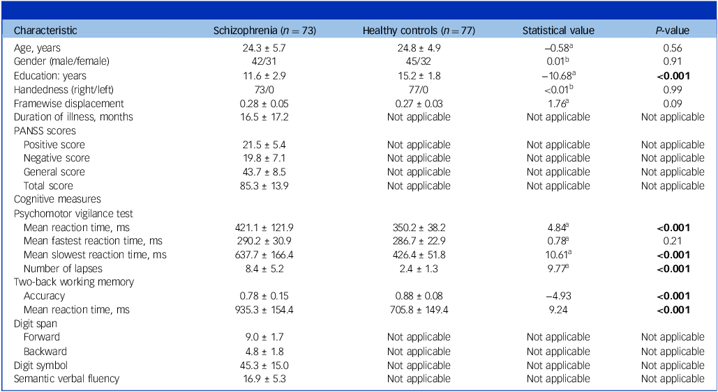
Table 1 presents the demographic and clinical characteristics of schizophrenia patients and healthy control participants. There were no significant differences between the two groups in terms of age, gender distribution or handedness, as both groups were predominantly right-handed. However, schizophrenia patients had significantly fewer years of education compared to the healthy control group.
Demographic and clinical characteristics of the healthy controls and schizophrenia patients

PANSS, Positive and Negative Syndrome Scale.
Bold values indicate a statistically significant difference with a p-value of less than 0.05.
a. Value from the independent-sample t-test.
b. Value from the chi-squared test.
In terms of cognitive performance, schizophrenia patients exhibited significant deficits in multiple cognitive domains. On the PVT, schizophrenia patients had significantly slower reaction times, including both mean and slowest reaction times, and they experienced a higher number of lapses during the task. In addition, on the 2-back working memory task, schizophrenia patients demonstrated lower accuracy and significantly slower response times compared to healthy control participants. These findings highlight significant cognitive impairments in schizophrenia patients, particularly in attention, working memory and reaction time, relative to healthy controls.
The tract strength differences between schizophrenia patients and healthy controls
The GLM analysis revealed notable differences in tract strength between the schizophrenia and healthy control groups, as shown in Fig. 2(a) and Supplementary Table 1. All p-values were corrected for multiple comparisons using the FDR across 20 tests. Specifically, (a) the schizophrenia group exhibited significantly lower tract strength in the left-hand dlPFC–striatum connection compared to the healthy control group (t = 2.770, p = 0.043, FDR-corrected), with a trend towards lower tract strength in the right-hand dlPFC–striatum connection (t = 2.486, p = 0.057, FDR-corrected); (b) the schizophrenia group showed significantly higher tract strength in the left-hand dACC–striatum (t = −2.668, p = 0.043, FDR-corrected), left-hand insula–striatum (t = −3.036, p = 0.043, FDR-corrected) and right-hand IFG–striatum connections (t = −2.690, p = 0.043, FDR-corrected), with a trend towards higher tract strength in the left-hand SMA–striatum connection (t = −2.417, p = 0.057, FDR-corrected). No significant group differences were found in the other striatal circuits (all p > 0.05, FDR-corrected).
Group differences in striatal tract strength between schizophrenia patients and healthy controls. (a) Box plots depict tract strength for connections between the striatum and cortical target regions. Compared with healthy control, schizophrenia patients showed significantly lower tract strength in the left-hand dorsolateral prefrontal cortex (dlPFC)–striatum connection, with a trend towards lower strength in the right-hand dlPFC–striatum connection. In contrast, schizophrenia patients demonstrated significantly higher tract strength in the left-hand dorsal anterior cingulate cortex (dACC)–striatum, left-hand insula–striatum and right-hand inferior frontal gyrus (IFG)–striatum connections, with a trend towards higher strength in the left-hand supplementary motor area (SMA)–striatum connection. No significant differences were observed in other striatal circuits (all p > 0.05, false discovery rate-corrected). (b) Box plots illustrate tract strength differences in striatal subregions. Schizophrenia patients exhibited significantly lower tract strength in the left-hand dlPFC–associative striatum and (dlPFC-AST) left-hand dlPFC–sensorimotor striatum (dlPFC-SMST) compared with healthy controls, but significantly higher tract strength in the left-hand dACC–associative striatum, right-hand IFG–associative striatum (IFG-AST) and left-hand insula–limbic striatum (Insula-LST).

In the subsequent analysis, the striatum connection was further subdivided into the dlPFC–limbic striatum, dlPFC–associative striatum and dlPFC–sensorimotor striatum for a more detailed comparison between schizophrenia patients and healthy controls. This analytical approach was similarly applied to the dACC–striatum, insula–striatum and IFG–striatum connections, as shown in Fig. 2(b). All p-values for these subregion comparisons were FDR-corrected across 12 tests (4 cortical regions × 3 striatal subregions). This analysis identified that, compared to healthy controls, schizophrenia patients exhibited significantly lower tract strength in the left-hand dlPFC–associative striatum (t = 3.789, p = 0.002, FDR-corrected) and the left-hand dlPFC–sensorimotor striatum (t = 4.214, p < 0.001, FDR-corrected) connections. Conversely, schizophrenia patients demonstrated significantly higher tract strength in the left-hand dACC–associative striatum (t = −3.294, p = 0.005, FDR-corrected), the right-hand IFG–associative striatum (t = −3.019, p = 0.009, FDR-corrected) and the left-hand insula–limbic striatum (t = −3.587, p = 0.003, FDR-corrected), connections. No significant differences were observed in other striatal subregion circuits between groups. Detailed statistical results are provided in Supplementary Table 2.
Relationship between tract strength of striatal circuits and clinical measurements in schizophrenia patients
The association between PANSS scores and tract strength in various striatal subregion circuits is illustrated in Supplementary Fig. 1. All p-values for these correlation analyses were FDR-corrected across 20 comparisons (5 tracts × 4 PANSS subscales). Results indicated that the tract strength of the dlPFC–associative striatum was negatively correlated with the PANSS positive score (r = −0.436, p < 0.001, FDR-corrected), general score (r = −0.318, p = 0.007, FDR-corrected) and total score (r = −0.295, p = 0.013, FDR-corrected). Similarly, the tract strength of the dlPFC–sensorimotor striatum showed a negative correlation with the PANSS positive score (r = −0.511, p < 0.001, FDR-corrected), general score (r = −0.268, p = 0.025, FDR-corrected) and total score (r = −0.253, p = 0.035, FDR-corrected). In contrast, the tract strength of the dACC–associative striatum and insula–limbic striatum demonstrated a significant positive correlation with the PANSS positive score (r = 0.552, p < 0.001; r = 0.303, p = 0.011, respectively, FDR-corrected). In addition, the tract strength of the IFG–associative striatum was positively correlated with the PANSS positive score (r = 0.278, p = 0.020, FDR-corrected), negative score (r = 0.318, p = 0.007) and total score (r = 0.373, p = 0.001, FDR-corrected).
Treatment effects on PANSS scores and tract strength in schizophrenia patients
A total of 39 schizophrenia patients completed the follow-up assessment, which included both PANSS evaluations and neuroimaging. The mean interval between baseline and follow-up assessments was 4.79 months (s.d. = 1.46 months). We compared baseline demographic and clinical characteristics, as well as head motion (mean framewise displacement), between patients who completed the follow-up and those who did not. No significant differences were observed between the two groups (all p > 0.05), suggesting no evidence of attrition bias (detailed in Supplementary Tables 3 and 4). The treatment resulted in significant changes in the PANSS scores for schizophrenia patients. As shown in Supplementary Fig. 2, there was a significant reduction in the positive score (p < 0.001), general score (p = 0.013) and total score (p < 0.001), indicating improvements in symptom severity over the course of treatment. Specifically, the positive score decreased from 21.3 ± 4.2 to 17.5 ± 3.1, the general score reduced from 45.1 ± 8.2 to 44.4 ± 8.2 and the total score decreased from 85.5 ± 11.7 to 80.7 ± 11.1. However, there was no significant change observed in the negative score, which decreased slightly from 19.2 ± 5.9 to 18.6 ± 5.4 (p = 0.052). Detailed results are shown in Supplementary Table 5. Cognitive measures, however, did not show significant changes after continued antipsychotic therapy.
Compared to baseline, the schizophrenia group showed significant changes in tract strength over the course of treatment, as shown in Fig. 3(a). Specifically, the tract strength of the left-hand dlPFC–associative striatum connection significantly increased from 0.073 ± 0.008 to 0.077 ± 0.015 (t = 3.26, p = 0.002), indicating an improvement in this pathway after continued antipsychotic therapy. In contrast, the tract strength of the left-hand dACC–associative striatum connection showed a nominally significant decrease from 0.071 ± 0.022 to 0.065 ± 0.015 (t = −2.11, p = 0.042), suggesting a reduction in the integrity of this connection after continued antipsychotic therapy. However, no significant changes were observed in the tract strength of the left-hand dlPFC–sensorimotor striatum (from 0.068 ± 0.012 to 0.067 ± 0.012, t = −0.39, p = 0.692), left-hand insula–limbic striatum (from 0.076 ± 0.015 to 0.080 ± 0.017, t = 1.68, p = 0.101) or right-hand IFG–associative striatum (from 0.067 ± 0.016 to 0.068 ± 0.022, t = 0.18, p = 0.857) connections. These p-values reflect exploratory analyses and are uncorrected.
Treatment-related changes in striatal tract strength and their associations with positive symptom improvement in schizophrenia patients. (a) Box plots depict changes in tract strength between baseline and follow-up under continued antipsychotic therapy for key striatal circuits, including the left-hand dorsolateral prefrontal cortex–associative striatum (dlPFC-AST), left-hand dlPFC–sensorimotor striatum (dlPFC-SMST), left-hand insula–limbic striatum (Insula-LST), left-hand dorsal anterior cingulate cortex–associative striatum (dACC-AST) and right-hand inferior frontal gyrus–associative striatum (IFG-AST). Significant changes are indicated by asterisks. The dlPFC-AST showed a significant increase in tract strength (t = 3.26, p = 0.002), while the dACC-AST showed a nominally significant decrease (t = –2.11, p = 0.042). No significant changes were observed in other connections. Note: p-values reflect exploratory analyses and are uncorrected for multiple comparisons. (b) Scatterplots show correlations between changes in tract strength of the left dlPFC-AST and dACC-AST with reductions in Positive and Negative Syndrome Scale positive scores over the follow-up period. Increased tract strength of the dlPFC-AST was significantly associated with greater reductions in positive symptoms (r = –0.398, p = 0.012), while decreased tract strength of the dACC-AST was significantly associated with greater reductions in positive symptoms (r = 0.378, p = 0.018). Solid lines represent regression lines; dotted lines represent 95% confidence intervals.

Given the significant differences observed in the left-hand dlPFC–associative striatum and dACC–associative striatum connections at baseline and after continued antipsychotic therapy, we primarily focused on the correlation between changes in tract strength for these connections and the reduction in PANSS positive scores over the course of treatment. The results indicated that the increase in tract strength of the left-hand dlPFC–associative striatum was significantly correlated with a greater reduction in PANSS positive scores (r = −0.398, p = 0.012). Conversely, the reduction in tract strength of the left-hand dACC–associative striatum was significantly correlated with a greater reduction in PANSS positive scores (r = 0.378, p = 0.018), as shown in Fig. 3(b). We further examined whether cumulative antipsychotic dose was associated with changes in tract strength. No significant correlations were found between cumulative dose and tract strength changes across the examined circuits (all p > 0.05).
Discussion
This study aimed to examine the structural connectivity of the striatum’s subregions in individuals with FE-SCZ, focusing on the relationship between white matter tract strength and positive symptoms, as well as the impact of antipsychotic treatment on these circuits. Using advanced DTI and probabilistic tractography, we identified significant differences in the connectivity of striatal subregions, particularly the associative and the limbic subregions. In addition, we found that changes in these structural connections were associated with reductions in positive symptoms following antipsychotic treatment, providing further insight into the neurobiological underpinnings of schizophrenia.
One of the key findings of this study was the lower tract strength observed in the left-hand dlPFC–striatum connection in the schizophrenia group compared to controls. This result supports previous research highlighting the role of the dlPFC in cognitive control and its interactions with the striatum in regulating goal-directed behaviour. Reference Smucny, Hanks, Lesh, O’Reilly and Carter20,Reference Slifstein, van de Giessen, Van Snellenberg, Thompson, Narendran and Gil21 Our analysis further revealed that the reduction in tract strength was most pronounced in the associative subregions of the striatum. The associative striatum, which is involved in planning, inhibition and cognitive flexibility, has been implicated in the persistence of delusions and hallucinations in schizophrenia. Reference Vano, McCutcheon, Rutigliano, Kaar, Finelli and Nordio22,Reference Avram, Brandl, Knolle, Cabello, Leucht and Scherr23 Disruptions in this connection could hinder the brain’s ability to suppress inappropriate or intrusive thoughts, which is consistent with the positive symptoms observed in schizophrenia. These findings align with emerging models of schizophrenia that suggest impairments in executive function and cognitive control as critical contributors to psychotic experiences. Reference Kanchanatawan, Thika, Anderson, Galecki and Maes24,Reference Yang, Yang, Lan, Liu and Chou25
In contrast, we observed significantly higher tract strength in several key striatal circuits, including the left-hand dACC–striatum, the left-hand insula–striatum and the right-hand IFG–striatum. Further analysis indicated that these changes were especially pronounced in the associative and the limbic subregions of the striatum. The dACC and insula are components of the brain’s salience network, involved in processing stimuli deemed personally relevant or significant. Reference Uddin26,Reference Limongi, Jeon, Mackinley, Das, Dempster and Théberge27 Increased connectivity between these regions and the striatum may reflect an exaggerated attribution of salience to irrelevant or non-threatening stimuli, a hallmark of delusions and hallucinations. Reference Palaniyappan, Simmonite, White, Liddle and Liddle12,Reference Winton-Brown, Fusar-Poli, Ungless and Howes28 These findings suggest that dysfunction in the limbic and associative striatum and its connections to the salience network may contribute to the misinterpretation of both external and internal events in schizophrenia. Reference Howes and Nour5,Reference Koller and Cannon29 In addition, the increased connectivity between the striatum and IFG may reflect attempts to regulate abnormal salience processing, but ultimately, the underlying structural impairments may contribute to the persistence of psychotic symptoms. Reference Heller, Steinmann, Levitt, Makris, Antshel and Fremont30
Our longitudinal analysis of changes in tract strength following antipsychotic treatment revealed that the improvement in symptom severity was associated with restoration of connectivity in specific circuits. Notably, we observed a significant increase in the tract strength of the left-hand dlPFC–associative striatum connection, alongside a decrease in the tract strength of the left-hand dACC–associative striatum connection. These changes in structural connectivity were correlated with a reduction in positive symptoms, as measured by the PANSS positive subscale. The increase in tract strength between the dlPFC and associative striatum suggests that antipsychotic treatment may promote a recovery of cognitive control and inhibitory function, which may help reduce the intensity of delusions and hallucinations. Conversely, the reduction in tract strength between the dACC and the associative striatum could reflect a normalisation of salience processing, where stimuli are less likely to be perceived as aberrantly significant. Along with our previous studies, these findings highlight the potential of structural connectivity as a marker for treatment response and underscore the complex interaction between cognitive control and salience attribution in the pathophysiology of schizophrenia. Reference Xi, Wu, Cui, Bai, Gan and Jia17,Reference Liu, Cai, Chang, Zhu, Cai and Xu19
While our results provide compelling evidence for the role of striatal connectivity in schizophrenia, several limitations must be considered. First, although our sample size is comparable to prior studies focusing on FE-SCZ, it may still be underpowered to detect subtle effects in some striatal subregions, because of the clinical and logistical challenges inherent in recruiting and assessing this patient population. Second, because of the exploratory nature and modest size of the longitudinal subsample, we did not apply FDR correction to within-group comparisons or correlations. As a result, findings such as the observed reduction in dACC–associative striatum tract strength should be interpreted with caution and require replication in larger cohorts. Third, although this study included longitudinal imaging and clinical assessments, the follow-up duration (∼4.80 months) may be insufficient to capture the longer-term trajectory of white matter connectivity changes. Future research with longer follow-up periods is needed to characterise how these structural changes evolve over the course of the illness and treatment. In addition, while DTI is a powerful tool for studying white matter integrity, it does not provide direct information on axonal density or microstructure, which could be informative for understanding the mechanisms underlying connectivity changes. Future research using advanced imaging techniques, such as high-angular resolution diffusion imaging (HARDI) or tractography with more sophisticated models, may provide a more refined view of the microstructural alterations in schizophrenia. Finally, although the striatum is known to play a key role in cognitive processes, cognitive performance in our sample did not show significant change during the follow-up period, limiting the relevance of connectivity–cognition associations in this cohort. Therefore, the present study focused specifically on striatal connectivity in relation to positive symptomatology, consistent with our a priori hypotheses.
In conclusion, this study provides critical insights into the role of striatal connectivity in the positive symptoms of schizophrenia, emphasising how disruptions in specific striatal subregions – particularly those linked to the dlPFC, dACC, insula and IFG – are associated with the severity of psychotic experiences. Our findings underscore the potential of structural connectivity as a biomarker for symptom severity and treatment response, offering a foundation for future work to investigate how these connectivity changes evolve over time, and how antipsychotic treatments influence these circuits. Longer longitudinal studies exploring these dynamics will be crucial in refining our understanding of the neurobiological underpinnings of schizophrenia’s positive symptoms and in developing more targeted therapeutic strategies.
Supplementary material
The supplementary material is available online at https://doi.org/10.1192/bjp.2025.10434
Data availability
The data-sets used and/or analysed during the current study are available from the corresponding author upon reasonable request. The analysis code supporting the findings has been provided in the Supplementary Materials. No additional materials were generated or analysed in this study.
Author contributions
W.L. drafted the manuscript; C.W. and K.Y. contributed to data preprocessing and statistical analyses; M.C. provided access to clinical and imaging data; F.G. contributed to the preparation of the main text and responses to reviewers; Y.Z. contributed substantially to study design, data interpretation and critical revision of the manuscript; M.G. and R.S. assisted with clinical assessments and data collection. All authors reviewed and approved the final version of the manuscript.
Funding
This study was supported by the Fast Response Program of Air Force Medical University (Grant No. 2023KXKT050), the Xijing Hospital-Air Force Medical University Joint Fund (Grant No. LHJJ24WQ05), the National Natural Science Foundation of China (Grant Nos. 82371500 and 82571745) and the Xidian University Specially Funded Project for Interdisciplinary Exploration (Grant No. TZJH2024011).
Declaration of interest
None.





eLetters
No eLetters have been published for this article.