INTRODUCTION
Toxoplasma gondii is an opportunistic pathogen that causes significant disease in immune-compromised hosts. An obligate intracellular parasite, T. gondii actively invades its target cells, and forms a non-fusogenic parasitophorous vacuole (PV) within which it replicates (Sibley et al. Reference Sibley, Weidner and Krahenbuhl1985; Suss-Toby et al. Reference Suss-Toby, Zimmerberg and Ward1996; Mordue et al. Reference Mordue, Hakansson, Niesman and Sibley1999). One striking feature of the PV is its rapid association with host ER and mitochondria (Jones and Hirsch, Reference Jones and Hirsch1972; Sinai et al. Reference Sinai, Webster and Joiner1997). The function of this phenomenon is unclear; however, the ER and mitochondria are important calcium-handling organelles, and this observation led to the hypothesis that Toxoplasma infection could perturb calcium handling in host cells. Indeed, in support of this possibility, prolonged infection with T. gondii is associated with accumulation of calcium within the PV (Pingret et al. Reference Pingret, Millot, Sharonov, Bonhomme, Manfait and Pinon1996), and soluble Toxoplasma antigen (STAg) is reported to induce a transient calcium elevation in cells transfected with CCR5 (Aliberti et al. Reference Aliberti, Valenzuela, Carruthers, Hieny, Andersen, Charest, Reis E. Sousa, Fairlamb, Ribeiro and Sher2003). In addition, Toxoplasma induces calcium-dependent signalling events in macrophages, but short-term imaging of parasitized cells does not detect changes in intracellular calcium that can be temporally related to these events (Masek et al. Reference Masek, Fiore, Leitges, Yan, Freedman and Hunter2006). Nevertheless, the studies presented here show that longer-term imaging of macrophages exposed to live parasites reveals robust changes in intracellular calcium that derive from extracellular sources, and these fluxes occur independently of MyD88 or CCR5, two molecules involved in distinct pathways required for innate recognition of T. gondii.
MATERIALS AND METHODS
Macrophages
Bone marrow-derived macrophages were cultured as previously described (Caamano et al. Reference Caamano, Alexander, Craig, Bravo and Hunter1999) from female, 4 to 6-week-old C57BL/6, CCR5−/− (Jackson laboratories, Bar Harbor, ME), or MyD88−/− mice (generously gifted from A. Gelman and L. Turka, University of Pennsylvania (Gelman et al. Reference Gelman, Zhang, Choi and Turka2004)). Macrophages were harvested on day 7 of culture, plated at a density of 1×10⁶ on sterilized 22 mm cover-slips (Fisher Scientific, Pittsburgh, PA), then primed for 16 h with 100 u/ml rmIFN-γ (BD Pharmingen, San Diego, CA) in macrophage medium consisting of DMEM (Invitrogen Life Technologies, Carlsbad, CA) supplemented with 10% heat-inactivated fetal calf serum (HIFCS; U.S. Bio-Technologies Inc. Parkerford, PA), 1% penicillin and streptomycin, 25 mm HEPES, and 1 mm sodium pyruvate (Invitrogen).
Parasites
T. gondii tachyzoites (RH strain) were maintained in vitro by passage through monolayers of human foreskin fibroblasts cultured in DMEM media supplemented with 10% HIFCS, 1% penicillin, streptomycin and gentamicin (Invitrogen). For use in experiments, intracellular parasites were liberated by shearing cells through a 22-gauge needle, then washed twice with calcium-free, magnesium-free dPBS (Cellgro, Mediatech Inc., Herndon, VA). Tachyzoites were then resuspended in normal bath buffer (140 mm NaCl, 5 mm KCl, 2 mm MgCl2, 2·5 mm CaCl2, 10 mm glucose, 10 mm NaOH-HEPES, pH 7·3, Osm 290–300) or calcium-free medium (145 mm NaCl, 5 mm KCl, 2 mm MgCl2, 10 mm Glucose, 10 mm NaOH-HEPES, pH 7·3, Osm 290–300) immediately prior to infection. Soluble Toxoplasma antigen (STAg) was prepared from RH tachyzoites as previously described (Sharma et al. Reference Sharma, Mullenax, Araujo, Erlich and Remington1983) and tested for absence of LPS and Mycoplasma contaminants by the University of Pennsylvania Cell Center (Philadelphia, PA). Heat-killed parasites were prepared by incubation at 60°C for 30 min (Sibley et al. Reference Sibley, Weidner and Krahenbuhl1985).
Intracellular calcium imaging
Ratiometric live cell imaging with Fura-2 loaded macrophages was as previously described (Masek et al. Reference Masek, Fiore, Leitges, Yan, Freedman and Hunter2006). Briefly, macrophages (1×10⁶) plated on sterilized 22 mm coverslips (Fisher Scientific, Pittsburgh, PA) and primed overnight with rm-IFN-γ were loaded with the calcium-sensitive indicator Fura-2 AM (5 μm; Invitrogen) in macrophage medium containing 0·2% Pluronic-F127 (Sigma-Aldrich) for 60 min at 37°C. Following loading, cover-slips were immediately transferred to a microscope recording chamber and perfused with normal buffer or calcium-free buffer to remove extracellular Fura-2. For ratiometric live cell imaging of intracellular calcium, Fura-2 loaded cells were excited by discrete bandwidth light (340±10 nm, 380±10 nm) from a xenon light source through a computer-controlled monochromator (TILL, Applied Scientific Imaging, Eugene, OR). Emitted fluorescence was passed through a 470 nm long pass filter and images were obtained with an intensified charge coupled video-camera (Hammamatsu Model C2400-68, Japan) connected to a Nikon inverted microscope. Four fluorescent video images collected at intervals of 400 ms were averaged and digitized (0·5 Hz) using a custom macro written for ImagePro Plus software (Media Cybernetics, Bethesda, MD). Stored images were analysed off-line. Within cursor defined areas of interest, paired 340/380 images were background subtracted and the ratio was calculated. The absolute ratio values within the cursor-defined areas were exported into Microsoft Excel (Microsoft Corporation, Redmond, WA) and converted to [Ca2+] as previously described (Grynkiewicz et al. Reference Grynkiewicz, Poenie and Tsien1985). Calibration factors, Rmax (340/380 ratio obtained in the presence of 5 mm ionomycin and 10 mm Ca2+), Rmin (340/380 ratio in the presence of 5 mm ionomycin and excess EGTA) (Ionomycin and EGTA from Sigma-Aldrich), and F380max and F380 min (minimum 510 nm emission with 380 nm excitation) were determined in situ. Across experiments, the number of cells recorded in the imaging field ranged from 55 to 75. Images were captured at 5 frames per min for the durations indicated and data from all cells in the imaging field are expressed as the intracellular calcium concentration, based on calibration as described above. In all experiments, live tachyzoites (5:1), STAg (50 μg/ml), or heat-killed parasites (10:1) were added in normal or calcium-free buffer as indicated after 3 min of baseline recording. Experiments were ended by addition of 1 μm thapsigargin (Sigma-Aldrich) in normal buffer to determine responsiveness.
RESULTS
Toxoplasma induces calcium elevations in macrophages
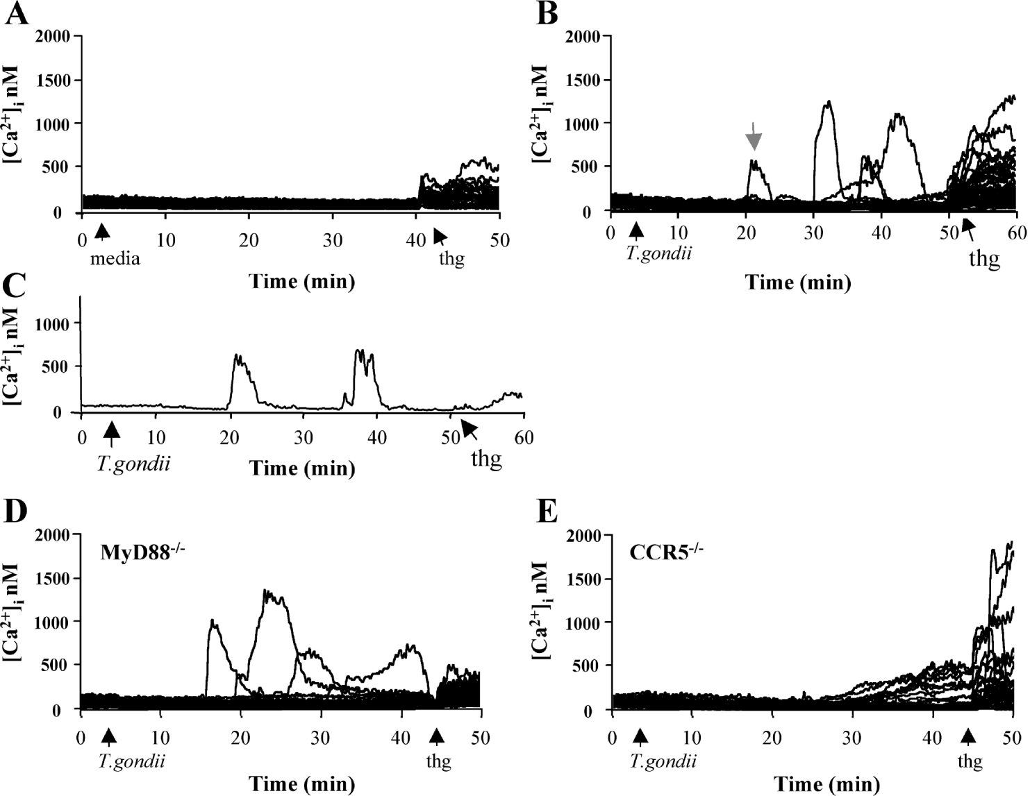
There is a general consensus that for macrophages to produce pro-inflammatory cytokines in response to T. gondii they must be pre-activated with IFN-γ. Therefore, as part of studies to understand the role of calcium in innate sensing of this pathogen, bone-marrow derived macrophages were primed with IFN-γ overnight, and then loaded with the calcium indicator Fura-2 for use in ratiometric, live-cell calcium imaging. During the imaging period, intracellular calcium remained constant in untreated cells (Fig. 1A), whereas cytoplasmic calcium elevations were recorded in macrophages exposed to live T. gondii tachyzoites (Fig. 1B). These elevations, which were not observed in unprimed cells (data not shown), typically began 15–20 min after addition of parasites, and occurred in up to 20% of macrophages in an imaging field. The microscopy equipment available for these experiments is not configured to simultaneously measure host cell calcium and visualize intracellular parasites, thus we were unable to confidently determine whether calcium responses occurred in infected cells. However, across experiments, the percentage of infected cells in the culture after 60 min ranged from 70–90%, whereas calcium fluxes were detected in up to 20% of cells, indicating that not all infected cells exhibited a calcium spike. The mechanism behind this response is unknown, but one possibility is that the elevations in intracellular calcium result from membrane damage generated by parasite egress. This appears unlikely given that most cells survive calcium mobilization, as evidenced by their ability to maintain intracellular Fura-2 loading, and respond to thapsigargin treatment, which elevates cytoplasmic calcium (Fig. 1B). Moreover, some of these cells respond to parasite challenge with multiple rounds of calcium mobilization (Fig. 1C), suggesting a specific and proximal effect that is likely associated with parasite invasion.
Toxoplasma gondii induces calcium elevations in macrophages. Fura-2 loaded macrophages were treated with normal bath buffer (A, media) or infected with live T. gondii at a ratio of 5:1 (B) following 3 min of baseline recording in normal bath buffer and changes in intracellular calcium ([Ca2+]i nm) were recorded as described in the Materials and Methods section. The grey arrowhead in (B) denotes an example of a macrophage that exhibits multiple rounds of calcium mobilization following exposure to T. gondii; this recording is isolated from the other cells in the imaging field and depicted in (C). Data in A–C represent at least 6 experiments. MyD88 (D) or CCR5 (E) deficient macrophages were loaded with Fura-2 and infected with T. gondii (5:1). Thapsigargin (thg; 1 μm) was added where indicated to demonstrate a positive response. Results in D–E are representative of 2–3 experiments each.

Calcium elevations do not require MyD88 or CCR5
The TLR-adaptor MyD88 is a key molecule involved in recognition of many pathogens, and it is required for macrophage production of IL-12 following T. gondii challenge (Scanga et al. Reference Scanga, Aliberti, Jankovic, Tilloy, Bennouna, Denkers, Medzhitov and Sher2002; Del Rio et al. Reference Del Rio, Butcher, Bennouna, Hieny, Sher and Denkers2004; Kim and Denkers, Reference Kim and Denkers2006). To determine whether MyD88 is required for generating calcium responses in macrophages exposed to T. gondii, BMMφ from wild-type (WT) and knock-out mice were treated with parasites and monitored for changes in intracellular calcium levels. Similar to WT controls (Fig. 1B), elevations in intracellular calcium were recorded in MyD88−/− cells following exposure to parasites (Fig. 1D), indicating that this adaptor protein is dispensable for this effect. In addition to MyD88, CCR5 has been implicated as an important host recognition molecule for Toxoplasma (Aliberti et al. Reference Aliberti, Valenzuela, Carruthers, Hieny, Andersen, Charest, Reis E. Sousa, Fairlamb, Ribeiro and Sher2003). Similar to WT (Fig. 1B) and MyD88−/− cells (Fig. 1D), BMMφ derived from CCR5−/− mice responded to parasites with changes in intracellular calcium (Fig. 1E). However, whereas calcium elevations in WT or MyD88−/− cells began abruptly, came to a peak, and then returned to baseline within a few min, the calcium changes that occurred in CCR5−/− macrophages were slower to develop and remained elevated for a longer period of time. This finding suggests that signalling through CCR5, while not required for parasite-induced changes in host cell calcium, may modulate this response. In addition to robust fluxes in calcium, we have also observed that a minority of cells in parasite-treated cultures responds to thapsigargin with excessive release of calcium relative to untreated controls (Fig. 1A, B, Fig. 2A). This finding further supports the conclusion that Toxoplasma perturbs calcium handling in macrophages.
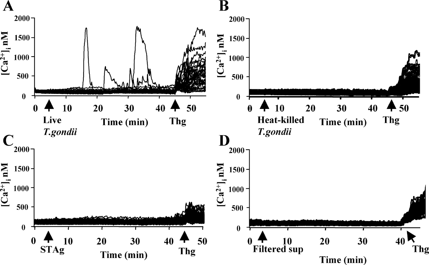
Live Toxoplasma gondii, but not heat-killed parasites or STAg, induce calcium elevations in macrophages. Fura-2 loaded macrophages were infected with live (A, 5:1) or heat-killed (B, 10:1) T. gondii, or STAg (C, 50 μg/ml) and intracellular calcium ([Ca2+]i nm) was monitored for the time-periods indicated. (D) Filtered supernatants from macrophages infected for 2 h at a ratio of 5:1 were applied to fresh Fura-2 loaded macrophages and the intracellular calcium was recorded over time. Thapsigargin (thg; 1 μm) was added at the end of each experiment to demonstrate responsiveness. Data represent 2–5 experiments each.

Calcium elevations result from live parasites
To characterize the basis for these changes in intracellular calcium, experiments were performed to distinguish whether they result from live infection, phagocytosis of organisms, or from exposure to parasite-expressed proteins, such as C-18 or profilin (Aliberti et al. Reference Aliberti, Valenzuela, Carruthers, Hieny, Andersen, Charest, Reis E. Sousa, Fairlamb, Ribeiro and Sher2003; Yarovinsky et al. Reference Yarovinsky, Zhang, Andersen, Bannenberg, Serhan, Hayden, Hieny, Sutterwala, Flavell, Ghosh and Sher2005). Whereas live parasites induced calcium elevations (Fig. 2A), stimulation with heat-killed parasites or STAg prepared from RH tachyzoites did not (Fig. 2B–C). These findings suggest that live parasites are required for this effect. Moreover, given that macrophages readily phagocytose heat-killed parasites (Sibley et al. Reference Sibley, Weidner and Krahenbuhl1985 and our unpublished observation), this finding argues against phagocytosis as the cause of calcium elevations in parasite-treated cells. In addition, when supernatants from infected cells were collected, filtered to remove parasites and cell debris, and immediately applied to Fura-2 loaded macrophages, they had no effect on intracellular calcium in these cells (Fig. 2D), indicating that calcium elevation induced by live T. gondii is a cell-associated effect arising from direct parasite contact or infection. However, it cannot be ruled out that a short-lived paracrine or secreted product may be involved.
Parasite-induced calcium elevations derive from extracellular sources
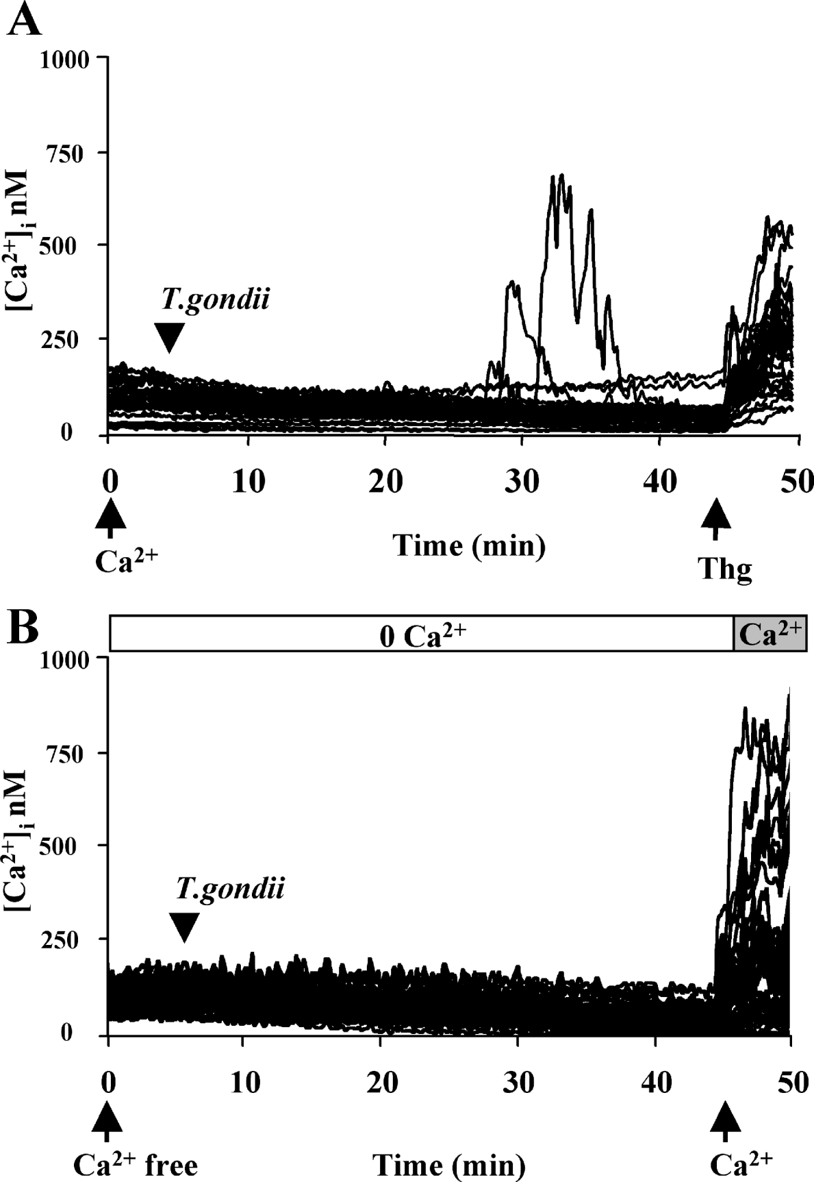
The size of the calcium elevations induced by parasites suggested that they could result from influx from extracellular sources. Indeed, the calcium elevations observed in normal bath buffer (Fig. 3A) were not detected in cells infected in calcium-free medium until calcium was added back in the form of normal bath buffer (Fig. 3B). This finding was not a secondary consequence of the experimental procedure, as the percentages of infected macrophages were comparable in both experiments, and changes in calcium were not observed in media-treated cells under the same conditions (data not shown). These observations suggest that parasite-induced calcium responses predominantly result from influx of extracellular calcium.
Extracellular calcium is required for Toxoplasma gondii-induced calcium elevations. (A) Fura-2 loaded macrophages were infected with live T. gondii at a ratio of 5:1 in calcium-containing normal bath buffer, then stimulated with thapsigargin (thg; 1 μm) to demonstrate releasable calcium stores. (B) Fura-2 loaded macrophages were infected with live parasites (5:1) in calcium-free media (0 Ca2+) for the period indicated, then 0 Ca2+ was replaced with normal-calcium bath buffer and changes in intracellular calcium ([Ca2+]i nm) were recorded over time. Infection rates were comparable between experiments and each experiment was repeated twice with similar results.

DISCUSSION
Extensive efforts to image calcium changes in host cells during acute Toxoplasma invasion have failed to detect an immediate response (Lovett and Sibley, Reference Lovett and Sibley2003; Masek et al. Reference Masek, Fiore, Leitges, Yan, Freedman and Hunter2006). However, we now report that longer-term imaging of live macrophages challenged with T. gondii reveals that parasites initiate robust changes in cytoplasmic calcium in some cells. Thus, these delayed calcium elevations likely result from changes in the host cell that occur after invasion. The mechanism underlying this effect is unknown; however, parasite-induced fluxes require the presence of extracellular calcium, suggesting a process that involves surface calcium channels and influx from the extracellular space. One possibility is that infection directly alters calcium homeostasis in parasitized cells, perhaps by direct interaction with calcium-mobilizing machinery, as has been reported to occur in macrophages infected with Listeria monocytogenes (Goldfine and Wadsworth, Reference Goldfine and Wadsworth2002). An alternative possibility is that parasite association with ER and mitochondria (Sinai et al. Reference Sinai, Webster and Joiner1997) could perturb calcium handling by these organelles, leading to discharge in some cells.
T. gondii produces several secreted products, and it is reported that STAg induces a calcium transient when applied to cells transfected with the CCR5 receptor (Aliberti et al. Reference Aliberti, Valenzuela, Carruthers, Hieny, Andersen, Charest, Reis E. Sousa, Fairlamb, Ribeiro and Sher2003). However, despite robust changes induced by live T. gondii, neither STAg nor heat-killed parasites induce changes in cytoplasmic calcium in BMMφ, indicating that live parasites are required for this effect. In this study, we assessed the effects of parasite exposure on intracellular calcium using macrophages primed with IFN-γ. We repeated these studies using unprimed macrophages and found that Toxoplasma failed to induce measurable calcium changes in these cells (data not shown). However, we interpret this finding with caution because we have found that, in general, unprimed macrophages exhibit inconsistent and less robust responses to calcium-mobilizing stimuli (e.g. thapsigargin) relative to primed cells (unpublished observation). This may be related to our consistent observation that unprimed macrophages load to a lesser extent with Fura-2 than primed cells; however, the possibility that IFNγ treatment itself may affect macrophage calcium homeostasis cannot be ruled out. In macrophages, IFNγ acts synergistically with TNF-α to restrict replication of intracellular parasites. The mechanism underlying this effect is poorly understood, but recent work has implicated roles for various IFN-γ-inducible p47 GTPases (Taylor et al. Reference Taylor, Collazo, Yap, Nguyen, Gregorio, Taylor, Eagleson, Secrest, Southon, Reid, Tessarollo, Bray, McVicar, Komschlies, Young, Biron, Sher and Vande Woude2000; Collazo et al. Reference Collazo, Yap, Sempowski, Lusby, Tessarollo, Woude, Sher and Taylor2001; Butcher et al. Reference Butcher, Greene, Henry, Annecharico, Weinberg, Denkers, Sher and Taylor2005; Martens et al. Reference Martens, Parvanova, Zerrahn, Griffiths, Schell, Reichmann and Howard2005). In addition, one of the major functions of IFN-γ in macrophages is to activate the process of phagocytosis, which has been linked to changes in intracellular calcium through Fcγ receptor ligation (Myers and Swanson, Reference Myers and Swanson2002). Thus, we ruled out phagocytosis as a cause for Toxoplasma-induced calcium changes by treating primed macrophages with heat-killed parasites. Despite parasite uptake, no changes in intracellular calcium were recorded in IFN-γ primed macrophages. This finding suggests it is not phagocytosis, but rather live parasites, that induce changes in intracellular calcium in these cells.
Due to technical limitations, we did not determine whether calcium responses occurred as a direct result of infection. However, greater than 70% of the cells in the culture were infected by the end of each imaging experiment, whereas calcium elevations were detected in fewer than 20%, indicating that not all infected cells exhibit a calcium spike. This observation suggests something unique about the infection process in the responding, minority population. One possibility is that calcium elevations may be recorded as a result of damage during invasion or egress in a portion of cells. The finding that extracellular calcium is important for parasite-induced changes in intracellular calcium supports this. However, we observed that most of the cells appeared to acutely survive the calcium elevations, and some showed multiple rounds of mobilization, arguing against non-specific membrane damage as a cause. A second possibility is that the calcium elevations arose in the uninfected population; however, this too seems unlikely given that uninfected cultures do not exhibit spontaneous calcium spikes. A more likely hypothesis is that single parasites induce imperceptible changes in intracellular calcium in most cells, whereas these changes merge into detectable elevations in cells that are co-infected with multiple parasites. Subjectively, this idea is supported by the observation that the likelihood of observing calcium elevations increases with parasite numbers and with time. In addition, because infections were unsynchronized in these studies, it should be noted that the number of macrophages responding to parasite challenge with changes in intracellular calcium could be underestimated. Thus, whereas the majority of cells became infected within 10 min of parasite exposure (data not shown), the prolonged period between invasion and generation of detectable calcium elevations could preclude detection of calcium changes in cells that are infected at later points in the experiments.
Previous work has shown that T. gondii parasites contain intracellular calcium stores that fluctuate during the course of host infection (Vieira and Moreno, Reference Vieira and Moreno2000; Lovett and Sibley, Reference Lovett and Sibley2003), and it is recognized that intra-parasitic calcium is important in multiple functions during the infection cycle, including parasite motility, cytoskeletal rearrangements, secretion of key proteins involved in PV formation, and egress (Arrizabalaga and Boothroyd, Reference Arrizabalaga and Boothroyd2004). Here we report that T. gondii also induces calcium elevations in the host cell; however, the consequences and functions of these changes are unknown. One possibility is that these calcium fluxes may facilitate apoptosis in infected cells (Hajnoczky et al. Reference Hajnoczky, Davies and Madesh2003). Apoptosis is an important component of innate immunity, and while several reports have established that T. gondii acts to suppress host apoptotic pathways (Goebel et al. Reference Goebel, Luder, Lugert, Bohne and Gross1998, Reference Goebel, Luder and Gross1999; Nash et al. Reference Nash, Purner, Leon, Clarke, Duke and Curiel1998; Payne et al. Reference Payne, Molestina and Sinai2003; Kim and Denkers, Reference Kim and Denkers2006), little is known about the mechanisms that initiate this process in parasitized cells. Alternatively, intracellular T. gondii replicates within its PV for up to 3 days before lysing the cell, and little is known about the interactions that occur between parasite and host cell at later time-points. Thus, it is possible that these calcium elevations could activate calcium-sensitive machinery that directs transcription of late genes involved in innate resistance to this organism.
We would like to thank Kevin Foskett for his intellectual input. We also appreciate the generous gift of MyD88−/− mice from Andy Gelman and Larry Turka at the University of Pennsylvania. This work was supported by NIH AI46288, T32 AI055400, T32 RR074063-10, and NIGMS T32-07229.