Introduction
The vibratory, membranous portion of the human vocal fold mucosa is connected to the thyroid cartilage anteriorly via the intervening anterior macula flava and anterior commissure tendon.Reference Hirano and Sato1 Posteriorly, it is joined to the vocal process of the arytenoid cartilage via the intervening posterior macula flava.Reference Hirano and Sato1 The vocal ligament runs between the anterior and posterior maculae flavae.Reference Hirano and Sato1 Many vocal fold stellate cells, which store vitamin A in their lipid droplets, are distributed in the human maculae flavae.Reference Sato, Hirano and Nakashima2, Reference Sato, Hirano and Nakashima3
Human maculae flavae located at both ends of the vocal fold mucosa are involved in the metabolism of extracellular matrices, which are essential for the viscoelastic properties of the lamina propria of the human vocal fold.Reference Sato, Umeno and Nakashima4 Human adult maculae flavae are inferred to be involved in the maintenance of the characteristic layered structure of human vocal fold mucosa.Reference Sato, Umeno and Nakashima4 Human newborn, infant and child maculae flavae are thought to be responsible for forming the characteristic layered structure of adult human vocal fold mucosa.Reference Sato, Hirano and Nakashima5–Reference Sato, Umeno and Nakashima7 Human maculae flavae are believed to be an important structure in the growth, development and ageing of human vocal fold mucosa.Reference Sato, Umeno and Nakashima4–Reference Sato, Hirano and Nakashima8
Vocal fold stellate cells are vitamin A storing cells and a member of the ‘diffuse stellate cell system’.Reference Sato, Umeno and Nakashima9 Vocal fold stellate cells in the maculae flavae are considered a new category of cells in the human vocal fold.
Adult tissue-specific stem cells (tissue stem cells) have the capacity to self-renew and to generate functionally differentiated cells that replenish lost cells throughout an organism's lifetime. There is growing evidence that the vocal fold stellate cells in the human maculae flavae are tissue stem cells or progenitor cells in the human vocal fold mucosa.Reference Sato, Umeno and Nakashima10 The human maculae flavae are a candidate for a stem cell niche, which is a microenvironment nurturing a pool of stem cells which, in this case, are vocal fold stellate cells.Reference Sato, Umeno and Nakashima10 Investigations concerning how to regulate these cells contained in the human maculae flavae are challenging but important in regenerative medicine of the human vocal fold. Artificial manipulations of these cells (e.g. via chemical biology) can lead to novel developments in vocal fold regeneration.
A proper microenvironment in the maculae flavae of human vocal fold mucosa is necessary for the maculae flavae to be effective as a stem cell niche that maintains the stemness of the contained tissue stem cells. This study investigated the role of microenvironment in the maculae flavae of the human vocal fold mucosa.
Materials and methods
The authors assert that all procedures contributing to this work complied with the ethical standards of the relevant national and institutional guidelines on human experimentation (Ethical Committee of Kurume University), and with the Helsinki Declaration of 1975, as revised in 2008.
Six normal human adult vocal folds from surgical specimens were used. Any larynges which had diseases that could possibly affect the tissue of the vocal fold were excluded from the study.
After extraction of the anterior maculae flavae of the human vocal fold mucosae from surgical specimens under microscope, the maculae flavae were minced, cultured and proliferated in two types of culture mediums. One was Dulbecco's modified Eagle's medium (Nissui, Tokyo, Japan) supplemented with 10 per cent fetal bovine serum containing 50 µg/ml each of penicillin G (1000 U/ml) and streptomycin (1000 µg/ml). The other was MF-start primary culture medium and MF-medium (mesenchymal stem cell growth medium; Toyobo, Osaka, Japan). After using MF-start primary culture medium, MF-medium was used to proliferate the cells.
Each tissue was cultured at 37°C in a humidified atmosphere of 5 per cent carbon dioxide. The maculae flavae containing the vocal fold stellate cells were subcultured and morphological features were assessed. Cells were observed with a phase-contrast microscope (Olympus, Tokyo, Japan).
Results
Culturing with Dulbecco's modified Eagle's medium
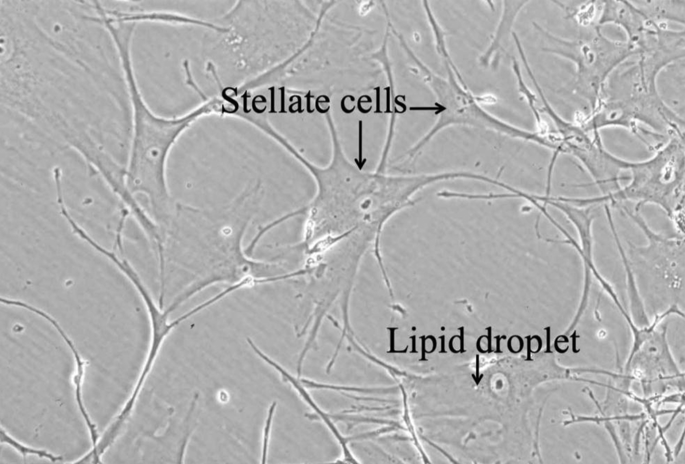
The cells cultured from the maculae flavae were stellate in shape and possessed slender cytoplasmic processes (Figure 1). The nuclei were oval, and the nucleus-to-cytoplasm ratio was low (Figure 1). In the primary culture, small lipid droplets were present in the cytoplasm; however, these disappeared in the second culture. The stellate cells proliferated by attaching their cytoplasmic processes to each other (Figure 1). These cells were morphologically similar to vocal fold stellate cells.
Primary culture of macula flava with Dulbecco's modified Eagle's medium. Stellate cells grew from the macula flava fragments.

During the subculture period, each cell continued to exhibit the same morphological characteristics.
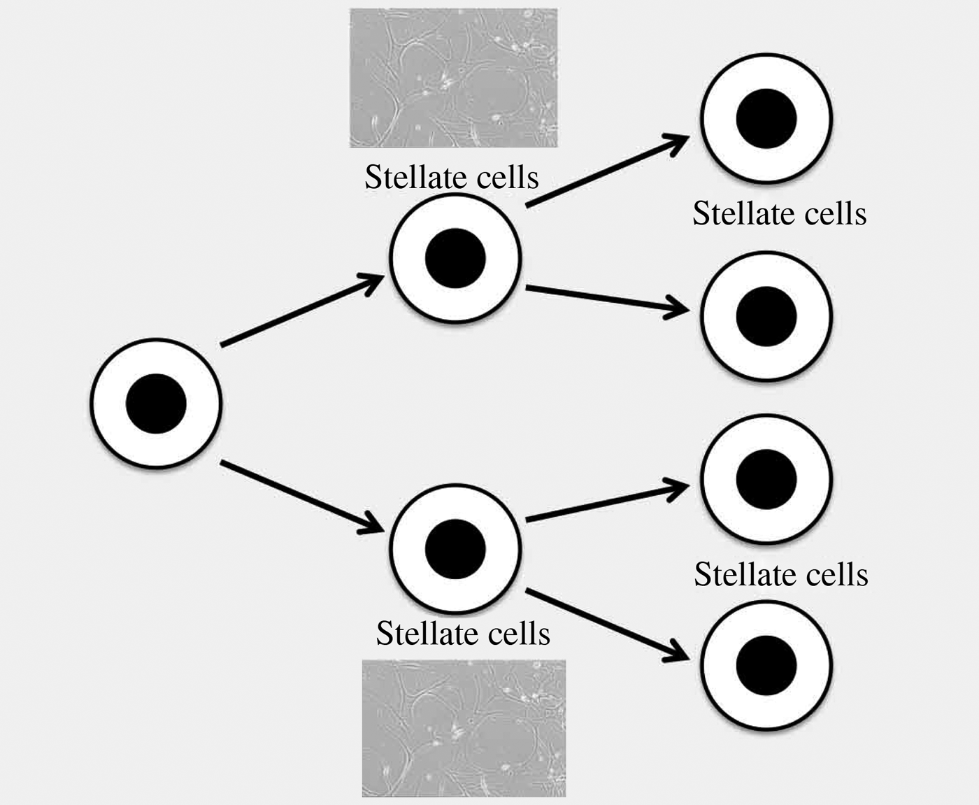
These phenomena suggest that cell division in the human maculae flavae with Dulbecco's modified Eagle's medium is reflective of symmetric self-renewal (Figure 2).
Symmetric cell division with Dulbecco's modified Eagle's medium. During the subculture period, each cell continued to exhibit stellate shape.
Culturing with mesenchymal stem cell growth medium
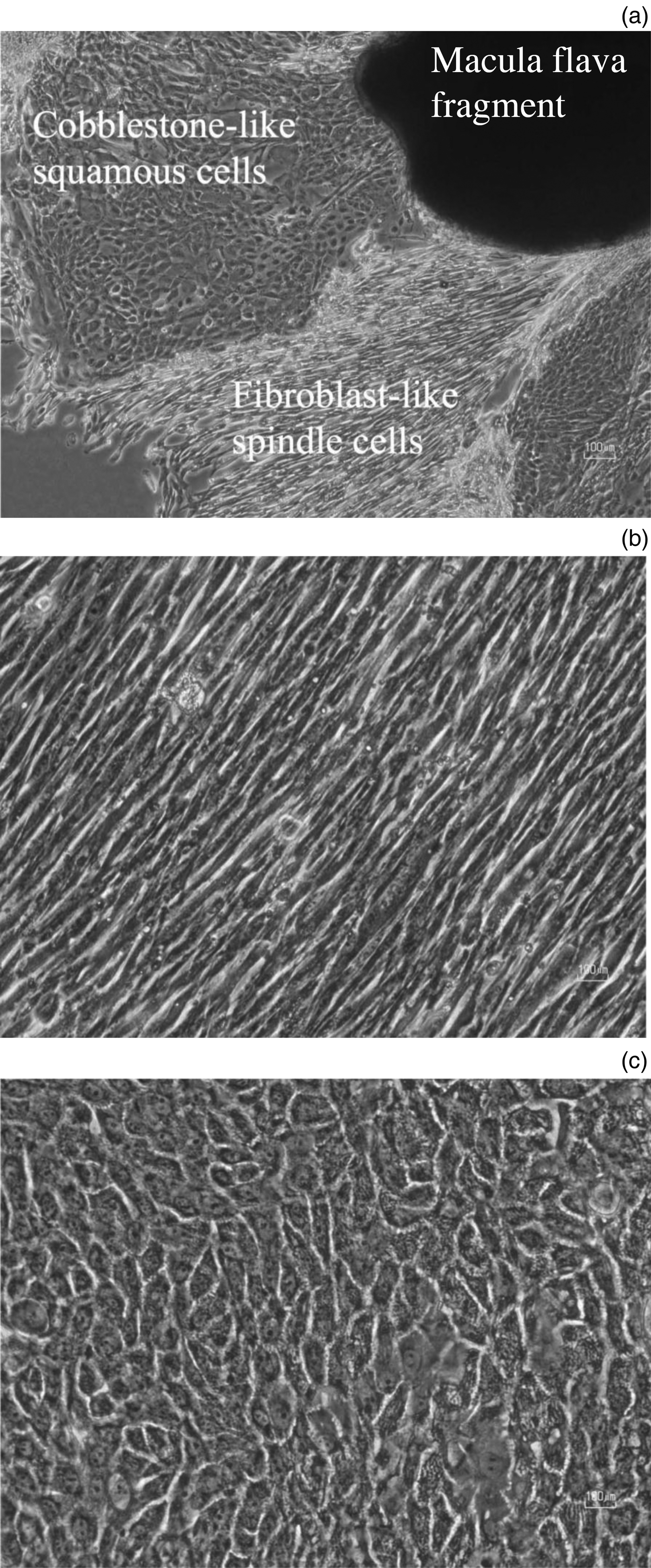
After a few weeks of primary culture in an MF-start primary culture medium (Toyobo), two types of cells, which were fibroblast-like spindle cells (group A) and cobblestone-like squamous cells (group B), grew out from the macula flava fragments (Figures 3a–3c). The cobblestone-like squamous cells were polygonal in shape and had oval-shaped nuclei (Figure 3c). The nucleus-to-cytoplasm ratio was high (Figure 3c).
Primary culture of macula flava with MF-start primary culture medium. (a) Two types of cells, cobblestone-like squamous cells and fibroblast-like spindle cells, grew from the macula flava fragments in the primary culture. (b) Fibroblast-like spindle cells. (c) Cobblestone-like squamous cells.
After removing the two types of cells by cell scraper, each cell was individually subcultured in an MF-medium (mesenchymal stem cell growth medium; Toyobo) to proliferate the cells.
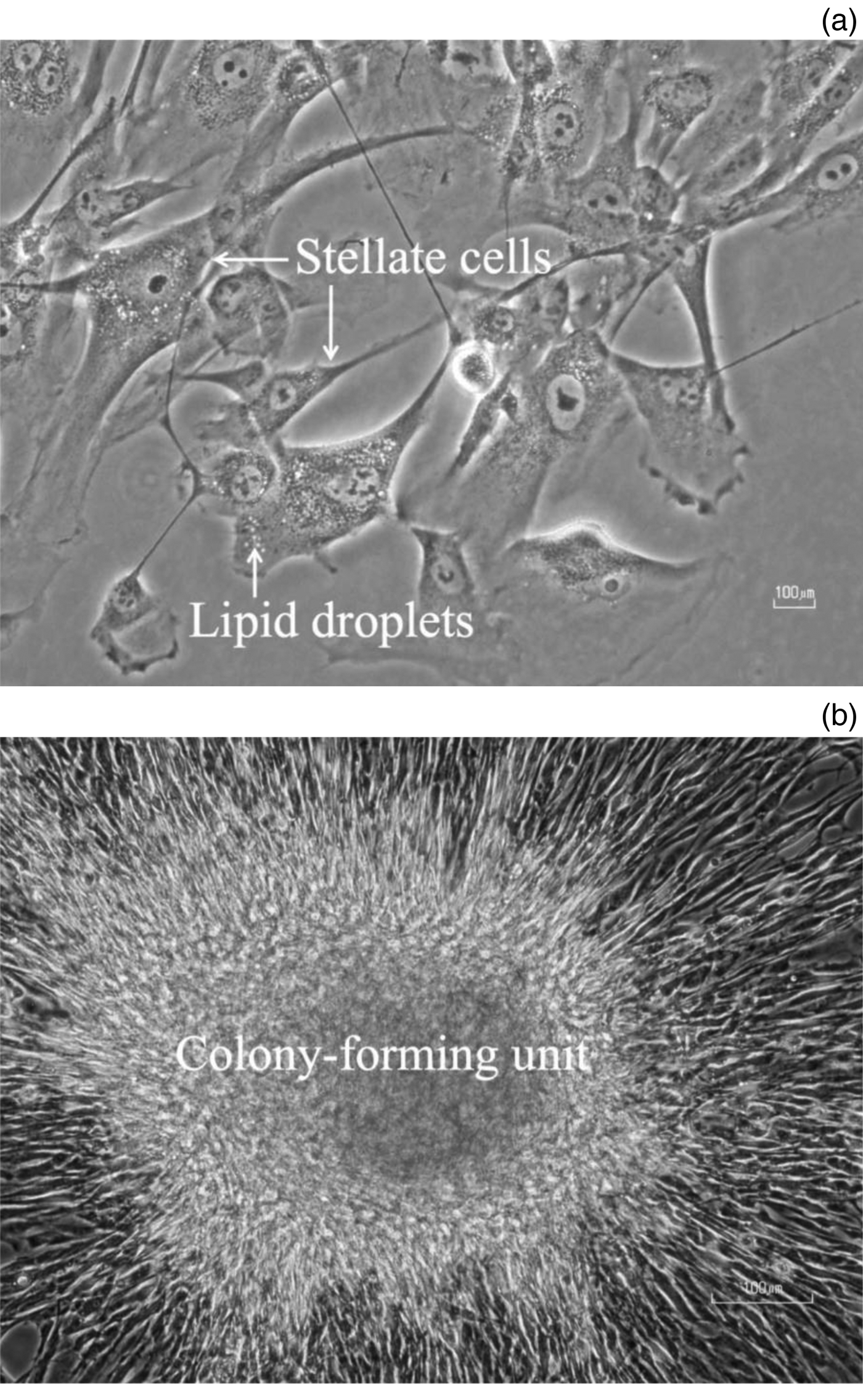
After a week of first subculture, subcultured group A cells became stellate in shape and possessed slender cytoplasmic processes (Figure 4a). Small lipid droplets were present in the cytoplasm (Figure 4a). The nuclei were oval in shape and their nucleus-to-cytoplasm ratios were low. These cells were morphologically similar to vocal fold stellate cells.
(a) Stellate cells. Fibroblast-like cells in the primary culture became stellate in shape and possessed slender cytoplasmic processes and small lipid droplets in the cytoplasm in the subculture. (b) Colony-forming unit. Cobblestone-like squamous cells in an MF-medium formed a colony-forming unit.
After a week of second subculture, subcultured group B cells formed a colony-forming unit (Figure 4b), indicating that these cells are mesenchymal stem cells or stromal stem cells in the bone marrow.
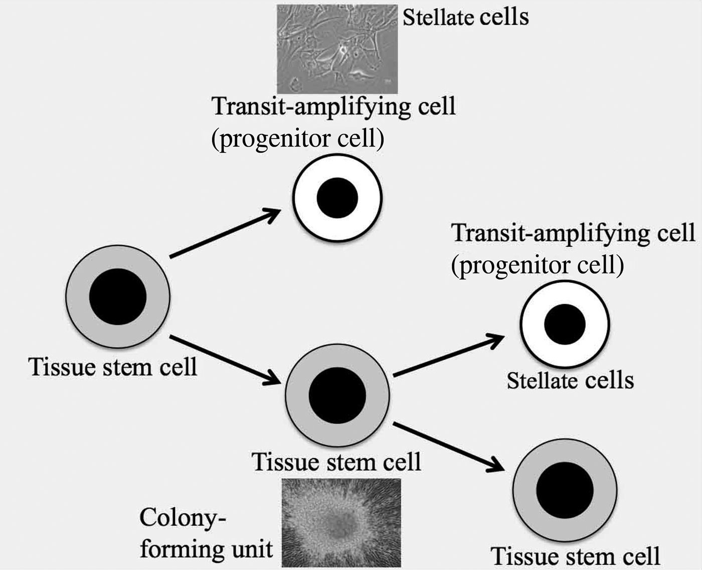
These phenomena suggest that cell division in the human maculae flavae is reflective of asymmetric self-renewal (Figure 5). Asymmetry in cell division gives rise to the possibility that the human maculae flavae contain tissue stem cells (Figure 5). The vocal fold stellate cells are possibly progenitor cells (transit-amplifying cells) (Figure 5).
Asymmetric cell division with MF-medium. Cell division in the human maculae flavae reflected asymmetric self-renewal. One type of cell was tissue stem cells (vocal fold stem cells), which formed a colony-forming unit. The other type was transit-amplifying cells (progenitor cells), whose shape was stellate and similar to the vocal fold stellate cells.
Discussion
Stem cells are a subset of cells that have the unique ability to replenish themselves through self-renewal and the potential to differentiate into different types of mature cells.Reference Xie and Li11 These characteristics therefore play essential roles in organogenesis during embryonic development and tissue regeneration.Reference Xie and Li11
There are two main types of stem cells: embryonic and adult.Reference Xie and Li11 As development proceeds, the need for organogenesis arises, and the embryo proper forms germline stem cells for reproduction and somatic stem cells for organogenesis.Reference Xie and Li11 After birth, adult stem cells, including both germline stem cells and somatic stem cells, reside in a specific microenvironment termed the ‘niche’, which varies in nature and location depending on the tissue type.Reference Xie and Li11 These adult stem cells are an essential component of tissue homeostasis; they support ongoing tissue regeneration, replacing cells lost as a result of natural cell death (apoptosis) or injury.Reference Xie and Li11
Macula flava in vocal fold as stem cell niche
The structural and biochemical microenvironment that confers stemness upon cells in multicellular organisms is referred to as the stem cell niche. The stem cell niche is composed of a group of cells in a special tissue location for the maintenance of stem cells.Reference Xie and Li11
Hyaluronan serves as an important niche component for numerous stem cell populations.Reference Nilsson and Haylock12, Reference Preston and Sherman13 After the discovery of hyaluronan, it was assumed that its major functions were in the biophysical and homeostatic properties of tissues. However, current studies indicate that hyaluronan also plays a crucial role in cell behaviour.Reference Toole and Hay14 A hyaluronan-rich matrix, which is composed of the glycosaminoglycan hyaluronan and its transmembrane receptors (cell surface hyaluronan receptors), are able to directly affect the cellular functions of stem cells in a stem cell niche.Reference Nilsson and Haylock12, Reference Preston and Sherman13
Our past investigation revealed that the vocal fold stellate cells in the human maculae flavae have cell surface hyaluronan receptors and are surrounded by a high concentration of hyaluronan, indicating that the maculae flavae are composed of a hyaluronan-rich matrix.Reference Sato, Umeno and Nakashima10 The maculae flavae can be a candidate for the stem cell niche, which is a microenvironment nurturing a pool of cells including vocal fold stellate cells.
Stemness of cells including vocal fold stellate cells in maculae flavae
Our past investigation revealed that vocal fold stellate cells expressed a mesenchymal stem cell marker.Reference Sato, Umeno and Nakashima10 The vocal fold stellate cells did not express haematopoietic (CD 133) and embryonic stem cell markers (Oct-4).Reference Sato, Umeno and Nakashima10 The vocal fold stellate cells were resting cells (G0 cell cycle phase) like other stem cells are.Reference Sato, Umeno and Nakashima10 Consequently, the cells including vocal fold stellate cells in the human maculae flavae may have multipotency but may not have pluripotency.
A special DNA polymerase called telomerase can catalyse the formation of additional copies of the telomeric repeat sequence, thereby compensating for the gradual shortening that occurs at both ends of the chromosome during DNA replication.Reference Becker, Kleinsmith and Hardin15 In multicellular organisms, telomerase resides mainly in the germ cells that give rise to sperm and eggs, and in a few other kinds of proliferating normal cells such as stem cells.Reference Becker, Kleinsmith and Hardin15 Because telomerase is not found in most cells, the chromosomal telomeres get shorter and shorter with each cell division.Reference Becker, Kleinsmith and Hardin15 The presence of telomerase allows cells to divide indefinitely without telomere shortening.Reference Becker, Kleinsmith and Hardin15 Our past investigation revealed that telomerase resides in the vocal fold stellate cells.Reference Sato, Umeno and Nakashima10
The expression of proteins in the intermediate filaments of the cytoplasm is specific to cell type and differentiation. Intermediate filaments containing cytokeratin (epithelium-associated protein), vimentin (mesenchymal cell associated protein), glial fibrillary acidic protein (neuron-associated protein) and desmin (muscle-associated protein) are distributed in the cytoplasm of the vocal fold stellate cells.Reference Sato, Umeno and Nakashima9, Reference Kurita, Sato, Chitose, Fukahori, Sueyoshi and Umeno16 Therefore, the cells including vocal fold stellate cells in the human maculae flavae are undifferentiated cells, and express the intermediate proteins of ectodermal and mesodermal germ layers.Reference Sato, Umeno and Nakashima9, Reference Kurita, Sato, Chitose, Fukahori, Sueyoshi and Umeno16
The cells including vocal fold stellate cells in the human maculae flavae arise not from resident interstitial cells but from the differentiation of bone marrow cells.Reference Kurita, Sato, Chitose, Fukahori, Sueyoshi and Umeno16 They are undifferentiated cells derived from the bone marrow.Reference Kurita, Sato, Chitose, Fukahori, Sueyoshi and Umeno16
Colony formation is one of the characteristics of stem cells. The colony-forming unit in vitro was first described by Friedenstein et al.Reference Friedenstein, Deriglasova, Kulagina, Panasuk, Rudakowa and Luria17 They established that adherent fibroblastic cells that form cell colonies in vitro in culture can be isolated from the bone marrow stroma. This colony-forming unit can differentiate into cartilage, bone and adipose tissue.Reference Pittenger, Mackay, Beck, Jaiswal, Douglas and Mosca18 Such a colony is also observed in embryonic stem cells,Reference Thomson, Itskovitz-Eldor, Shapiro, Waknitz, Swiergiel and Marshall19 induced pluripotent stem cells,Reference Takahashi, Tanabe, Ohnuki, Narita, Ichisaka and Tomoda20 and tissue stem or progenitor cells such as hepatic stem cellsReference Suzuki, Zheng, Kondo, Kusakabe, Takada and Fukao21 and renal progenitor cells.Reference Osafune, Takasato, Kispert, Asashima and Nishinakamura22 Therefore, the colony-forming phenomenon gives rise to the possibility that the cells, including the vocal fold stellate cells, in the human maculae flavae are tissue stem cells.
As mentioned above, these findings raise the possibility that cells including vocal fold stellate cells in the human maculae flavae are somatic stem cells (tissue-specific resident stem cells) such as mesenchymal stem cells or multipotent mesenchymal stromal cells.
Cell division in human maculae flavae
In the present study, the human macula flava was cultured and the cells were proliferated in either conventional Dulbecco's modified Eagle's medium or mesenchymal stem cell growth medium.
In the Dulbecco's modified Eagle's medium, each cell continued to exhibit the same morphological characteristics during the subculture period. These phenomena suggest that cell division in the human maculae flavae with Dulbecco's modified Eagle's medium is reflective of symmetric self-renewal.
In contrast, in the mesenchymal stem cell growth medium, two types of cells grew. One was stellate in shape, and possessed slender cytoplasmic processes and small lipid droplets in the cytoplasm. These characteristic cell structures show that these cells are vocal fold stellate cells. Another type of cell (cobblestone-like squamous cells) formed a colony-forming unit, indicating these cells are mesenchymal stem cells or stromal stem cells in the bone marrow. These phenomena indicate that the cell division in the maculae flavae with mesenchymal stem cell growth medium is reflective of asymmetric self-renewal.
Asymmetry in the stem cell niche indicates that daughter cells are different from each other.Reference Deasy, Rajasekhar and Vemuri23 There is significant evidence that many stem cell divisions result in one daughter cell that is similar to the parent cell and, hence, necessarily allows for self-renewal of the stem cell phenotype, whereas the other daughter cell is a differentiated or committed cell type.Reference Deasy, Rajasekhar and Vemuri23
Asymmetry in cell division gives rise to the possibility that the maculae flavae in the human vocal fold reflect a stem cell niche containing tissue stem cells.
Here, the question arises whether the vocal fold stellate cells are tissue stem cells or progenitor cells (transit-amplifying cells). The vocal fold stellate cells are possibly transit-amplifying (progenitor) cells. However, at the present stage of our investigation, it is difficult to determine whether the vocal fold stellate cells are tissue stem cells or progenitor cells.
• There is growing evidence that cells including vocal fold stellate cells in the maculae flavae are tissue stem cells of the human vocal fold mucosa
• The evidence suggests that maculae flavae are a candidate for a stem cell niche
• A proper microenvironment in the maculae flavae is necessary to be effective as a stem cell niche that maintains the stemness of the contained tissue stem cells
Future prospects
The manipulation not only of cells but also their microenvironment using chemical biology is one of the strategies in regenerative medicine. Understanding the mechanisms responsible for microenvironmental regulation of vocal fold stellate cells in the human maculae flavae will provide the tools needed to manipulate vocal fold stellate cells through their microenvironment for the development of therapeutic approaches to diseases and tissue injuries. Translational medicine concerning how to regulate cells and extracellular matrices (microenvironment) contained in the maculae flavae of vocal folds will contribute to our ability to restore and regenerate human vocal fold tissue.
Conclusion
The results of this study are consistent with the hypothesis that the cells including the vocal fold stellate cells in the human maculae flavae are tissue stem cells or progenitor cells (transit-amplifying cells) of the human vocal fold mucosa.
A proper microenvironment in the maculae flavae of the human vocal fold mucosa is necessary to be effective as a stem cell niche that maintains the stemness of the contained tissue stem cells.
Acknowledgement
This investigation was supported by a Grant-in-Aid for Scientific Research (number 24592612) from the Japanese Ministry of Education, Culture, Sports, Science and Technology.